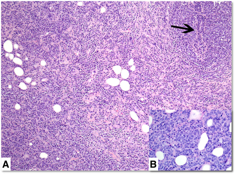
Figure 3. Hematoxylin-eosin stain of the core biopsy specimen.
Hematoxylin-eosin stain with original ×100 magnification (A) and right insert with original ×400 magnification (B). There is a dense neoplastic mononuclear infiltrate with small foci of residual benign ducts (arrow). Tumor cells have round to oval nuclei, dispersed chromatin, and occasional small nucleoli (B).

