Abstract
[Purpose] A case series is featured demonstrating reduction of curvature in three adult patients who presented with a mild to moderate severity of a uniquely high thoracic curvature clinical presentation. [Participants and Methods] Three adult patients who presented with an upper thoracic scoliosis deformity of mild to moderate severity underwent Chiropractic BioPhysics® treatment protocols to treat their deformity. Radiographic stress imaging was performed to correctly position and ascertain potential treatment effect of the Denneroll spinal orthotic device. Patients performed spinal traction for 10–20 minutes daily with intermittent spinal manipulative therapy. [Results] There was a 4.5° average reduction in computerized Cobb angle measurement after treatment. All patients reported reductions in spinal pain and also reported subjective improvements in sleep quality and quality of life. [Conclusion] Mild reductions in uniquely high thoracic curves can be reduced in adult scoliosis patients with mild to moderate (17°–26°) curve magnitudes by CBP treatment protocols. Stress X-ray images are recommended to properly place the fulcrum and assess correction potential.
Key words: Scoliosis, Rehabilitation, Thoracic spine
INTRODUCTION
Scoliosis is a lateral deviation of the spine in the coronal plane with rotation of the affected vertebrae1). The incidence of adolescent idiopathic scoliosis is up to 5%2); many patients with mild curvature go undiagnosed until later in life when discovered by X-ray for spine complaints. Any spinal displacement from symmetry will biomechanically induce asymmetrical loading of the spinal tissues. Therefore, even mild scoliosis deformity may, over time, result in pathological consequences including degenerative changes and pain3), thus, patients who present with mild scoliosis curves and spine complaints require treatment for their deformity.
Although severe spinal curvature may require surgical placement of Harrington rods for its reduction, smaller curves are treatable by non-surgical rehabilitation methods4). In recent literature, several reports have shown that scoliosis rehabilitation programs may provide successful outcome in those with scoliosis5,6,7,8,9). One method showing initial evidence of reducing spinal curvature is the procedures of Chiropractic BioPhysics® (CBP®) technique methods8,9,10). In one case series it was shown that lumbar and thoracolumbar spinal curves could be reduced in adults8). Another case showed the reduction of a thoracic curve in an adolescent9). The commonality of the CBP approach is to apply rehabilitation practices to reverse the spinal curve; that is, to stress the spine in the opposite direction (i.e. mirror image®)8,9,10).
CBP technique was invented and developed throughout the 1980s and is a full-spine approach to improving spine alignment and posture11, 12). CBP methods include mirror image postural adjusting, exercises and spinal traction techniques to remodel the spine towards its ideal position11, 12). Typically, spinal X-rays are performed prior to and after a series of treatments to document spine alignment improvements. CBP methods have been shown to increase cervical13) and lumbar lordosis14), reduce thoracic kyphosis15), reduce lateral thoracic16) and head17) translation postures as well as many other postural deformities (subluxations)18).
Here, we feature a case series demonstrating reduction of curvature in three adult patients who present with a mild to moderate severity of a uniquely high thoracic curvature clinical presentation.
PARTICIPANTS AND METHODS
A retrospective case series of three adult patients with scoliosis displaying Cobb angles19) ranging from 16° to 26° in the upper thoracic spine region are presented (Table 1). All three patients were female having ages of 51, 35 and 37, heights of 165 cm, 167.5 cm and 164 cm, and weights of 59.9 kg, 47.6 kg and 63.5 kg, respectively. The scoliosis affected vertebrae in all patients ranged from T1-T6. The treatment procedures in all three cases were in accordance with CBP protocols10). All three individuals were treated with limited cervical and thoracic spinal manipulative therapy (SMT) and prescribed daily treatment with a small lumbar Denneroll spinal traction block (Denneroll Spinal Orthotics, Wheeler Heights, NSW, Australia).
Table 1. Patient age, gender, vertebrae affected as well as Cobb angles.
| Age (years) | Gender | Vertebrae affected | Initial | f/u | Change | Stress view | |
| Case 1 | 51 | F | T1-T5 | 25.9° | 21.9° | 4.0° | 8.2° |
| Case 2 | 35 | F | T3-T6 | 21.0° | 16.2° | 4.8° | 8.5° |
| Case 3 | 37 | F | T1-T5 | 16.7° | 12.0° | 4.7° | −4.6° |
| Average | 41 | - | - | 21.2° | 16.7° | 4.5° | 4.0° |
F: female; f/u: follow-up.
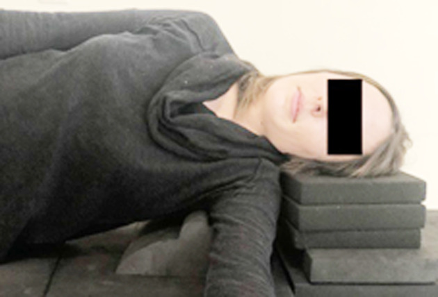
The CBP approach (Fig. 1) required the patients to lay on the Denneroll on the side of their scoliotic convexity so that the traction block would force a reversal of their corresponding upper thoracic curve (i.e. the Cobb angles would be stressed towards the reversed direction during the procedure). To avoid undue neck strain, patients were advised to place their head on a soft support such as a pillow if necessary. Patients were to lay on their side with their knees bent to 90° with their knees and ankles on top of each other. The lower arm was placed forward and the upper arm was simply relaxed on their upper side torso. The shoulders were allowed to be rotated slightly such that the upper shoulder was back and the lower shoulder was forward of vertical midline from the base of the neck. Instructions regarding the traction consisted of the following criteria: 1) Patients were to perform the traction once a day until their next scoliosis check-up and 2) the traction was to be performed for a minimum of 10 minutes (maximum of 20 minutes) due to achieving maximum viscoelastic creep in the ligamentous tissues20).
Fig. 1.
Mirror image traction blocking. The patient lays over the block placing it approximately in the armpit. The lower arm shoots forward, the legs are bent to 90° and the shoulders are slightly rotated back on the upper side. The head is relaxed, but supported only enough to remove any strain to the neck. Patient remains relaxed for 10–20 minutes per session.
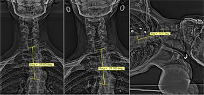
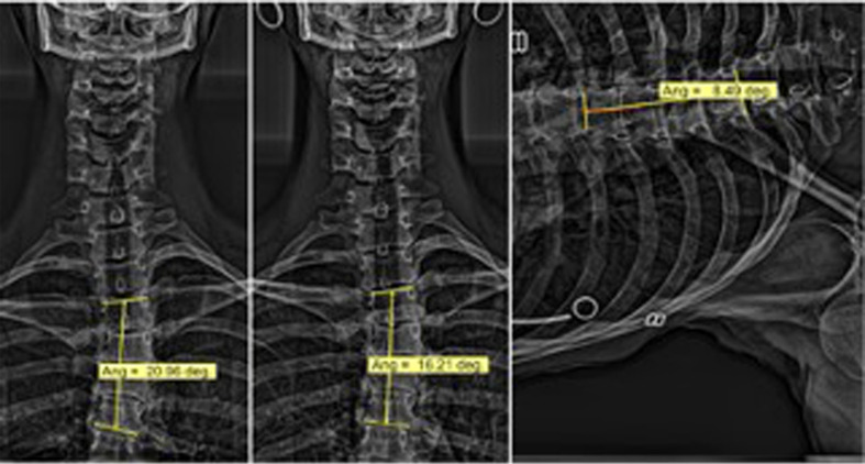
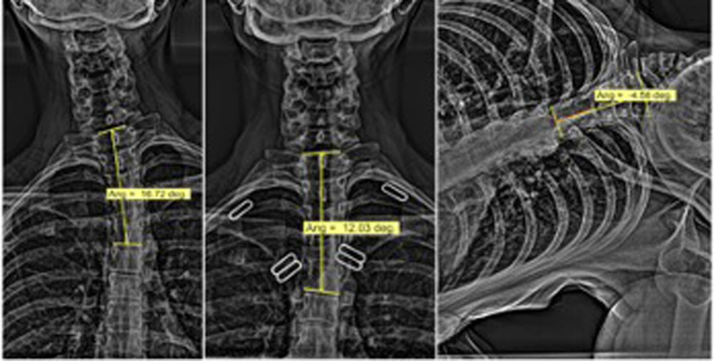
Three radiographic images are presented for each patient (Figs. 2–4). The first and second radiographic images represent the initial and follow-up anterior-posterior (AP) cervicothoracic views taken in the standing, bare foot position. The third image is a ‘stress view’ of the patient laying on the Denneroll orthotic in the desired position; that is, placing the spinal convexity downward over the block. Spinal curve measurement was made in the Metron software using the Cobb angle11) which measures the intersection of the lines drawn perpendicular to the upper involved vertebral endplate and the lower involved vertebral endplate. All three patients gave verbal and written consented to publish their clinical outcomes.
RESULTS
A 51 year-old female presented with neck and mid-thoracic back pain. Her primary complaint was her neck pain, which she rated an average of 5.5/10 on the numeric pain rating scale (NPRS: 0=no pain; 10=severe pain with the patient bed ridden); however, the pain could increase to a 9/10 on certain days. The patient’s second complaint was pain in the cervico-thoracic area, rated as a 4.5/10 NPRS. Consequently, the patient experienced pain related interrupted sleep which resulted in a lower quality of life. The patient experienced a reduced active cervical range of motion (ROM) in all directions with pain elicited with flexion-extension and bilateral rotation.
AP radiographic images of the cervico-thoracic vertebrae demonstrated a left scoliosis with a Cobb angle of 25.9° from T1-T5 with the apex at T3 (Fig. 2). No vertebral abnormalities were noted. A stress image was taken with the patient laying on their right over the small lumbar Denneroll traction orthotic to mirror-image the left convexity which reduced the angle to 8.2° (Fig. 2). A follow-up radiograph was taken after 3 months of treatment showing an angle of 21.9°, a 4° reduction in scoliosis (Fig. 2). Assessment at follow-up indicated the patient’s NPRS reduced to 3.5. In addition, the patient reported an improved quality of sleep and an overall better quality of life.
Fig. 2.
Anterior-posterior (AP) cervicothoracic views of Case 1. Left: A left T1-T5 scoliosis Cobb angle of 25.9°. Middle: Post-treatment Cobb angle of 21.9°. Right: A stress image of patient laying on their left to mirror-image their curve over the small lumbar Denneroll orthotic showing a Cobb angle reducing to 8.2°.
A 35 year-old female presented with chronic neck, upper and mid back pains. Notably, the primary complaint was at the base of the neck described as a numbing pain which was debilitating as it hindered the patient’s ability to sit and stand with proper posture (‘standing up straight’). The patient also suffered a reduction in her well-being and quality of life from having an interrupted sleep schedule which also led to midday exhaustion, irritability and depression. The patient demonstrated a reduction in active cervical ROM in all planes with pain elicited during sagittal plane movements.
AP radiographic images of the cervico-thoracic spine showed a moderate scoliotic curve measuring a Cobb angle of 21° from T3-T6 (Fig. 3). The patient’s scoliotic curve presented a leftward convexity correlated with an apex at T4. No vertebral abnormalities were present. A stress image featuring the patient laying with their left side on the denneroll to reduce the leftward convexity showed the Cobb angle reduced from 21° to 8.5° (Fig. 3). After one year, the patient showed a Cobb angle of 16.2°, demonstrating a 4.8° reduction.
Fig. 3.
Anterior-posterior (AP) cervicothoracic views of Case 2. Left: An initial left scoliosis Cobb angle of 21°. Middle: A post-treatment Cobb angle of 16.2°. Right: A stress image of patient laying on their left over the small lumbar Denneroll orthotic to mirror image their left convexity showing the Cobb angle reducing to 8.5°.
Assessment upon follow up showed the patient’s active cervical ROM had significantly improved. Proper posture (‘standing up straight’) was now easily attainable for the patient and the chronic back and neck pains were significantly reduced. The patient stated that on occasion she “forgot I even had scoliosis”. The symptoms of irritability, midday exhaustion and depression were eventually alleviated because she was able to sleep without interruption from pain.
A 37 year-old female patient presented with a primary complaint of right-sided chronic cervicothoracic pain. The patient rated her pain as a 6/10 NPRS. There was reported numbness and tingling of her fourth and fifth digits on her right hand and right sided stiffness in the upper and lower back. The patient experienced occasional neck pain which often transitioned into a long-lasting headache and a lack of balance. Due to the pain, the patient suffered from frequent sleep disturbances, irritability, depression and decreased productivity. Active cervical ROM was globally reduced.
AP radiographs displayed a left T1-T5 upper thoracic scoliosis with a measured Cobb angle of 16.7° with an apex at T4 (Fig. 4). Stress image of the patient showed a reversal of the scoliosis, and a 6-month follow-up image showed a 4.7° reduction in magnitude (16.7° to 12°) (Fig. 4).
Fig. 4.
Anterior-posterior (AP) cervicothoracic views of Case 3. Left: Initial view showing a Cobb angle of 16.7°. Centre: Post-treatment showing the reduction of the Cobb angle to 12°. Right: A stress image of patient laying on their left over the small lumbar Denneroll orthotic to mirror image their left convexity showing the Cobb angle reducing to −4.6° or showing a reversed convexity.
At follow-up assessment, the patient’s active ROM had significantly increased in all directions as well as a reduction of pain upon movement. The frequency of numbness and tingling in the fourth and fifth digits had also been significantly reduced. Due to the reduction of her neck and back pains the patient experienced improved sleep, increased productivity and a lack of depression and irritability.
DISCUSSION
The case series has demonstrated that mild reductions in uniquely high thoracic curves can be reduced in adult scoliosis patients with mild to moderate (17°–26°) curve magnitudes by CBP treatment protocols. All patients reported reductions in spinal pain and also reported subjective improvements in sleep quality and quality of life. This case series adds to the limited evidence that scoliosis-specific treatment approaches may lead to reduction of scoliosis curves in adult patients. Regarding the treatment of high thoracic deformities, we were unable to find a single case describing its reduction by spine rehabilitative methods in adults. Thus, this series serves as the first evidence of improving scoliosis curves in adults with a high thoracic presentation that we are aware of.
Importantly, it should be noted that smaller scoliosis curves are not immune to progression, even in adults21). In fact, Stokes22) argues that if scoliotic curves progress from asymmetric biomechanical loading, then the shearing force component would be the driving factor for progression. Therefore, any magnitude of reduction to a spinal curve that lessens the shearing force vector would result in the spine becoming more robust to progression. We concur with Harrison and Oakley that “any conservative approach to successfully better balance the scoliotic spine would be beneficial regardless of age, at least biomechanically”8).
The CBP treatment protocol employed here uses a mirror image concept; that is, to stress the spine opposite of its presenting deformity. This has been pointed out as a common theme in successful scoliosis treatment strategies23).
In fact, as proven in clinical trials on adolescent idiopathic scoliosis patients6, 7), ‘patient-specific’ customized exercise programs always outperform ‘conventional’ exercise programs that do not prescribe treatment that is specific to the spine deformity. This trend is also observed in the treatment of other spine deformities such as increasing cervical and lumbar hypolordosis, where customized rehabilitation treatment that is radiographically-guided results in better long-term patient outcomes13, 14).
The reliability of measuring scoliosis curves on an X-ray is critical in the management of scoliosis patients24); thus, measurement error considerations are important. Although the Cobb method for measuring scoliosis has been traditionally reported as having large variability,25) this stems from early studies using the 4-line manual method. Computer-based mensuration affords a means for much more precise measures, for example <3°26) or <2°27). In fact, Srinivasalu et al. found the intra-observer variability in digitizing 318 radiographs was 1.3°27). Further, one study found that diurnal variation throughout the day may lead to changes in scoliotic measures within the same patient28), but this was in very large curves (65°) and others did not find that occurring in patients with smaller curves.29) One last point is that intra-observer reliability in Cobb angle use is very high and most variability in measuring scoliosis curves are introduced by using different endpoint vertebra30), of which neither was a factor in this study. Thus, our use of the Cobb method of measurement documenting modest but significant reductions in scoliosis patients (average change=4.5°) are valid.
One important consideration in treating high thoracic scoliotic curves is the differentiation from lateral head translation postures which creates an S-curve in the cervicothoracic spine or a so-called ‘pseudo-scoliosis’31). Lateral head translation causes a lateral bend of the upper thoracic spine towards the head shift and an opposite lateral bend of the upper cervical spine towards the vertical, however, the patients included in this report did not demonstrate this classic spinal coupling and were determined to have a mild upper thoracic scoliosis with simultaneous lateral head translation away from the convexity of the curve.
Limitations to this case series include a lack of long-term follow-up. Another limitation is whether the results are generalizable due to the small sample size, limited age group and that only females were treated. Further study of the effects of reversing upper thoracic scoliotic deformities are warranted as smaller curves that are of mild to moderate magnitudes of severity are non-surgical cases that are amenable to rehabilitation methods. Since most of the scoliosis treatment literature focuses on children and adolescents, there is a need for further research on the treatment efficacy in adults.
Conflicts of interest
Dr. Paul Oakley (PAO) is a paid consultant for CBP NonProfit, Inc.; Dr. Deed Harrison (DEH) teaches chiropractic rehabilitation methods and sells products to physicians for patient care as described in this manuscript.
REFERENCES
- 1.Berven S, Bradford DS: Neuromuscular scoliosis: causes of deformity and principles for evaluation and management. Semin Neurol, 2002, 22: 167–178. [DOI] [PubMed] [Google Scholar]
- 2.Konieczny MR, Senyurt H, Krauspe R: Epidemiology of adolescent idiopathic scoliosis. J Child Orthop, 2013, 7: 3–9. [DOI] [PMC free article] [PubMed] [Google Scholar]
- 3.Harrison DD, Troyanovich SJ, Harrison DE, et al. : A normal sagittal spinal configuration: a desirable clinical outcome. J Manipulative Physiol Ther, 1996, 19: 398–405. [PubMed] [Google Scholar]
- 4.Oakley PA, Ehsani NN, Harrison DE: The scoliosis quandary: are radiation exposures from repeated x-rays harmful? Dose Response, 2019, 17: 1559325819852810. [DOI] [PMC free article] [PubMed] [Google Scholar]
- 5.Kuru T, Yeldan İ, Dereli EE, et al. : The efficacy of three-dimensional Schroth exercises in adolescent idiopathic scoliosis: a randomised controlled clinical trial. Clin Rehabil, 2016, 30: 181–190. [DOI] [PubMed] [Google Scholar]
- 6.Noh DK, You JS, Koh JH, et al. : Effects of novel corrective spinal technique on adolescent idiopathic scoliosis as assessed by radiographic imaging. J Back Musculoskeletal Rehabil, 2014, 27: 331–338. [DOI] [PubMed] [Google Scholar]
- 7.Monticone M, Ambrosini E, Cazzaniga D, et al. : Active self-correction and task-oriented exercises reduce spinal deformity and improve quality of life in subjects with mild adolescent idiopathic scoliosis. Results of a randomised controlled trial. Eur Spine J, 2014, 23: 1204–1214. [DOI] [PubMed] [Google Scholar]
- 8.Harrison DE, Oakley PA: Scoliosis deformity reduction in adults: a CBP® Mirror Image® case series incorporating the ‘non-commutative property of finite rotation angles under addition’ in five patients with lumbar and thoraco-lumbar scoliosis. J Phys Ther Sci, 2017, 29: 2044–2050. [DOI] [PMC free article] [PubMed] [Google Scholar]
- 9.Haggard JS, Haggard JB, Oakley PA, et al. : Reduction of progressive thoracolumbar adolescent idiopathic scoliosis by chiropractic biophysics® (CBP®) mirror image® methods following failed traditional chiropractic treatment: a case report. J Phys Ther Sci, 2017, 29: 2062–2067. [DOI] [PMC free article] [PubMed] [Google Scholar]
- 10.Harrison DE, Betz JW, Harrison DD, et al. : CBP structural rehabilitation of the lumbar spine. Harrison Chiropractic Biophysics Seminars, Inc. 2007.
- 11.Harrison DD, Janik TJ, Harrison GR, et al. : Chiropractic biophysics technique: a linear algebra approach to posture in chiropractic. J Manipulative Physiol Ther, 1996, 19: 525–535. [PubMed] [Google Scholar]
- 12.Oakley PA, Harrison DD, Harrison DE, et al. : Evidence-based protocol for structural rehabilitation of the spine and posture: review of clinical biomechanics of posture (CBP) publications. J Can Chiropr Assoc, 2005, 49: 270–296. [PMC free article] [PubMed] [Google Scholar]
- 13.Oakley PA, Ehsani NN, Moustafa IM, et al. : Restoring cervical lordosis by cervical extension traction methods in the treatment of cervical spine disorders: a systematic review of controlled trials. J Phys Ther Sci, 2021, 33: 784–794. [DOI] [PMC free article] [PubMed] [Google Scholar]
- 14.Oakley PA, Ehsani NN, Moustafa IM, et al. : Restoring lumbar lordosis: a systematic review of controlled trials utilizing Chiropractic Bio Physics® (CBP®) non-surgical approach to increasing lumbar lordosis in the treatment of low back disorders. J Phys Ther Sci, 2020, 32: 601–610. [DOI] [PMC free article] [PubMed] [Google Scholar]
- 15.Oakley PA, Harrison DE: Reducing thoracic hyperkyphosis subluxation deformity: a systematic review of chiropractic BioPhysics® methods employed in its structural improvement. J Contemp Chiropr, 2018, 1: 59–66. [Google Scholar]
- 16.Oakley PA, Harrison DE: A review of CBP methods applied to reduce lateral thoracic translation (pseudo-scoliosis) postures. J Contemp Chiro, 2022, 5: 13–18. [Google Scholar]
- 17.Oakley PA, Harrison DE: A review of CBP methods applied to reduce lateral head translation postures. J Contemp Chiro, 2022, 5: 19–24. [Google Scholar]
- 18.CBP NonProfit Publications. https://www.cbpnonprofit.com (Accessed May 5, 2022)
- 19.Cobb JR: Outline for the study of scoliosis. Instr Course Lect, 1948, 5: 261–275. [Google Scholar]
- 20.Oliver MJ, Twomey LT: Extension creep in the lumbar spine. Clin Biomech (Bristol, Avon), 1995, 10: 363–368. [DOI] [PubMed] [Google Scholar]
- 21.Stehbens WE, Cooper RL: Regression of juvenile idiopathic scoliosis. Exp Mol Pathol, 2003, 74: 326–335. [DOI] [PubMed] [Google Scholar]
- 22.Stokes IA: Analysis of symmetry of vertebral body loading consequent to lateral spinal curvature. Spine, 1997, 22: 2495–2503. [DOI] [PubMed] [Google Scholar]
- 23.Oakley PA: Is early treatment for mild adolescent idiopathic scoliosis superior over the traditional ‘watch & wait’ approach? A case report with long-term follow-up. J Phys Ther Sci, 2018, 30: 680–684. [DOI] [PMC free article] [PubMed] [Google Scholar]
- 24.Carman DL, Browne RH, Birch JG: Measurement of scoliosis and kyphosis radiographs. Intraobserver and interobserver variation. J Bone Joint Surg Am, 1990, 72: 328–333. [PubMed] [Google Scholar]
- 25.Harrison DE, Betz JW, Cailliet R, et al. : Radiographic pseudoscoliosis in healthy male subjects following voluntary lateral translation (side glide) of the thoracic spine. Arch Phys Med Rehabil, 2006, 87: 117–122. [DOI] [PubMed] [Google Scholar]
- 26.Morrissy RT, Goldsmith GS, Hall EC, et al. : Measurement of the Cobb angle on radiographs of patients who have scoliosis. Evaluation of intrinsic error. J Bone Joint Surg Am, 1990, 72: 320–327. [PubMed] [Google Scholar]
- 27.Srinivasalu S, Modi HN, Smehta S, et al. : Cobb angle measurement of scoliosis using computer measurement of digitally acquired radiographs-intraobserver and interobserver variability. Asian Spine J, 2008, 2: 90–93. [DOI] [PMC free article] [PubMed] [Google Scholar]
- 28.Beauchamp M, Labelle H, Grimard G, et al. : Diurnal variation of Cobb angle measurement in adolescent idiopathic scoliosis. Spine, 1993, 18: 1581–1583. [DOI] [PubMed] [Google Scholar]
- 29.Zetterberg C, Hansson T, Lindström J, et al. : Postural and time-dependent effects on body height and scoliosis angle in adolescent idiopathic scoliosis. Acta Orthop Scand, 1983, 54: 836–840. [DOI] [PubMed] [Google Scholar]
- 30.Gstoettner M, Sekyra K, Walochnik N, et al. : Inter- and intraobserver reliability assessment of the Cobb angle: manual versus digital measurement tools. Eur Spine J, 2007, 16: 1587–1592. [DOI] [PMC free article] [PubMed] [Google Scholar]
- 31.Harrison DE, Harrison DD, Cailliet R, et al. : Cervical coupling during lateral head translations creates an S-configuration. Clin Biomech (Bristol, Avon), 2000, 15: 436–440. [DOI] [PubMed] [Google Scholar]


