Correction to: World Journal of Pediatrics (2020) 16:502–513 10.1007/s12519-019-00268-w
The originally published version of this article contained some errors in (1) the graphs of Figs. 2c, 2f–j, 4, and 6b; (2) the legends of Figs. 1–4, and 6. The corrected figures and legends are given in this Correction.
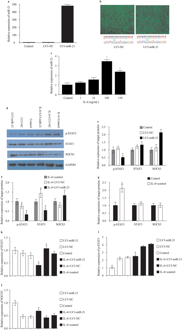
Fig. 2 Transfection of LV3-miR-21 suppressed p-STAT3/STAT3 protein expression. a, b The titer of miR-21 in RASFs after LV3-miR-21 transfection; c OD value of RASFs with the simulation of IL-6 (*P < 0.05, IL-6 100 ng/mL vs. control group, IL-6 150 ng/mL vs. control group); d–j The expression of STAT3/p-STAT3/SOCS3 in RASFs after transfection of LV3-miR-21 with the simulation of IL-6 (d
P < 0.05, IL-6 + LV3-miR-21 vs. IL-6 + LV3-NC group; e *P < 0.05, LV3-miR-21 vs. LV3-NC group, f *P < 0.05, IL-6 + LV3-NC vs. IL-6 + control group, †P < 0.05, IL-6 + LV3-miR-21 vs. IL-6 + LV3-NC group; g *P < 0.05, IL-6 + control vs. control group; h *P < 0.05, IL-6 + LV3-miR-21 vs. IL-6 + control group, IL-6 + LV3-NC vs. IL-6 + control group; i *P < 0.05, LV3-miR-21 vs. control group, †P < 0.05, IL-6 + LV3-miR-21 vs. LV3-miR-21 group; j *P < 0.05, IL-6 + LV3-miR-21 vs. LV3-miR-21 group). STAT3 signal transducers and activators of transcription 3, SOCS3 suppressor of cytokine signaling 3, IL-6 interleukin-6, RASF rheumatoid arthritis fibroblast-like synovial cell, NC negative control.
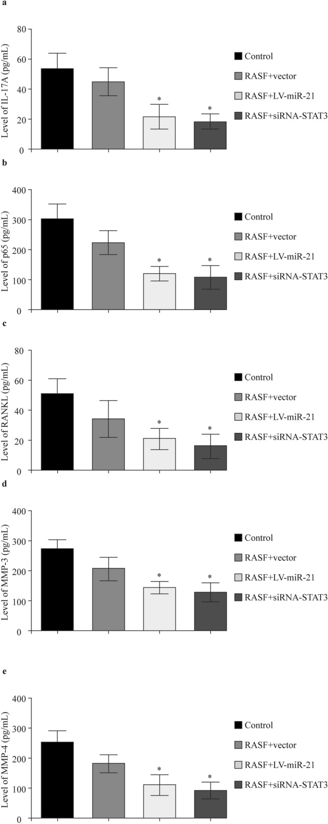
Fig. 4 Enhancing miR-21 or silencing STAT3 could suppress the expression of IL-17A (a), p65 (b), RANKL (c), MMP-3 (d), and MMP-4 (e). STAT3 signal transducers and activators of transcription 3, IL interleukin, RASF rheumatoid arthritis fibroblast-like synovial cell, MMP matrix metalloproteinase, RANKL receptor activator of nuclear factor-κB ligand. *P < 0.05, RASF + LV-miR-21 vs. RASF + vector group, RASF + siRNA-STAT3 vs. RASF + vector group
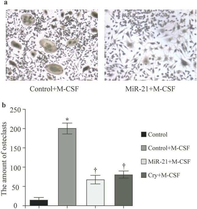
Fig. 6 M-CSF was used for inducing the RASFs to osteoclasts. a The TRAP staining results showed that the amounts of osteoclasts in miR-21 mimics group were significantly lower than the control group (P < 0.05, miR-21 + M-CSF vs. control + M-CSF group); b Both miR-21 mimics and cryptotanshinone (cry) could decrease the amount of osteoclasts with no difference between these two groups (*P < 0.05, control + M-CSF vs. control group, †P < 0.05, miR-21 + M-CSF vs. control + M-CSF group, cry + M-CSF vs. control + M-CSF group). RASF rheumatoid arthritis fibroblast-like synovial cell, M-CSF macrophage colony-stimulating factor
The corrected legends of Figs. 1 and 3 are as follows:
Fig. 1 Expression of miR-21 (a), STAT3 (b) and SOCS3 (c) mRNA of PBMCs and their correlation (d, e) in JIA. sJIA systemic juvenile idiopathic arthritis, pJIA polyarticular juvenile idiopathic arthritis, STAT3 signal transducers and activators of transcription 3, SOCS3 suppressor of cytokine signaling 3, PBMCs peripheral blood mononuclear cells. *P < 0.05, sJIA vs. control group, pJIA vs. control group
Fig. 3 Enhancing miR-21 or silencing STAT3 could suppress the IL-6 induced regulation of MMP-3 (a), MMP-4 (b), RANKL (c), and NF-κb (d). STAT3 signal transducers and activators of transcription 3, IL-6 interleukin-6, RASF rheumatoid arthritis fibroblast-like synovial cell, MMP matrix metalloproteinase, RANKL receptor activator of nuclear factor-κB ligand, NF-κb nuclear factor-κB. *P < 0.05, RASF + LV-miR-21 vs. RASF + vector group, RASF + siRNA-STAT3 vs. RASF + vector group
In the paragraph under the section “Results—Transfection of LV3-miR-21 suppressed p-STAT3/STAT3 protein expression”: The sentence “Then RASFs were stimulated with IL-6 (0, 10, 100, 150 µg/mL) for 4 h (Fig. 2c)” should read as “Then RASFs were stimulated with IL-6 (0, 10, 100, 150 ng/mL) for 4 h (Fig. 2c)”.


