Abstract
The 2019 coronavirus disease (COVID-19) pandemic has demonstrated the importance of predicting, identifying, and tracking mutations throughout a pandemic event. As the COVID-19 global pandemic surpassed one year, several variants had emerged resulting in increased severity and transmissibility. Here, we used PCR as a surrogate for viral load and consequent severity to evaluate the real-world capabilities of a genome-based clinical severity predictive algorithm. Using a previously published algorithm, we compared the viral genome-based severity predictions to clinically derived PCR-based viral load of 716 viral genomes. For those samples predicted to be “severe” (probability of severe illness >0.5), we observed an average cycle threshold (Ct) of 18.3, whereas those in in the “mild” category (severity probability <0.5) had an average Ct of 20.4 (P=0.0017). We also found a nontrivial correlation between predicted severity probability and cycle threshold (r = −0.199). Finally, when divided into severity probability quartiles, the group most likely to experience severe illness (≥75% probability) had a Ct of 16.6 (n = 10), whereas the group least likely to experience severe illness (<25% probability) had a Ct of 21.4 (n = 350) (P=0.0045). Taken together, our results suggest that the severity predicted by a genome-based algorithm can be related to clinical diagnostic tests and that relative severity may be inferred from diagnostic values.
1. Introduction
A classic model of disease causation is the epidemiologic triad of host, agent, and environment. Stated differently, the severity of an illness is based on an interplay between the method of exposure (environment), pathogenicity of the organism (agent), and the host (host susceptibility and host response to the infectious agent). The recent SARS-CoV-2 pandemic has demonstrated that substantial diversity in both the host and virus can lead to a wide spectrum of clinical outcomes. Early research into symptom severity largely focused on host phenotypes, such as blood type [1], age [2], and gender [3]. As the scale of the pandemic grew [4], the role of the geographic region and viral mutations in severe clinical outcomes began to emerge [5], followed by additional insights on host genetic susceptibility [6]. Finally, efforts to predict a patient's outcomes using computational models developed with phenotypic, genetic, and demographic data are being pursued in order to tailor patient care and manage resources [7–10].
Early identification of patients at the increased risk of developing severe symptoms can help preserve life and health. Estimates of viral load upon admission have been shown to be correlated with higher mortality [11, 12]. Using real-time PCR data, Choudhuri and colleagues demonstrated that increased cycle thresholds were associated with 9% reduction in the odds of in-hospital mortality [11]. The greatest difference was found for those patients reporting to the hospital with a cycle threshold below 23; these patients encountered 3.9-fold increased odds of in-hospital mortality compared to patients with cycle thresholds above 33. Overall, using PCR cycle thresholds as a predictor of outcome, the area under the curve was found to be 0.68, suggesting an effective but limited discriminative ability for severity classification by cycle threshold at the time of admission.
The use of SARS-CoV-2 genome-wide sequencing has identified numerous variants that the World Health Organization has subsequently declared “variants of concern” [13]. Many of these variants are in the spike protein necessary for host recognition [14], but many are also scattered throughout the remainder of the viral genome [15]. Using data from the first year of the pandemic, we previously developed an algorithm to predict severity based on viral mutations [7]. In addition, we reported 17 variants associated with severe clinical outcomes (odds ratio (OR) ≥ 2) and 67 variants associated with mild clinical outcomes (OR ≤ 0.5). The area under the curve for our predictive algorithm was 0.91, suggesting a strong discriminative ability for classifying severe patients. Here, we present results comparing the computationally predicted probability of a severe outcome to real-world laboratory-measured PCR values.
2. Materials and Methods
2.1. Patient Sample Selection
This study was reviewed and approved by the Air Force Research Laboratory's Institutional Review Board (FWR20190037N). Genome sequences were downloaded from the public access GISAID database [16, 17] restricted to sequences uploaded by the US Air Force School of Aerospace Medicine (USAFSAM) as of 16 April 2021 (see Supplementary Materials for a complete list of accession numbers and references). Only the USAFSAM sequences were used as we also have access to PCR cycle thresholds for those data. rt-PCR diagnostic tests were performed using US Centers for Disease Control and Prevention assay as described elsewhere [18], and the cycle threshold (Ct) for the N1 gene was used as the comparator.
2.2. Laboratory Validation
2.2.1. Severity Predictions
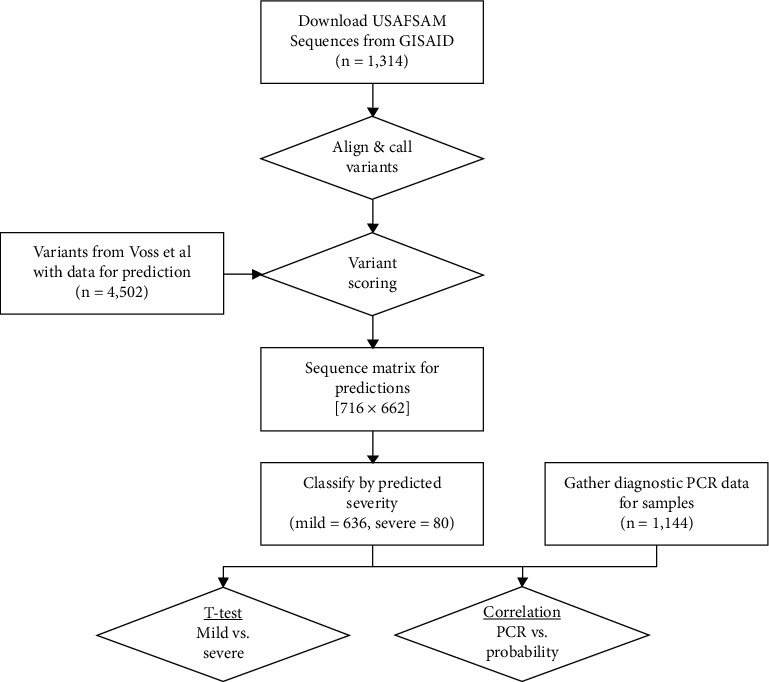
For each specimen, we generated a probability of severe illness using an algorithm that considers variants in SARS-CoV-2 genomic sequences, the geographic region of collection, and the patient's age and gender [7]. The genomic sequences were obtained from GISAID and matched to specimens for PCR as described above. Once downloaded, the sequences were aligned to the Wuhan reference strain (NCBI:NC_045512.2; GISAID:EPI_ISL_402125) using “MiniMap2” (version 2.17) [19] and “MAFFT” [20], while subsequent variants were called using “SNP sites” [21]. Variants present in the algorithm but not in a genomic sequence were scored as “0,” whereas observed variants were scored as “1.” Collectively, we observed 662 of the 4,502 variants used in the algorithm (Figure 1).
Figure 1.

Process flowchart for orthogonal validation of a severity prediction algorithm. Sequences were downloaded from GISAID, processed through reference alignment and variant calling, predicted based upon variants identified by Voss et al., and compared to observed PCR threshold data by the t-test and correlations. The matrix used for predictions was 716 samples with 662 variants overlapping the Voss et al. variant list (all other variants were assigned 0 for Python predictions).
2.3. Statistical Comparisons with PCR Values
The sample IDs were divided into classes based upon severity prediction probabilities generated here for analysis. The mean observed Ct values for each class were compared using the unequal variance unpaired t-test. We used Welch's corrections because of unequal sample sizes. A second approach tested for correlation between the predicted probability of a viral sequence belonging to the severe class and the observed Ct value. No modification to the original algorithm using PCR measurements was performed. Linear correlation was tested using Pearson's correlation. We used GraphPad Prism 7.0c for performing the t tests and Pearson's correlation.
3. Results
3.1. Sample Statistics
We obtained 1,314 SARS-CoV-2 sequences published to GISAID by the USAFSAM Epidemiology Laboratory (collection dates between 22 February 2020 and 11 June 2021). Of those sequences, 716 had corresponding N gene PCR data and were used for analysis.
3.1.1. Comparing PCR Values of Predicted “Mild” to Predicted “Severe”
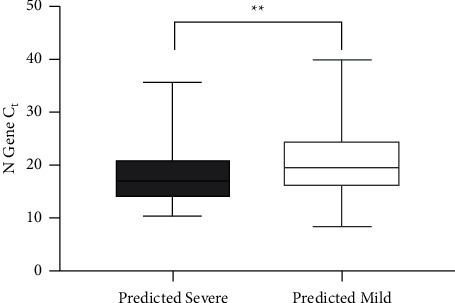
Using the previously published SARS-CoV-2 genome severity prediction model [7], 636 (89%) specimens had a ≤50% chance of being severe (“predicted mild” group) and 80 (11%) had a probability of >50% (“predicted severe” group). The “predicted severe” group had a significantly lower observed N gene Ct value (18.3 ± 0.6) as compared to the “predicted mild” group (20.4 ± 0.2). This difference of 2.1 ± 0.7 Ct (95% CI: 0.81–3.4) is consistent with differences seen in other studies signifying highly transmissible variants of concern [22] and is significant based on the unpaired t-test (Figure 2, P=0.0017).
Figure 2.

PCR Ct values for viral specimens predicted to be from patients with mild (open box, n = 636) or severe (dotted box, n = 80) outcomes. ∗∗P=0.0017.
3.1.2. Comparing PCR Values of Upper and Lower Prediction Quartiles
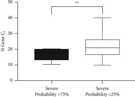
A similar analysis on the upper and lower quartiles of severity prediction revealed a more substantial difference (Figure 3). Those specimens with the greatest probability of severe outcomes (≥75% probability) had a Ct of 16.6 ± 1.3 (n = 10, 1.4% of samples), whereas those least likely to be associated with severe outcomes (<25%) had a Ct of 21.4 ± 0.3 (n = 350, 49% of samples) (P=0.0045). The difference between the means and 95% confidence interval was 4.9 (CI: 1.9–7.8).
Figure 3.

PCR Ct values for viral specimens least likely (probability < 25%, open box, n = 350) or most likely (probability ≥ 75%, dotted box, n = 10) outcomes. ∗∗P=0.0045.
4. Discussion
4.1. Principal Results
Thoroughly validating predictions from machine learning models is necessary for establishing the credibility and power of analytics for public health. For biology and medicine, this often means finding a testable measurement that represents the predicted outputs. In our case, we used an outgroup sample with an orthogonal severity marker to provide strong evidence that the algorithm can identify strains that are biologically unique and present meaningful clinical differences.
Research on the MERS coronavirus showed that viral load, as inferred from PCR cycle threshold, is associated with risk of severe disease and death, with a 17% higher risk of death for each 1 point drop in Ct after adjusting for age and underlying illness [23]. In the context of SARS-CoV-2, data from Brazil also suggest cycle threshold can predict severity in a manner that is not explained by age [24]. Indeed, the relationship between cycle threshold and COVID-19 severity has also been identified in a prior systematic review of 18 studies with a positive association in a majority [25], while also being described in a more recent narrative review [26]. Typically, a Ct value of less than 20 is considered “highly infective” [27]. Here, our results are consistent with these previous observations in which those viral genomes with the greatest predicted probability of severe clinical outcomes had, on average, a 5 Ct difference. This difference corresponds to viral titers of approximately 1.1 million viable viral particles per mL for those most likely to be severe versus only 33,000 particles per mL for those least probable to be severe.
The practical implications of Ct differences are critical to consider when determining future public health responses. In a recent study of military basic trainees, Marcus and colleagues found three interesting differences for index patients: those index patients who were symptomatic had a 7 cycle lower Ct than those who were asymptomatic (20 vs. 27.2 cycles), symptomatic index patients had a 4 cycle lower Ct than symptomatic patients who did not occur as part of a cluster (20 vs. 24.4 cycles), and those who were asymptomatic had a 9 cycle difference (27.2 for asymptomatic index patients vs. 36.4 for asymptomatic nonindex patients) [28]. Another study found a significant difference in viral loads in a cohort of military beneficiaries associated with obesity and inpatient or outpatient status [29]. When inpatients and outpatients were combined, the authors did not observe any significant difference in viral load. The greatest difference observed by the authors was in severely obese outpatients, who had 77-fold higher viral loads than nonobese patients, suggesting that in mildly ill patients, those who are more obese can tolerate greater viral loads. Consequently, it becomes apparent that viral load alone is not the most powerful predictor of disease severity.
Ultimately, while it is likely that there are other factors (biological or methodological) contributing to severity, it is possible that these balance out in a large enough population or where these factors are less likely to be different. For example, if there are measurement errors in the Ct associated with random differences in sample acquisition techniques, time of day a participant was tested, or sample type (nasopharyngeal vs. anterior nasal vs. nasal wash vs. oropharyngeal), these factors are likely to make the association between Ct and predicted risk appear smaller than it is. Alternatively, it remains plausible that the predicted severity from the genome does not differ by time of collection or bodyweight category, as both the biological and methodological confounders primarily bias toward the null [30]. Consequently, it is possible that the difference we found is explained by factors like those mentioned above, especially considering the constrained metadata regarding the lack of patient outcomes in our dataset. Regardless, because we observed, in an independent patient sample set, a decrease in cycle thresholds (a surrogate for increased disease severity) between groups of patients predicted to experience severe clinical outcomes like hospitalization, it remains compelling that the predictions generated from the computational algorithm are reflecting biological mechanisms and are clinically meaningful for patients, providers, and public health leaders. Future work is required to take the results of these two studies from statistically significant research findings to practical tools for use in clinical practice and public health management.
4.2. Limitations
One limitation of this study is the use of PCR cycle threshold as a surrogate for clinical severity. As mentioned in the main text, the ability of PCR to predict clinical outcome only has an area under the curve of 0.68, making it a fair, but not ideal analog for clinical outcomes. Clinical outcome data were not available for this study. Another limitation related to using the PCR Ct value is the variability in sample collection per patient as some patients report to clinics early in illnesses, whereas others may have reported only after symptoms became more severe. Regardless of these limitations, the high predictive ability of the algorithm along with the significant correlation with PCR data suggest that our results can be replicated in other populations and by other laboratories.
Another limitation is the relatively small number of viral genotypes. A post hoc power analysis for the predicted severe (Ct mean = 18.33 ± 5.514, n = 80) versus predicted mild (20.44 ± 5.633, n = 636) with a 5% type I error rate revealed 89.6% power to detect the observed difference, indicating that though the sample sizes were relatively small, the study had sufficient power to draw the conclusions from the results. Similarly, the post hoc power analysis for the most and least likely to be severe groups had a 95.3% power.
5. Conclusions
In conclusion, here, we report a correlation between a computationally predicted severity and a clinically measured surrogate for severity, PCR cycle threshold. Our results show that using viral genetic information combined with patient demographics could aid clinical triage and public health surveillance. While whole viral genome sequencing of every patient upon admission is cost prohibitive and results are unlikely to be timely enough to improve patient treatment, rapid diagnostic tests developed to identify variants critical to severity may provide insights to improve outcomes. Additionally, using sequence data along with laboratory validated, in silico-derived severity markers could help prioritize new variant vaccine development and in cases where there is clinical equipoise (for example, in cases where monoclonal antibody treatment is being considered or where postexposure presymptomatic prophylaxis is being considered for known contacts, among others). Finally, public health officials employing large-scale surveillance methods could benefit from these in silico markers by watching for the emergence of markers and gain a few days' lead time for preparedness actions.
Acknowledgments
The authors thank the laboratory technicians of the USAFSAM Applied Technology and Genomics Division for their support in generating the SARS-CoV-2 genomic sequences and the staff of the USAFSAM Public Health Epidemiology Laboratory for their support and coordination of sample acquisition. This work was performed with intramural funding within the US Air Force and the Defense Health Agency.
Data Availability
The PCR data used to support the findings of this study are included within the Supplementary Materials. The genomic data used to support the findings in this study are available from GISAID with the identifiers provided in the Supplementary Materials.
Disclosure
A preprint has previously been published [31]. Neither funding organization had any influence in the drafting or publication of this manuscript.
Conflicts of Interest
The authors declare that there are no conflicts of interest.
Supplementary Materials
Supplementary File 1: list of GISAID identifiers used. Supplementary File 2: results of the prediction (binary), probability of severity, and observed Ct values (if any).
References
- 1.Göker H., Aladağ Karakulak E., Demiroğlu H., et al. The effects of blood group types on the risk of COVID-19 infection and its clinical outcome. Turkish Journal of Medical Sciences . 2020;50(4):679–683. doi: 10.3906/sag-2005-395. [DOI] [PMC free article] [PubMed] [Google Scholar]
- 2.O’Driscoll M., Ribeiro Dos Santos G., Wang L., et al. Age-specific mortality and immunity patterns of SARS-CoV-2. Nature . 2020;590(7844):140–145. doi: 10.1038/s41586-020-2918-0. [DOI] [PubMed] [Google Scholar]
- 3.Pradhan A., Olsson P. E. Sex differences in severity and mortality from COVID-19: are males more vulnerable? Biology of Sex Differences . 2020;11(1):p. 53. doi: 10.1186/s13293-020-00330-7. [DOI] [PMC free article] [PubMed] [Google Scholar]
- 4.Khurshid Z., Asiri F. Y. I., Al Wadaani H. Human saliva: non-invasive fluid for detecting novel coronavirus (2019-nCoV) International Journal of Environmental Research and Public Health . 2020;17(7):p. 2225. doi: 10.3390/ijerph17072225. [DOI] [PMC free article] [PubMed] [Google Scholar]
- 5.Zhou B., Thao T. T. N., Hoffmann D., et al. SARS-CoV-2 Spike D614G variant confers enhanced replication and transmissibility. 2020. https://www.biorxiv.org/content/10.1101/2020.10.27.357558v1 .
- 6.COVID-19 Host Genetics Initiative. The COVID-19 Host Genetics Initiative, a global initiative to elucidate the role of host genetic factors in susceptibility and severity of the SARS-CoV-2 virus pandemic. European Journal of Human Genetics . 2020;28(6):715–718. doi: 10.1038/s41431-020-0636-6. [DOI] [PMC free article] [PubMed] [Google Scholar]
- 7.Voss J. D., Skarzynski M., McAuley E. M., et al. Variants in SARS-CoV-2 associated with mild or severe outcome. Evolution, Medicine, and Public Health . 2021;9(1):267–275. doi: 10.1093/emph/eoab019. [DOI] [PMC free article] [PubMed] [Google Scholar]
- 8.Nagy Á, Ligeti B., Szebeni J., Pongor S., Gyrffy B. COVIDOUTCOME-estimating COVID severity based on mutation signatures in the SARS-CoV-2 genome. Database The Journal of Biological Databases and Curation . 2021;2021 doi: 10.1093/database/baab020. [DOI] [PMC free article] [PubMed] [Google Scholar]
- 9.Dong Y., Zhou H., Li M., et al. A novel simple scoring model for predicting severity of patients with SARS-CoV-2 infection. Transboundary and Emerging Disease . 2020;67(6):2823–2829. doi: 10.1111/tbed.13651. [DOI] [PMC free article] [PubMed] [Google Scholar]
- 10.Gallo Marin B., Aghagoli G., Lavine K., et al. Predictors of COVID-19 severity: a literature review. Reviews in Medical Virology . 2021;31(1):1–10. doi: 10.1002/rmv.2146. [DOI] [PMC free article] [PubMed] [Google Scholar]
- 11.Choudhuri J., Carter J., Nelson R., et al. SARS-CoV-2 PCR cycle threshold at hospital admission associated with patient mortality. PLoS One . 2020;15(12) doi: 10.1371/journal.pone.0244777.e0244777 [DOI] [PMC free article] [PubMed] [Google Scholar]
- 12.Pujadas E., Chaudhry F., McBride R., et al. SARS-CoV-2 viral load predicts COVID-19 mortality. The Lancet Respiratory Medicine . 2020;8(9):p. e70. doi: 10.1016/S2213-2600(20)30354-4. [DOI] [PMC free article] [PubMed] [Google Scholar]
- 13.Mercatelli D., Giorgi F. M. Geographic and genomic distribution of SARS-CoV-2 mutations. Frontiers in Microbiology . 2020;11:p. 1800. doi: 10.3389/fmicb.2020.01800. [DOI] [PMC free article] [PubMed] [Google Scholar]
- 14.Becerra-Flores M., Cardozo T. SARS-CoV-2 viral spike G614 mutation exhibits higher case fatality rate. International Journal of Clinical Practice . 2020;74(8) doi: 10.1111/ijcp.13525.e13525 [DOI] [PMC free article] [PubMed] [Google Scholar]
- 15.Nazario-Toole A. E., Xia H., Gibbons T. F. Whole-genome sequencing of SARS-CoV-2: using phylogeny and structural modeling to contextualize local viral evolution. Military Medicine . 2021;187 doi: 10.1093/milmed/usab031. [DOI] [PMC free article] [PubMed] [Google Scholar]
- 16.Elbe S., Buckland-Merrett G. Data, disease and diplomacy: GISAID’s innovative contribution to global health. Global Challenges . 2017;1(1):33–46. doi: 10.1002/gch2.1018. [DOI] [PMC free article] [PubMed] [Google Scholar]
- 17.Shu Y., McCauley J. GISAID: global initiative on sharing all influenza data – from vision to reality. Euro Surveillance . 2017;22(13):p. 30494. doi: 10.2807/1560-7917.es.2017.22.13.30494. [DOI] [PMC free article] [PubMed] [Google Scholar]
- 18.Chapleau R. R., Christian M., Connors B., et al. Early identification of SARS-CoV-2 emergence in the department of defense via retrospective analysis of 2019-2020 upper respiratory illness samples. Inside MS . 2021;28(6):2–5. [PubMed] [Google Scholar]
- 19.Li H. Minimap2: pairwise alignment for nucleotide sequences. Bioinformatics . 2018;34(18):3094–3100. doi: 10.1093/bioinformatics/bty191. [DOI] [PMC free article] [PubMed] [Google Scholar]
- 20.Katoh K., Misawa K., Kuma K., Miyata T. MAFFT: a novel method for rapid multiple sequence alignment based on fast Fourier transform. Nucleic Acids Research . 2002;30(14):3059–3066. doi: 10.1093/nar/gkf436. [DOI] [PMC free article] [PubMed] [Google Scholar]
- 21.Page A. J., Taylor B., Delaney A. J., et al. SNP-sites: rapid efficient extraction of SNPs from multi-FASTA alignments. Microbial Genomics . 2016;2(4) doi: 10.1099/mgen.0.000056.e000056 [DOI] [PMC free article] [PubMed] [Google Scholar]
- 22.Roquebert B., Haim-Boukobza S., Trombert-Paolantoni S., et al. SARS-CoV-2 variants of concern are associated with lower RT-PCR amplification cycles between january and march 2021 in France. 2021. https://www.medrxiv.org/content/10.1101/2021.03.19.21253971v1 . [DOI] [PMC free article] [PubMed]
- 23.Feikin D. R., Alraddadi B. M., Qutub M., et al. Association of higher MERS-CoV virus load with severe disease and death, Saudi arabia, 2014. Emerging Infectious Diseases . 2014;21(11):2029–2035. doi: 10.3201/eid2111.150764. [DOI] [PMC free article] [PubMed] [Google Scholar]
- 24.Faíco-Filho K. S., Passarelli V. C., Bellei N. Is higher viral load in SARS-CoV-2 associated with death? The American Journal of Tropical Medicine and Hygiene . 2020;103(5):2019–2021. doi: 10.4269/ajtmh.20-0954. [DOI] [PMC free article] [PubMed] [Google Scholar]
- 25.Rao S. N., Manissero D., Steele V. R., Pareja J. A systematic review of the clinical utility of cycle threshold values in the context of COVID-19. Infectious Disease and Therapy . 2020;9(3):573–586. doi: 10.1007/s40121-020-00324-3. [DOI] [PMC free article] [PubMed] [Google Scholar]
- 26.Rabaan A. A., Tirupathi R., Sule A. A., et al. Viral dynamics and real-time RT-PCR Ct values correlation with disease severity in COVID-19. Diagnostics (basel) . 2021;11(6):p. 1091. doi: 10.3390/diagnostics11061091. [DOI] [PMC free article] [PubMed] [Google Scholar]
- 27.Engelmann I., Alidjinou E. K., Ogiez J., et al. Preanalytical Issues and Cycle Threshold Values in SARS-CoV-2 Real-Time RT-PCR Testing: Should Test Results Include These? ACS Omega . 2021;6(10):6528–6536. doi: 10.1021/acsomega.1c00166. [DOI] [PMC free article] [PubMed] [Google Scholar]
- 28.Marcus J. E., Frankel D. N., Pawlak M. T., et al. Risk factors associated with COVID-19 transmission among US Air Force trainees in a congregate setting. JAMA Network Open . 2021;4(2) doi: 10.1001/jamanetworkopen.2021.0202.e210202 [DOI] [PMC free article] [PubMed] [Google Scholar]
- 29.Epsi N. J., Richard S. A., Laing E. D., et al. Clinical, immunological and virological SARS-CoV-2 phenotypes in obese and non-obese military health system beneficiaries. The Journal of Infectious Diseases . 2021;224:1462–1472. doi: 10.1093/infdis/jiab396. [DOI] [PMC free article] [PubMed] [Google Scholar]
- 30.Fosgate G. T. Non-differential measurement error does not always bias diagnostic likelihood ratios towards the null. Emerging Themes in Epidemiology . 2006;3(1):p. 7. doi: 10.1186/1742-7622-3-7. [DOI] [PMC free article] [PubMed] [Google Scholar]
- 31.Skarzynski M., McAuley E. M., Maier E. J., Fries A. C., Voss J. D., Chapleau R. R. SARS-CoV-2 genome-based severity predictions correspond to lower qPCR values and higher viral load. 2021. https://www.medrxiv.org/content/10.1101/2021.11.22.21266688v1 . [DOI] [PMC free article] [PubMed]
Associated Data
This section collects any data citations, data availability statements, or supplementary materials included in this article.
Supplementary Materials
Supplementary File 1: list of GISAID identifiers used. Supplementary File 2: results of the prediction (binary), probability of severity, and observed Ct values (if any).
Data Availability Statement
The PCR data used to support the findings of this study are included within the Supplementary Materials. The genomic data used to support the findings in this study are available from GISAID with the identifiers provided in the Supplementary Materials.


