

Abstract
目的
探究IL-17A在原发性高血压大鼠肾上皮间质转化(EMT)中的作用。
方法
4周龄的SPF级自发性高血压大鼠(SHR)(实验组)、京都维斯塔尔大鼠(WKY)(对照组)各20只,分别随机分为4组,每组5只,在4、6、10、30周龄进行实验观察。采用无创尾动脉血压测量仪动态检测大鼠的血压情况;流式细胞技术分析大鼠脾淋巴细胞中Th17细胞的频数;RT-PCR法及免疫组化技术检测大鼠肾脏IL-17A、iNOS、Arg-1、E-cadherin、α-SMA mRNA和蛋白的表达水平;ELISA法检测大鼠血浆中IL-17A的水平。
结果
从6周龄始SHR大鼠血压显著高于同期的WKY组(P < 0.05或P < 0.01),SHR大鼠Th17细胞频数以及肾脏和血浆中IL-17A的表达亦显著高于同期WKY组(P < 0.05或P < 0.01);在30周龄时SHR大鼠肾脏中E-cadherin mRNA及蛋白的表达显著低于WKY组(P < 0.01),而Arg-1 mRNA及蛋白的表达显著高于WKY组(P < 0.01);iNOS mRNA及蛋白的表达在6、10周龄时显著高于WKY组(P < 0.05或P < 0.01),α-SMA mRNA及蛋白从10周龄始显著高于WKY组(P < 0.05或P < 0.01)。SHR大鼠肾脏中IL-17A mRNA及蛋白表达水平从10周龄始与E-cadherin mRNA及蛋白表达水平呈显著负相关(r=-0.731,P < 0.05;r=-0.827,P < 0.01),而与α-SMA mRNA及蛋白表达水平呈显著正相关(r=0.658,P < 0.05;r=0.968,P < 0.01)。
结论
IL-17A与SHR大鼠肾EMT的过程密切相关,可能是通过介导肾脏浸润的巨噬细胞M1/M2极化发挥作用。
Keywords: IL-17A, 高血压, 上皮间质转化, 巨噬细胞
Abstract
Objective
To explore the role of interleukin-17A (IL-17A) in renal epithelial- mesenchymal transition (EMT) in essential hypertensive nephropathy.
Methods
Four-week-old spontaneously hypertensive rats (SHR) and Wistar-Kyoto (WKY) rats (control group) were both randomized into 4 groups (n=5) for observation at 4, 6, 10 and 30 weeks of age. Blood pressure of the rats was monitored using a noninvasive tail artery blood pressure measurement instrument. The percentage of Th17 cells in the splenocytes was analyzed using flow cytometry. The mRNA and protein expression levels of IL-17A, iNOS, Arg-1, E-cadherin, and α-SMA in the kidneys of the rats were detected using RT-PCR and immunohistochemical staining, respectively, and plasma levels of IL-17A were regularly detected using ELISA.
Results
At the age of 6 weeks, the SHRs began to show significantly higher blood pressure with greater Th17 cell percentage in the splenocytes and high renal expression and plasma level of IL-17A than WKY rats (P < 0.05 or P < 0.01). At 30 weeks, renal expression of E-cadherin mRNA and protein was significantly lower and the expression of Arg-1 mRNA and protein was significantly higher in SHR than in WKY rats (P < 0.01). Compared with the WKY rats, the SHRs showed significantly higher mRNA and protein expressions of iNOS at 6 and 10 weeks (P < 0.05 or 0.01) and higher α-SMA mRNA and protein expressions since 10 weeks of age (P < 0.05 or 0.01). In SHRs older than 10 weeks, renal IL-17A mRNA and protein expression levels were negatively correlated with those of E-cadherin (r=-0.731, P < 0.05; r=-0.827, P < 0.01) and positively correlated with those of α-SMA (r=0.658, P < 0.05; r=0.968, P < 0.01).
Conclusion
IL-17A is closely correlated with the progression of renal EMT in SHR and plays its role possibly by mediating M1/M2 polarization of renal infiltrating macrophages.
Keywords: interleukin-17A, high blood pressure, epithelial-mesenchymal transition, macrophages
原发性高血压(EH)是一种以血压升高为主要临床表现而病因尚不明确的疾病。调查发现全球有超过11.3亿高血压患者,预计到2025年底,全球将有15.6亿人患有高血压[1]。2020年我国发布的《中国居民营养与慢性病状况报告(2020年)》数据显示,成年人高血压患病率为27.5%,总人数高达3亿。因此,高血压仍然是全球主要的公共卫生问题。
肾脏作为调节血压的重要器官,是EH靶器官损害的主要器官[2]。EH引起的肾损伤主要包括肾纤维化、肾小管肥厚和肾小球的改变[3]。研究表明,上皮间充质转化(EMT)在高血压诱导的肾纤维化发展中发挥着重要作用[4, 5]。EMT在胚胎组织和器官分化过程中首次被发现,是伤口愈合、癌症和纤维化的关键过程,但在高血压肾病中EMT的作用机制仍不清楚[6]。近年来,越来越多的证据表明免疫系统参与高血压的病理生理过程以及终末器官的损伤,细胞因子对高血压炎症反应的影响也一直是研究的热点[7, 8]。IL-17A作为一种促炎因子,主要由Th17细胞极化产生。研究发现,在高血压患者血浆中IL-17A的水平升高,并且在高血压动物模型诱导的肾损伤中IL-17A的表达亦显著升高[9-11]。但对IL-17A是否参与高血压肾EMT及相关机制暂未发现相关报道。因此,本研究采用国际公认的最接近人类EH的动物模型自发性高血压大鼠(SHR),初步探究IL-17A在SHR大鼠肾EMT中的作用,为免疫干预治疗高血压肾损伤提供初步的实验依据。
1. 材料和方法
1.1. 主要材料与试剂
PE标记的抗大鼠IL-17A单克隆抗体、FITC标记的抗大鼠CD4单克隆抗体及相应同型对照抗体、抗大鼠IL-17A单克隆抗体(eBioscience)、E-cadherin多克隆抗体(BOSTER)、α-SMA单克隆抗体(CST)、iNOS多克隆抗体(BOSTER)、Arg-1单克隆抗体(ABclonal); 抗兔/鼠通用型免疫组化试剂盒(上海基因科技); 逆转录试剂盒(Thermo Scientific); Trizol试剂(上海生工); 大鼠IL-17A ELISA试剂盒(eBioscience)。
1.2. 方法
1.2.1. 实验动物
4周龄SPF级雄性WKY大鼠(对照组)、SHR大鼠(实验组)各20只,购自北京维通利华公司,动物许可证号为SCXK(京)2016-0006;按简单随机法将两种大鼠各分为4组,每组5只,分别在4、6、10、30周龄(相当于EH的0期、初始期、形成期、靶器官损害期)进行实验观察。实验动物的使用和管理均经安徽理工大学生物医学研究伦理委员会批准(编号:2021019)。
1.2.2. 血压测量
使用BP-6A型全自动无创血压测量仪于清晨8点在大鼠安静状态时测量尾动脉收缩压,每只大鼠测量3次,每次间隔5 min,取3次平均值作为该大鼠的收缩压值。
1.2.3. 大鼠脾淋巴细胞IL-17A表达检测
无菌环境下取各期大鼠脾脏,将脾脏置于含10%胎牛血清的RPMI 1640培养液中。用眼科剪将脾脏包膜剪破后用研磨棒在300目不锈钢滤网上将脾脏研磨成细胞匀浆,然后通过静置、离心、破红、洗涤、再次离心、重悬等步骤制备脾淋巴细胞悬液。每个流式管内加入大鼠脾淋巴细胞1× 10⁶(经PMA预先刺激4 h),流式管中加入FITC标记的抗大鼠CD4单克隆抗体(0.5 μL/管)及PE标记的抗大鼠IL-17A单克隆抗体(1.0 μL/管),并设置同型对照。加入固定/破膜工作液1 mL,旋涡震荡,重悬细胞,4 ℃避光孵育30 min。1200 r/min×5 min,洗2次,重悬细胞,经流式细胞仪测定细胞各种荧光强度,用CellQuest软件进行分析。
1.2.4. 大鼠肾脏IL-17A、iNOS、Arg-1、E-cadherin、α- SMA mRNA的表达
取各期大鼠左肾组织,每次取约50 mg用Trizol法提取肾脏总RNA,其余放-80 ℃冰箱备用。按照逆转录试剂盒说明书将提取的总RNA于20 µL反应体系中进行逆转录合成第一链cDNA。PCR体系为Taq PCR Master Mix 12.5 µL、上游引物1 µL、下游引物1 µL、cDNA模板2 µL、加水至25 µL,以GAPDH mRNA作为内参。引物设计与合成:在基因数据库中检索目的基因序列,采用Primer 6.0软件设计引物(表 1)。PCR扩增反应条件为95 ℃预变性5 min,94 ℃变性30 s,54.2 ℃退火30 s,72 ℃延伸30 s,循环34次,72 ℃终末延伸10 min。PCR产物应用Gel-Pro Analyzer分析系统测量相对表达量。
1.
RT-PCR引物序列
RT-PCRprimerssequence
| Gene | Primer sequence 5'-3' | Length of product (bp) |
| F: Forward primer; R: Reverse primer. | ||
| GAPDH | F: GTGAAGGTCGGTGTGAA R: GGAGTTGCTGTTGAAGTC |
861 |
| IL-17A | F: GTGCCTGATGCTGTTGCTGCTA R: GTGAAGTGGAACGGTTGAGGTAGTC |
200 |
| iNOS | F: CTTGGAGCGAGTTGTGGATTGTTCT R: AACCTCTGCCTGTGCGTCTCTT |
216 |
| Arg-1 | F: CCAAGCCAAAGCCCATAGAGATTATCG R: TCAGCGGAGTGTTGATGTCAGTGT |
393 |
| E-cadherin | F: TCCGCAAGACCAGTGACAGAGG R: GGAGATGAGCACAGGCAGGTAGT |
452 |
| α-SMA | F: CGATAGAACACGGCATCATCACCAA R: TCCAGAGTCCAGCACAATACCAGTT |
260 |
1.2.5. 大鼠肾脏IL-17A、iNOS、Arg-1、E-cadherin、α-SMA蛋白的表达
取各期大鼠右肾组织用10%甲醛液固定,常规脱水、石蜡包埋3µm厚连续切片,应用二步法免疫组织化学法对上述切片进行染色。阳性判定标准为境界清晰,突出于背景的棕黄色或棕褐色颗粒。每张切片随机选取5个不同的视野,采用Image Pro Plus6.0图像分析系统进行半定量分析。
1.2.6. 大鼠血浆中IL-17A的水平
收集各期大鼠血浆,按ELISA试剂盒说明书测定收集好的血浆中IL-17A的水平。按照每块酶标板上的标准品的A450值绘制标准曲线,计算血浆中IL-17A的含量。
1.2.7. 统计学分析
采用SPSS13.0软件进行统计学分析,实验结果数据以均数±标准差表示,组间比较采用两独立样本t检验,若方差不齐采用t'检验; 两变量间的相关性采用Pearson相关性分析。若P < 0.05表示差异具有统计学意义。
2. 结果
2.1. 大鼠血压的变化
血压测量结果显示,在大鼠6、10、30周龄时,SHR组的收缩压显著高于对照组(P < 0.05或P < 0.01,图 1)。
1.

大鼠血压的变化
Changes in blood pressure of the rats with age. *P < 0.05, **P < 0.01 vs WKY group.
2.2. 大鼠脾淋巴细胞中Th17细胞的频数变化
流式细胞术分析结果显示SHR大鼠脾淋巴细胞中Th17细胞的频数逐渐增加,在6、10、30周龄均显著高于WKY组,两者差异具有统计学意义(P < 0.05或P < 0.01,图 2)。
2.

大鼠脾淋巴细胞中Th17细胞的频数变化
Frequency of Th17 cells in splenocytes of the rats. A: Flow cytometry. B: Quantitative analysis of the results. *P < 0.05, **P < 0.01 vs WKY group.
2.3. 大鼠肾脏中IL-17A、iNOS、Arg-1、E-cadherin、α-SMA mRNA的表达
IL-17A在6、10、30周龄时SHR组显著高于WKY组,差异具有统计学意义(P < 0.05或P < 0.01)。M1型巨噬细胞标志物iNOS在6、10周龄时SHR组显著高于WKY组(P < 0.05或P < 0.01); 而M2型巨噬细胞标志物Arg-1在30周龄时SHR组显著高于WKY组(P < 0.01)。上皮标志物E-cadherin在4、6、10周龄时WKY组、SHR组的表达均上调,差异无统计学意义; 而在30周龄时SHR组的表达水平显著低于同期WKY组(P < 0.01); 间充质标志物α-SMA在10、30周龄时SHR组显著高于WKY组(P < 0.05或P < 0.01,图 3)。
3.

大鼠肾脏中IL-17A、iNOS、Arg-1、E-cadherin、α-SMA mRNA的表达变化
Expression of IL-17A, iNOS, Arg-1, E-cadherin, and α-SMA mRNA in the kidneys of the rats. A: RT-PCR. W: WKY. S: SHR. B: Quantitative analysis of the results. *P < 0.05, **P < 0.01 vs WKY group.
2.4. 大鼠肾脏中IL- 17A、iNOS、Arg-1、E-cadherin、α- SMA蛋白的表达
IL-17A在10、30周龄时SHR组显著高于WKY组(P < 0.05或P < 0.01); iNOS在6、10周龄时SHR组显著高于WKY组(P < 0.05或P < 0.01); 而Arg-1在30周龄时SHR组显著高于WKY组(P < 0.01)。E-cadherin在4、6、10周龄时WKY组与SHR组均升高,差异无统计学意义,而在30周龄时SHR组显著低于WKY组,且差异具有统计学意义(P < 0.01); α-SMA在10、30周龄时SHR组显著高于WKY组(P < 0.05或P < 0.01,图 4)。
4.
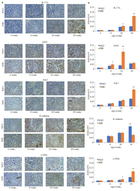
大鼠肾脏中IL-17A、iNOS、Arg-1、E-cadherin、α-SMA的表达水平
Expression of IL-17A, iNOS, Arg-1, E-cadherin and α-SMA proteins in the kidneys of the rats. A: Immunohistochemical staining of the kidney tissue (Original magnification: ×400). B: Quantitative analysis of the results. *P < 0.05, **P < 0.01 vs WKY group.
2.5. 血浆中IL-17A的水平
ELISA结果显示,SHR组IL-17A血浆表达水平在6、10、30周龄时显著高于WKY组,两者差异均具有统计学意义(P < 0.05或P < 0.01,图 5)。
5.
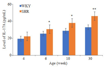
大鼠血浆IL-17A表达水平
Changes of plasma levels of IL-17A in the rats with age. *P < 0.05, **P < 0.01 vs WKY group.
2.6. IL-17A与大鼠肾脏中E-cadherin、α-SMA的相关性分析
随着病程时间的延长,从10周龄至30周龄SHR组大鼠肾脏中IL-17A mRNA及蛋白表达水平与E-cadherin mRNA及蛋白表达水平呈显著负相关(相关系数r=-0.731,P < 0.05;r=-0.827,P < 0.01); 与α-SMA mRNA及蛋白表达水平呈显著正相关(相关系数r= 0.658,P < 0.05;r=0.968,P < 0.01,图 6)。WKY组IL-17A表达水平与大鼠肾脏中E-cadherin、α-SMA的表达水平则皆无相关性。
6.

SHR大鼠肾脏中E-cadherin、α-SMA蛋白及mRNA的表达与IL-17A表达相关性分析(从10周龄始)
Correlation analysis of the changes of E-cadherin and α-SMA protein and mRNA expression with IL-17A expression in the kidneys of SHR (after 10 weeks of age). A: IL-17A vs E-cadherin. B: IL-17A vs α-SMA.
3. 讨论
最新研究发现,高血压与免疫炎症和慢性肾病之间的关系密切[11, 12]。持续性高血压将导致肾纤维化及终末器官损害,是继糖尿病肾病之后导致终末期肾病的第二大病因[13]。IL-17A主要是由Th17细胞产生的一种多效性细胞因子,其主要功能是通过释放促炎和中性粒细胞动员的细胞因子介导组织炎症反应。相关研究发现IL-17A在高血压肾病中扮演着重要角色[14-16]。Orejudo等[15, 17]在血管紧张素Ⅱ(AngⅡ)建立的高血压模型小鼠中发现,应用IL-17A单克隆抗体阻断的AngⅡ模型小鼠肾脏炎症反应明显降低,结果表明IL-17A可能参与小鼠血压升高以及高血压引起的肾脏炎症反应。Amador等[11, 18]实验数据也表明Th17细胞及其效应细胞因子IL-17A在高血压发病机制中的作用。本实验结果显示,SHR大鼠脾淋巴细胞中Th17细胞的频数明显高于WKY组,SHR大鼠外周血与肾脏中IL-17A的浓度和蛋白的表达水平显著高于对照组,且其血压亦显著高于对照组。结果提示IL-17A可能在SHR大鼠血压升高和肾脏损伤的过程中发挥重要作用。此外,在小鼠肾脏缺血再灌注的模型中注射抗IL-17A单克隆抗体后,小鼠肾脏的损伤减轻,肾脏组织和血浆中的促炎介质浓度也随之降低[19]。
在肾脏疾病中,巨噬细胞是慢性炎症反应的重要介质,其浸润程度与肾脏损伤的严重程度密切相关[20]。根据组织微环境的不同,人原代单核细胞来源的巨噬细胞(M0)可被极化为M1和M2两种不同的表型[21]。其中M1巨噬细胞产生大量的促炎介质,介导抗肿瘤免疫等;M2巨噬细胞具有抗炎功能,持续表达易导致组织纤维化[22]。常见的M1型巨噬细胞标志物有iNOS、CD86等;M2型巨噬细胞标志物有Arg-1、CD163、CD206等[23]。Liu等[24]研究表明巨噬细胞分泌的补体C3导致IL-17A介导的炎症细胞浸润到肾脏,从而导致肾损伤。此外,在对动物模型和高血压患者的研究中发现,巨噬细胞极化在高血压肾损伤中扮演着重要角色[25, 26]。本实验研究发现,iNOS mRNA及蛋白的表达在SHR大鼠肾脏组织6、10周龄时显著升高,而在30周龄时显著降低;同时Arg-1mRNA及蛋白在30周龄时显著表达。这些结果证实了巨噬细胞参与SHR大鼠肾损伤,这与前述结果一致。然而,本实验还发现在巨噬细胞浸润的过程中,SHR大鼠在6、10、30周龄时IL-17A血浆中的浓度和肾脏中mRNA的表达亦显著高于WKY组;这些数据表明在SHR大鼠肾脏组织中M1/M2巨噬细胞浸润与IL- 17A的表达呈一致性变化趋势。提示在SHR大鼠肾脏中IL-17A上调的同时伴随着M1/M2巨噬细胞极化偏移。另有研究表明,在肺癌、子宫内膜异位症等疾病中IL-17A通过其受体IL-17AR诱导M2巨噬细胞极化,此结果亦验证了IL-17A在巨噬细胞极化的过程中发挥着重要作用[27, 28]。
最新研究表明,EMT与高血压肾纤维化的发生密切相关[6, 29, 30]。根据其发生的生物学背景,EMT可分为三种亚型,Ⅰ型与器官发育相关;Ⅱ型继发于炎症,与组织再生和器官纤维化相关;Ⅲ型促进恶性肿瘤细胞的转移[31, 32]。在EMT发生的过程中,最关键的一步是下调Ecadherin的表达,随后细胞分裂浸入基底膜向间质移动;而后获得间质表型标志物,如α-SMA[30, 33]。本实验结果发现,在SHR大鼠30周龄时肾组织E-cadherin mRNA和蛋白的表达显著低于对照组;α-SMA mRNA和蛋白的表达在10、30周龄时显著高于对照组。相关研究发现,在注射AngⅡ的高血压大鼠中未检测到明显蛋白尿以及肾纤维化之前,肾小管周围就能检测到EMT的发生[34];此外,在饮食盐摄入诱导的高血压大鼠模型中,肾小管上皮细胞周围亦检测到E-cadherin表达降低,α-SMA表达增强[35]。在这些高血压动物模型中肾脏都发生了EMT,但其发生的机制需要进一步阐明。最新研究发现,人近端小管上皮细胞(HK-2)与M2巨噬细胞共培养48 h后,HK-2细胞α-SMA的表达明显上升,E-cadherin的表达明显下降[36]。此外,Yu等[37]研究发现M1/M2巨噬细胞极化可以调节顺铂处理的肾小管上皮细胞EMT的发生。我们的研究结果表明,在SHR大鼠6、10、30周龄M1/M2巨噬细胞极化的过程中SHR大鼠肾脏亦发生了EMT,与前述结果一致。本研究结果还发现,SHR大鼠从10周龄至30周龄时肾脏中IL-17A mRNA和蛋白的表达与E-cadherin mRNA和蛋白的表达呈显著负相关,与α-SMA mRNA和蛋白的表达呈显著正相关。提示IL-17A在SHR大鼠肾脏EMT的过程中发挥着重要作用。
因此,IL-17A参与高血压肾EMT的发生,其机制可能与其介导巨噬细胞极化有关。
Biography
向茂翠,在读硕士研究生,E-mail: 2533040254@qq.com
Funding Statement
安徽高校省级自然科学重点研究项目(KJ2017A098);安徽理工大学博士科研基金(11046);安徽理工大学中青年学术骨干培养工程(10167)
Contributor Information
向 茂翠 (Maocui XIANG), Email: 2533040254@qq.com.
王 瑜 (Yu WANG), Email: yuwang326@163.com.
References
- 1.Tiruneh SA, Bukayaw YA, Yigizaw ST, et al. Prevalence of hypertension and its determinants in Ethiopia: a systematic review and meta-analysis. PLoS One. 2020;15(12):e0244642. doi: 10.1371/journal.pone.0244642. [Tiruneh SA, Bukayaw YA, Yigizaw ST, et al. Prevalence of hypertension and its determinants in Ethiopia: a systematic review and meta-analysis[J]. PLoS One, 2020, 15(12): e0244642.] [DOI] [PMC free article] [PubMed] [Google Scholar]
- 2.Krebs CF, Lange S, Niemann G, et al. Deficiency of the interleukin 17/23 axis accelerates renal injury in mice with deoxycorticosterone Acetate + Angiotensin Ⅱ-induced hypertension. Hypertension. 2014;63(3):565–71. doi: 10.1161/HYPERTENSIONAHA.113.02620. [Krebs CF, Lange S, Niemann G, et al. Deficiency of the interleukin 17/23 axis accelerates renal injury in mice with deoxycorticosterone Acetate + Angiotensin Ⅱ-induced hypertension[J]. Hypertension, 2014, 63(3): 565-71.] [DOI] [PubMed] [Google Scholar]
- 3.Sun HJ. Current opinion for hypertension in renal fibrosis. Adv Exp Med Biol. 2019;1165:37–47. doi: 10.1007/978-981-13-8871-2_3. [Sun HJ. Current opinion for hypertension in renal fibrosis[J]. Adv Exp Med Biol, 2019, 1165: 37-47.] [DOI] [PubMed] [Google Scholar]
- 4.Hu HT, Hu S, Xu S, et al. miR-29b regulates Ang Ⅱ-induced EMT of rat renal tubular epithelial cells via targeting PI3K/AKT signaling pathway. Int J Mol Med. 2018;42(1):453–60. doi: 10.3892/ijmm.2018.3579. [Hu HT, Hu S, Xu S, et al. miR-29b regulates Ang Ⅱ-induced EMT of rat renal tubular epithelial cells via targeting PI3K/AKT signaling pathway[J]. Int J Mol Med, 2018, 42(1): 453-60.] [DOI] [PubMed] [Google Scholar]
- 5.Zhang Y, Peng W, Ao X, et al. TAK-242, a Toll-Like receptor 4 antagonist, Protects against aldosterone-induced cardiac and renal injury. PLoS One. 2015;10(11):e0142456. doi: 10.1371/journal.pone.0142456. [Zhang Y, Peng W, Ao X, et al. TAK-242, a Toll-Like receptor 4 antagonist, Protects against aldosterone-induced cardiac and renal injury[J]. PLoS One, 2015, 10(11): e0142456.] [DOI] [PMC free article] [PubMed] [Google Scholar]
- 6.Seccia T, Caroccia B, Piazza M, et al. The key role of epithelial to mesenchymal transition (EMT) in hypertensive kidney disease. Int J Mol Sci. 2019;20(14):3567. doi: 10.3390/ijms20143567. [Seccia T, Caroccia B, Piazza M, et al. The key role of epithelial to mesenchymal transition (EMT) in hypertensive kidney disease[J]. Int J Mol Sci, 2019, 20(14): 3567.] [DOI] [PMC free article] [PubMed] [Google Scholar]
- 7.Madhur MS, Elijovich F, Alexander MR, et al. Hypertension: do inflammation and immunity hold the key to solving this epidemic ? Circ Res. 2021;128(7):908–33. doi: 10.1161/CIRCRESAHA.121.318052. [Madhur MS, Elijovich F, Alexander MR, et al. Hypertension: do inflammation and immunity hold the key to solving this epidemic [J]? Circ Res, 2021, 128(7): 908-33.] [DOI] [PMC free article] [PubMed] [Google Scholar]
- 8.Zhang RM, McNerney KP, Riek AE, et al. Immunity and hypertension. Acta Physiol. 2021;231(1):e13487. doi: 10.1111/apha.13487. [Zhang RM, McNerney KP, Riek AE, et al. Immunity and hypertension[J]. Acta Physiol, 2021, 231(1): e13487.] [DOI] [PMC free article] [PubMed] [Google Scholar]
- 9.Saleh MA, Norlander AE, Madhur MS. Inhibition of interleukin- 17A, but not interleukin-17F, signaling lowers blood pressure, and reduces end-organ inflammation in angiotensin Ⅱ-induced hypertension. JACC Basic Transl Sci. 2016;1(7):606–16. doi: 10.1016/j.jacbts.2016.07.009. [Saleh MA, Norlander AE, Madhur MS. Inhibition of interleukin- 17A, but not interleukin-17F, signaling lowers blood pressure, and reduces end-organ inflammation in angiotensin Ⅱ-induced hypertension[J]. JACC Basic Transl Sci, 2016, 1(7): 606-16.] [DOI] [PMC free article] [PubMed] [Google Scholar]
- 10.王 瑜, 王 博士, 惠 旭, et al. 可诱导共刺激分子介导的Th17细胞极化对自发性高血压大鼠肾纤维化的影响. https://www.j-smu.com/CN/Y2018/V38/I05/534. 南方医科大学学报. 2018;38(5):534–40. doi: 10.3969/j.issn.1673-4254.2018.05.05. [王瑜, 王博士, 惠旭, 等. 可诱导共刺激分子介导的Th17细胞极化对自发性高血压大鼠肾纤维化的影响[J]. 南方医科大学学报, 2018, 38(5): 534-40.] [DOI] [PMC free article] [PubMed] [Google Scholar]
- 11.McMaster WG, Kirabo A, Madhur MS, et al. Inflammation, immunity, and hypertensive end-organ damage. Circ Res. 2015;116(6):1022–33. doi: 10.1161/CIRCRESAHA.116.303697. [McMaster WG, Kirabo A, Madhur MS, et al. Inflammation, immunity, and hypertensive end-organ damage[J]. Circ Res, 2015, 116(6): 1022-33.] [DOI] [PMC free article] [PubMed] [Google Scholar]
- 12.Madhur MS, Kirabo A, Guzik TJ, et al. From rags to riches. Hypertension. 2020;75(4):930–4. doi: 10.1161/HYPERTENSIONAHA.119.14612. [Madhur MS, Kirabo A, Guzik TJ, et al. From rags to riches[J]. Hypertension, 2020, 75(4): 930-4.] [DOI] [PMC free article] [PubMed] [Google Scholar]
- 13.Cavalcante PAM, Alenina N, Budu A, et al. Nephropathy in hypertensive animals is linked to M2 macrophages and increased expression of the YM1/Chi3l3 protein. Mediat Inflamm. 2019;2019:1–14. doi: 10.1155/2019/9086758. [Cavalcante PAM, Alenina N, Budu A, et al. Nephropathy in hypertensive animals is linked to M2 macrophages and increased expression of the YM1/Chi3l3 protein[J]. Mediat Inflamm, 2019, 2019: 1-14.] [DOI] [PMC free article] [PubMed] [Google Scholar]
- 14.Norlander AE, Madhur MS, Harrison DG. Correction: the immunology of hypertension. J Exp Med. 2018;215(2):719. doi: 10.1084/jem.2017177301022018c. [Norlander AE, Madhur MS, Harrison DG. Correction: the immunology of hypertension[J]. J Exp Med, 2018, 215(2): 719.] [DOI] [PMC free article] [PubMed] [Google Scholar]
- 15.Norlander AE, Saleh MA, Kamat NV, et al. Interleukin-17A regulates renal sodium transporters and renal injury in angiotensin Ⅱinduced hypertension. Hypertension. 2016;68(1):167–74. doi: 10.1161/HYPERTENSIONAHA.116.07493. [Norlander AE, Saleh MA, Kamat NV, et al. Interleukin-17A regulates renal sodium transporters and renal injury in angiotensin Ⅱinduced hypertension[J]. Hypertension, 2016, 68(1): 167-74.] [DOI] [PMC free article] [PubMed] [Google Scholar]
- 16.Davis GK, Fehrenbach DJ, Madhur MS. Interleukin 17A: key player in the pathogenesis of hypertension and a potential therapeutic target. Curr Hypertens Rep. 2021;23(3):1–9. doi: 10.1007/s11906-021-01128-7. [Davis GK, Fehrenbach DJ, Madhur MS. Interleukin 17A: key player in the pathogenesis of hypertension and a potential therapeutic target [J]. Curr Hypertens Rep, 2021, 23(3): 1-9.] [DOI] [PMC free article] [PubMed] [Google Scholar]
- 17.Orejudo M, Rodrigues-Diez RR, Rodrigues-Diez R, et al. Interleukin 17A participates in renal inflammation associated to experimental and human hypertension. Front Pharmacol. 2019;10:1015. doi: 10.3389/fphar.2019.01015. [Orejudo M, Rodrigues-Diez RR, Rodrigues-Diez R, et al. Interleukin 17A participates in renal inflammation associated to experimental and human hypertension[J]. Front Pharmacol, 2019, 10: 1015.] [DOI] [PMC free article] [PubMed] [Google Scholar]
- 18.Amador CA, Barrientos V, Peña J, et al. Spironolactone decreases DOCA- salt-induced organ damage by blocking the activation of T helper 17 and the downregulation of regulatory T lymphocytes. Hypertension. 2014;63(4):797–803. doi: 10.1161/HYPERTENSIONAHA.113.02883. [Amador CA, Barrientos V, Peña J, et al. Spironolactone decreases DOCA- salt-induced organ damage by blocking the activation of T helper 17 and the downregulation of regulatory T lymphocytes[J]. Hypertension, 2014, 63(4): 797-803.] [DOI] [PubMed] [Google Scholar]
- 19.Cortvrindt C, Speeckaert R, MoermanA, et al. The role of interleukin- 17A in the pathogenesis of kidney diseases. Pathology. 2017;49(3):247–58. doi: 10.1016/j.pathol.2017.01.003. [Cortvrindt C, Speeckaert R, MoermanA, et al. The role of interleukin- 17A in the pathogenesis of kidney diseases[J]. Pathology, 2017, 49 (3): 247-58.] [DOI] [PubMed] [Google Scholar]
- 20.Guiteras R, Flaquer M, Cruzado JM. Macrophage in chronic kidney disease. Clin Kidney J. 2016;9(6):765–71. doi: 10.1093/ckj/sfw096. [Guiteras R, Flaquer M, Cruzado JM. Macrophage in chronic kidney disease[J]. Clin Kidney J, 2016, 9(6): 765-71.] [DOI] [PMC free article] [PubMed] [Google Scholar]
- 21.Mantovani A, Sica A, Sozzani S, et al. The chemokine system in diverse forms of macrophage activation and polarization. Trends Immunol. 2004;25(12):677–86. doi: 10.1016/j.it.2004.09.015. [Mantovani A, Sica A, Sozzani S, et al. The chemokine system in diverse forms of macrophage activation and polarization[J]. Trends Immunol, 2004, 25(12): 677-86.] [DOI] [PubMed] [Google Scholar]
- 22.Murray PJ, Wynn TA. Protective and pathogenic functions of macrophage subsets. Nat Rev Immunol. 2011;11(11):723–37. doi: 10.1038/nri3073. [Murray PJ, Wynn TA. Protective and pathogenic functions of macrophage subsets[J]. Nat Rev Immunol, 2011, 11(11): 723-37.] [DOI] [PMC free article] [PubMed] [Google Scholar]
- 23.Cao Q, Harris DCH, Wang YP. Macrophages in kidney injury, inflammation, and fibrosis. Physiology (Bethesda) 2015;30(3):183–94. doi: 10.1152/physiol.00046.2014. [Cao Q, Harris DCH, Wang YP. Macrophages in kidney injury, inflammation, and fibrosis[J]. Physiology (Bethesda), 2015, 30(3): 183-94.] [DOI] [PubMed] [Google Scholar]
- 24.Liu YY, Wang K, Liang XJ, et al. Complement C3 produced by macrophages promotes renal fibrosis via IL-17A secretion. Front Immunol. 2018;9:2385. doi: 10.3389/fimmu.2018.02385. [Liu YY, Wang K, Liang XJ, et al. Complement C3 produced by macrophages promotes renal fibrosis via IL-17A secretion[J]. Front Immunol, 2018, 9: 2385.] [DOI] [PMC free article] [PubMed] [Google Scholar]
- 25.Kneedler SC, Phillips LE, Hudson KR, et al. Renal inflammation and injury are associated with lymphangiogenesis in hypertension. Am J Physiol Renal Physiol. 2017;312(5):F861–9. doi: 10.1152/ajprenal.00679.2016. [Kneedler SC, Phillips LE, Hudson KR, et al. Renal inflammation and injury are associated with lymphangiogenesis in hypertension[J]. Am J Physiol Renal Physiol, 2017, 312(5): F861-9.] [DOI] [PMC free article] [PubMed] [Google Scholar]
- 26.Harwani SC. Macrophages under pressure: the role of macrophage polarization in hypertension. Transl Res. 2018;191:45–63. doi: 10.1016/j.trsl.2017.10.011. [Harwani SC. Macrophages under pressure: the role of macrophage polarization in hypertension[J]. Transl Res, 2018, 191: 45-63.] [DOI] [PMC free article] [PubMed] [Google Scholar]
- 27.Liu LX, Ge DX, Ma L, et al. Interleukin-17 and prostaglandin E2 are involved in formation of an M2 macrophage-dominant microenvironment in lung cancer. J Thorac Oncol. 2012;7(7):1091–100. doi: 10.1097/JTO.0b013e3182542752. [Liu LX, Ge DX, Ma L, et al. Interleukin-17 and prostaglandin E2 are involved in formation of an M2 macrophage-dominant microenvironment in lung cancer[J]. J Thorac Oncol, 2012, 7(7): 1091- 100.] [DOI] [PMC free article] [PubMed] [Google Scholar]
- 28.Miller JE, Ahn SH, Marks RM, et al. IL-17A modulates peritoneal macrophage recruitment and M2 polarization in endometriosis. Front Immunol. 2020;11:108. doi: 10.3389/fimmu.2020.00108. [Miller JE, Ahn SH, Marks RM, et al. IL-17A modulates peritoneal macrophage recruitment and M2 polarization in endometriosis[J]. Front Immunol, 2020, 11: 108.] [DOI] [PMC free article] [PubMed] [Google Scholar]
- 29.Li Y, Song B, Ruan CC, et al. AdipoRon attenuates hypertensioninduced epithelial-mesenchymal transition and renal fibrosis via promoting epithelial autophagy. J Cardiovasc Transl Res. 2021;14(3):538–45. doi: 10.1007/s12265-020-10075-8. [Li Y, Song B, Ruan CC, et al. AdipoRon attenuates hypertensioninduced epithelial-mesenchymal transition and renal fibrosis via promoting epithelial autophagy[J]. J Cardiovasc Transl Res, 2021, 14(3): 538-45.] [DOI] [PubMed] [Google Scholar]
- 30.Cruz-Solbes AS, Youker K. Epithelial to mesenchymal transition (EMT) and endothelial to mesenchymal transition (EndMT): role and implications in kidney fibrosis[M]//Results and Problems in Cell Differentiation. Cham: Springer International Publishing, 2017: 345-372.
- 31.Lee K, Nelson CM. New insights into the regulation of epithelialmesenchymal transition and tissue fibrosis[M]//International Review of Cell and Molecular Biology. Amsterdam: Elsevier, 2012: 171-221.
- 32.Das V, Bhattacharya S, Chikkaputtaiah C, et al. The basics of epithelial-mesenchymal transition (EMT): a study from a structure, dynamics, and functional perspective. J Cell Physiol. 2019;234(9):14535–55. doi: 10.1002/jcp.28160. [Das V, Bhattacharya S, Chikkaputtaiah C, et al. The basics of epithelial-mesenchymal transition (EMT): a study from a structure, dynamics, and functional perspective[J]. J Cell Physiol, 2019, 234 (9): 14535-55.] [DOI] [PubMed] [Google Scholar]
- 33.Lamouille S, Xu J, Derynck R. Molecular mechanisms of epithelialmesenchymal transition. Nat Rev Mol Cell Biol. 2014;15(3):178–96. doi: 10.1038/nrm3758. [Lamouille S, Xu J, Derynck R. Molecular mechanisms of epithelialmesenchymal transition[J]. Nat Rev Mol Cell Biol, 2014, 15 (3): 178-96.] [DOI] [PMC free article] [PubMed] [Google Scholar]
- 34.Dussaule JC, Guerrot D, Huby AC, et al. The role of cell plasticity in progression and reversal of renal fibrosis. Int J Exp Pathol. 2011;92(3):151–7. doi: 10.1111/j.1365-2613.2011.00760.x. [Dussaule JC, Guerrot D, Huby AC, et al. The role of cell plasticity in progression and reversal of renal fibrosis[J]. Int J Exp Pathol, 2011, 92(3): 151-7.] [DOI] [PMC free article] [PubMed] [Google Scholar]
- 35.Wang Y, Mu JJ, Liu FQ, et al. Salt-induced epithelial-tomesenchymal transition in Dahl salt-sensitive rats is dependent on elevated blood pressure. Braz J Med Biol Res. 2014;47(3):223–30. doi: 10.1590/1414-431X20133554. [Wang Y, Mu JJ, Liu FQ, et al. Salt-induced epithelial-tomesenchymal transition in Dahl salt-sensitive rats is dependent on elevated blood pressure[J]. Braz J Med Biol Res, 2014, 47(3): 223- 30.] [DOI] [PMC free article] [PubMed] [Google Scholar]
- 36.Chen Z, Dong FM, Lu JM, et al. Polarized M2c macrophages have a promoting effect on the epithelial-to-mesenchymal transition of human renal tubular epithelial cells. Immunobiology. 2018;223(12):826–33. doi: 10.1016/j.imbio.2018.08.008. [Chen Z, Dong FM, Lu JM, et al. Polarized M2c macrophages have a promoting effect on the epithelial-to-mesenchymal transition of human renal tubular epithelial cells[J]. Immunobiology, 2018, 223 (12): 826-33.] [DOI] [PubMed] [Google Scholar]
- 37.Yu CC, Chien CT, Chang TC. M2 macrophage polarization modulates epithelial-mesenchymal transition in cisplatin-induced tubulointerstitial fibrosis. Bio Med. 2016;6(1):5. doi: 10.7603/s40681-016-0005-5. [Yu CC, Chien CT, Chang TC. M2 macrophage polarization modulates epithelial-mesenchymal transition in cisplatin-induced tubulointerstitial fibrosis[J]. Bio Med, 2016, 6(1): 5.] [DOI] [PMC free article] [PubMed] [Google Scholar]