

Abstract
This paper aims to investigate the preparation and characterisation of the alginate nanoparticles (NPs) as antigen delivery system loaded by diphtheria toxoid (DT). For this purpose, both the loading capacity (LC) and Loading efficiency (LE) of the alginate NPs burdened by DT are evaluated. Moreover, the effects of different concentrations of sodium alginate and calcium chloride on the NPs physicochemical characteristics are surveyed in addition to other physical conditions such as homogenization time and rate. To do so, the NPs are characterised using particle size and distribution, zeta potential, scanning electron microscopy, encapsulation efficiency, in vitro release study and FT‐IR spectroscopy. Subsequently, the effects of homogenization time and rate on the NPs are assessed. At the meantime, the NPs LC and efficiency in several DT concentrations are estimated. The average size of the NPs was 400.7 and 276.6 nm for unloaded and DT loaded, respectively. According to the obtained results, the zeta potential of the blank and DT loaded NPs are estimated as −23.7 mV and −21.2 mV, respectively. Whereas, the LC and LE were >80% and >90%, in that order. Furthermore, 95% of the releasing DT loaded NPs occurs at 140 h in the sustained mode without any bursting release. It can be concluded that the features of NPs such as morphology and particle size are strongly depended on the calcium chloride, sodium alginate concentrations and physicochemical conditions in the NPs formation process. In addition, appropriate concentrations of the sodium alginate and calcium ions would lead to obtaining the desirable NPs formation associated with the advantageous LE, LC (over 80%) and sustained in vitro release profile. Ultimately, the proposed NPs can be employed in vaccine formulation for the targeted delivery, controlled and slow antigen release associated with the improved antigen stability.
Keywords: antigen delivery system, diphtheria toxoid, loading capacity, loading efficiency, nanoparticles, sodium alginate
1. INTRODUCTION
Over the last decades, using of nanoparticles (NPs) such as polymeric and liposomal NPs have been increased as new adjuvants and drug or antigen delivery systems [1, 2]. The most commonly used are the aluminium‐based adjuvants apart from directing immune system, have some disadvantages [3]. They are not equally efficient for all antigens due to their restricted immune‐enhancement effects and local reactions such as redness, swelling and pain at the injection site, or systemic reactions such as fever, chills and body aches associated with unfavourable immunoglobulin E responses [4, 5, 6]. Considering the above‐mentioned weak points for alum‐based adjuvants, biodegradable NPs as an adjuvant were developed by numerous procedures [7, 8, 9, 10, 11]. One of the important challenges in therapeutics is to deliver drug to the target site and preserve the necessary dosage in a sufficient period to achieve the desired medical outcome [12]. Nanoparticles used as the delivery systems have shown a protective impact on drug or antigen from degradation in physiological conditions, control release of drug, improve stability, antigen delivery to the antigen‐presenting cells (APCs), controlled‐release properties for the encapsulated antigens and as adjuvant role to improve the immune responses including the immunoglobulin type and subtypes depending on the helper T lymphocyte activation [2, 13, 14, 15, 16, 17].
It is worth noting that NPs used for antigen or drug delivery usually consist of three different aspects as (1) constituent composition of NPs such as natural polymers, synthetic polymers, lipids, etc.; (2) immunogenic or immune‐modulatory agents such as antigens, siRNA, etc. (3) immune‐stimulatory ligands attached to the particle surface like immune specific ligands, pathogen associated with the molecular patterns, etc [18, 19].
Regarding the size of cellular components, particles smaller than 10 μm are recognized by phagocytic cells in cellular endocytosis mechanism specifically pinocytosis, promoting antigen recognition and presenting to the APCs [14, 20] which consequently elicits a higher serum IgG response [4, 20, 21].
It should be mentioned that the natural polymeric NPs including chitosan [22, 23], gelatin [24], hyaluronic acid [25], dextran [26], pullulan [27, 28], inulin [29, 30] and sodium alginate [31] can induce cytokines and antibody responses and can also be employed as drug and antigen delivery system [18, 32]. Among natural polymeric NPs, alginate salt has growingly attracted attention for drug and antigen delivery system because of high stability in gastrointestinal tract, good moisture absorption, possible surface modification, easy fabrication, biocompatibility, biodegradability, high oxygen permeability, the lack of immunogenicity and toxicity, high loading capacity (LC) for antigen, gelation obstructive, moco‐adhesion capability, adjuvanticity, adsorption of metal ions, solubility in water and can be used to form hydrogels under cytocompatible conditions [33, 34, 35]. Alginates (ALG) are anionic polysaccharides extracted from brown algae cell walls including Macrocystis pyrifera, Laminaria hyperborea, Ascophyllum nodosum [36, 37] as well as several bacterial strains such as Azotobacter and Pseudomonas [38]. ALG is linear biopolymers consisting of 1,4‐linked β‐D‐mannuronic acid (M) and 1,4‐linked α‐L‐guluronic acid (G) residues arranged in homogenous (poly‐G, poly‐M) or heterogenous (MG) block‐like patterns [39]. Additionally, in M‐blocks, mannuronic moieties can be found in the 4C1 conformation, whereas guluronic residues in G‐blocks are in the 1C4 conformation based upon which chain hardness is reduced by changing MG sequences [40, 41, 42]. Concerning the composition of alginate and relative molecular mass, alginates can be classified into low viscosity, moderate viscosity and high viscosity [33]. Alginate NPs can be generated by ionic gelation method via calcium ions as well as other divalent cations. This method contains many advantages such as simple preparation process without using the organic solvent and low cost [43, 44, 45, 46]. This method forms calcium alginate complexes and interactions between the calcium ions and the glucuronic sequences of alginate polymer which leads or redounds to the egg‐box structure formation [47]. In this method, polymers are led to create hydrogels and generate a network of cross‐linked polymer chains under different diameters that can entrap different amounts of drugs and biomolecules. Moreover, the properties of the obtained gels are related to the physical condition, alginate composition in particular on the length of the G‐blocks, alginate polymer concentration and cross‐linking agent [41, 48, 49].
Based on the above‐mentioned description, this study was designed to survey the applicability of the alginate NPs as antigen delivery system loaded by diphtheria. To do so, the LC and efficiency of alginate NPs as antigen delivery system loaded by diphtheria toxoid (DT) were explored. Finally, the effect of various concentrations of sodium alginate and calcium chloride were surveyed in addition to some physical conditions such as homogenization time and rate on the NPs physicochemical characteristics.
1.1. Contributions
In short, the major contributions of this project can be summarised as follows:
-
✓
Preparation and optimization of the alginate NPs as antigen delivery system loaded by DT.
-
✓
Investigation of the various concentrations effect of sodium alginate and calcium chloride on NPs formation.
For this purpose, this project attempted to answer two main questions arisen as follows:
Does the concentration of alginate solution and CaCl2 cross‐linker agent have an efficient effect on the particle size formation?
Does the DT concentration increment may increase the Loading efficiency (LE) and LC?
This paper is organised as Section 1 for Introduction, Section 2 provides the Material and Method employed such as materials, characterisation of DT, preparation and characterisation of Alg‐NPs, loading DT in Alg‐NPs, LE and capacity, in vitro DT release and Fourier Transform Infrared (FTIR) spectroscopy. Section 3 describes the results obtained. Finally, the discussion, and the conclusions and suggestions are described in Sections 4 and 5, respectively.
2. MATERIALS AND METHODS
Low molecular weight sodium alginate (medium viscosity‐3500 cps) was purchased from Sigma‐Aldrich, USA. Also, calcium chloride dihydrate, Coomassie Brilliant blue G‐250, methanol 95% and phosphoric acid 85% were obtained from Merck, Germany. In addition, DT (2000 Lf/ml) was prepared from Human Bacterial Vaccine Department of Razi Vaccine and Serum Research Institute. The aqueous solutions were prepared by water for injection. It should be mentioned that all materials used were at analytical grade.
2.1. Characterisation of diphtheria toxoid
Before loading the DT on Alg‐NPs, the properties of toxoid such as concentration, antigenic titre and molecular weight were estimated. Afterwards, the toxoid titre and protein concentration of DT were determined by Ramon flocculation test [50] and Lowry protein assay [51]. Furthermore, the DT molecular weight was assessed by the Sodium Dodecyl Sulfate‐Polyacrylamide Gel Electrophoresis (SDS‐PAGE) method [52]. Four micrograms of DT along with the standard protein markers were briefly loaded onto 4%–12% Tris‐Glycine gels and run at 1300 mA for 5 h at room temperature and then stained with 0.1% Coomassie brilliant blue in 10% acetic acid using a solution of methanol: water at 1:1 ratio.
2.2. Preparation of Alg‐NPs
The preparation of Alg‐NPs was performed by ionic gelation method. Then, different concentrations of the CaCl2 solution (0.1%, 0.2%, 0.3% w/v) were added to various concentrations of sodium alginate solution (0.1%, 0.2%, 0.3% w/v) drop‐wisely under different homogenization rates (800, 1000, 1500 and 2000 rpm) as well as time (15, 30, 45 and 60 min) at room temperature. Next, Alg‐NPs suspensions were isolated by centrifugation (Eppendorf 5417R, Germany) at 10,000 rpm for 20 min at 4°C.
2.3. Alg‐NPs characterisation
The NPs were prepared and characterised for their morphology and surface appearance by SEM (LEO Electron Microscopy/Leo 440i). In addition, the NPs zeta potential and size distribution were evaluated by zeta‐sizer (Malvern Instruments) using dynamic light scattering (DLS) technique. It is worthwhile mentioning that the zeta potential (or ζ potential) denotes the electrical charge at the surface of the hydrodynamic shear surrounding the colloidal particles. Zeta potential determination is a significant characterisation technique of nano‐crystals to estimate the surface charge, which can be employed for understanding the physical stability of nano‐suspensions.
2.4. Diphtheria toxoid loading on Alg‐NPs
The calcium chloride aqueous solution (0.1% w/v) was added drop wise to the sodium alginate solution (0.2% w/v) containing different DT concentrations (0.5, 1, 1.5, 2, 2.5, 3 mg/ml) under 2000 rpm homogenization rate. Then, the Alg‐NPs suspension was homogenized for 30 min at room temperature.
2.5. Loading efficiency and loading capacity
Loading efficiency and LC were measured to assess the Alg‐NPs ability to DT entrapment in different DT concentrations, indirectly, by free DT determination in the supernatant. For this purpose, NPs suspension was centrifuged at 10,000 rpm for 20 min and then the amount of DT in the supernatant was estimated by Bradford protein assay [53]. In this approach, DT concentration was determined using a calibration curve, where the LE and LC values were calculated using the following equations [54]:
| (1) |
| (2) |
2.6. In vitro release of diphtheria toxoid
In vitro behaviour release of the DT from Alg‐NPs was determined in phosphate buffer saline (PBS) at 37°C. To do so, 3 mg of DT entrapped in NPs were equally divided in several test tubes. Afterwards, PBS (0.01 M, pH 7.4) was added to each test tube. Then, the tubes were incubated in the shaker‐incubator (Jaltajhiz, JSH20LURS, Iran) (100 cycles/min) under the temperature adjusted to 37°C. At the scheduled time intervals, samples were taken and then centrifuged at 10,000 rpm for 20 min at 4°C. Next, the amount of the DT in the supernatant was determined by Bradford assay.
2.7. Fourier Transform Infrared spectroscopy
In order to analyse the interactions between polymer, CaCl2 and DT in blank Alg‐NPs and DT‐loaded NPs, the samples were evaluated using FTIR (ALPHA II, BRUKER) at room temperature [55].
3. RESULTS
3.1. Diphtheria toxoid characteristics
The total DT concentration and antigenic titre were obtained at 9.141 mg/ml and 2000 Lf/ml, using Bradford and Ramon assays, respectively. In addition, the molecular weight of DT was 58.3 KDa based on the SDS‐PAGE pattern, (as depicted in Figure 1).
FIGURE 1.

Determination of molecular weight of diphtheria toxoid by SDS‐PAGE (lane 1 and 2)
3.2. Effects of different factors on NPs preparation
Several sets under the alginate concentration increment and CaCl2 volumes were prepared to investigate the highest concentration limit of sodium alginate in the gel‐forming in the presence of calcium ions. Table 1 shows the final composition of each set and their macroscopic evaluations. Table 2 indicates that 0.2% w/v of sodium alginate and 1.8 ml of CaCl2 (0.1% w/v) were the maximum limit to obtain a suitable homogeneous gel. According to Tables 3 and 4, 0.2% w/v of sodium alginate with 0.1% w/v of CaCl2 were achieved as the optimized condition for appropriate Alg‐NPs preparation at magnetically homogenization rate of 2000 rpm for 30 min at room temperature.
TABLE 1.
The effect of sodium alginate and calcium chloride concentrations on the formation of nanoparticles
| Calcium chloride %w/v | |||
|---|---|---|---|
| Sodium alginate %w/v | 0.1 | 0.2 | 0.3 |
| 0.1 | CS | CS | μG |
| 0.2 | HG | μG | μG |
| 0.3 | μG | TG | Gel with lumps |
Abbreviations: μG, microgel; CS, clear solution; HG, homogeneous gel; TG, thick gel.
TABLE 2.
Gelation of sodium alginate with CaCl2: macroscopic and microscopic evaluations and different total solid contents in different sodium alginate concentrations
| No. | Cross linker 0.1%w/v | Concentration of sodium alginate %w/v (2 ml) | Total solid content (mg/ml) | Macroscopic evaluation |
|---|---|---|---|---|
| 1 | 1.4 ml | 0.1 | 2.1 | Clear solution |
| 2 | 1.8 ml | 0.2 | 4.4 | Homogeneous gel |
| 3 | 2 ml | 0.3 | 6 | Microgel |
TABLE 3.
Effect of the homogenization time and rate on the formation of nanoparticles
| Homogenisation time (min) | 15 | Microgel |
| 30 | Homogeneous gel | |
| 45 | Homogeneous gel | |
| 60 | Homogeneous gel | |
| Homogenisation speed (rpm) | 800 | Thick gel |
| 1000 | Microgel | |
| 1500 | Microgel | |
| 2000 | Homogeneous gel |
TABLE 4.
Optimised conditions to produce desirable blank Alg‐NPs
| Alginate concentration (%w/v) | CaCl2 concentration (%w/v) | Homogenisation time (min) | Homogenisation rate (rpm) | Total solid content (mg) | Macroscopic evaluation |
|---|---|---|---|---|---|
| 0.2 | 0.1 | 30 | 2000 | 4.4 | Homogeneous gel |
3.3. NPs characterisation
As illustrated in Figure 2, the morphological characteristics of NPs were surveyed using SEM. The obtained SEM image indicated that NPs were smooth and spherical in desirable distribution. Meanwhile, results of the DLS showed that the optimized blank alginate NPs had the average size of 400.7 nm with 0.584 polydispersity index (PDI). The zeta potential of alginate NPs was also calculated as −23.7 mv (see Table 5 and Figure 3 for more details). The NPs average size was estimated 276.6 nm where the zeta potential varied to −21.2 mV after loading with DT toxoid. These NPs showed PDI of 0.493 (Table 5 and Figure 4).
FIGURE 2.
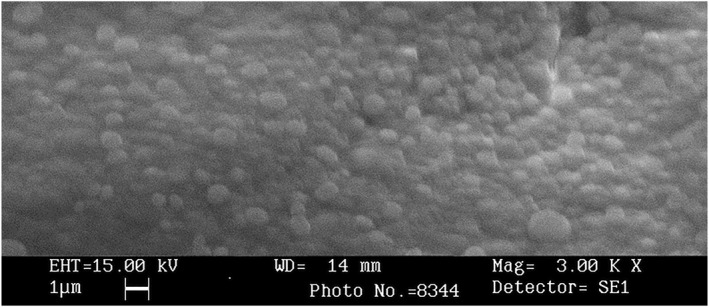
SEM image of nanoparticles
TABLE 5.
Particle size, zeta potential and PDI of the blank and DT loaded NPs
| Formulations | Z‐ average mean diameter (nm) | Intensity mean diameter (nm) | Volume mean diameter (nm) | Number mean diameter (nm) | PDI | Zeta potential (mV) |
|---|---|---|---|---|---|---|
| Optimised blank Alg‐NPs | 400.7 | 202.5 | 202.9 | 189.4 | 0.584 | −23.7 |
| DT loaded nanoparticles | 276.6 | 149.8 | 154.7 | 149.1 | 0.493 | −21.2 |
Abbreviations: DT, diphtheria toxoid; NPs, nanoparticles; PDI, polydispersity index.
FIGURE 3.

Size distribution records of blank Alg nanoparticles by (a) intensity, (b) volume, (c) number and (d) zeta potential distribution
FIGURE 4.

Size distribution record of diphtheria toxoid loaded Alg nanoparticles by (a) intensity, (b) volume, (c) number and (d) zeta potential distribution
3.4. Loading efficiency and loading capacity
Different concentrations of DT were loaded in Alg‐NPs to explore the effect of antigen amount on LE and LC. As illustrated in Figure 5, by increasing the DT concentration, the LE and LC increased as well. The obtained results demonstrated that the amount of LE and LC increased significantly using 3 mg/ml DT concentration.
FIGURE 5.

Effect of different diphtheria toxoid (DT) concentrations on the loading efficiency (LE) and loading capacity (LC) of Alg‐NPs
3.5. In vitro DT release
Release of the DT loaded Alg‐NPs was carried out using the phosphate buffer (PBS, pH 7.4) at 37°C for 190 h. According to Figure 6, the release behaviour of DT loaded Alg‐NPs was slow and continuous on the first 32 h, which had a steep slope and carried on in a constant slope until 140 h. Considering that, 95% of DT was approximately released in 140 h.
FIGURE 6.

In vitro release profile of diphtheria toxoid from alginate nanoparticles
3.6. Fourier transform infra‐red (FTIR) measurements
The free and loaded NPs spectra investigation showed some changes in the amino and carboxyl groups and amide peaks, indicating an ionic interaction between the carbonyl group of alginate and the amino group of DT. The obtained FTIR results confirmed the encapsulation of DT in NPs. The FTIR spectra of alginate and DT loaded NPs are demonstrated in Figure 7.
FIGURE 7.
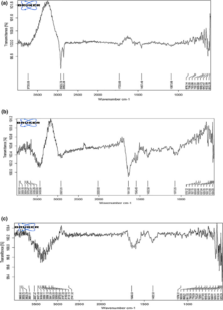
FTIR spectra of alginate and diphtheria toxoid (DT) loaded nanoparticles (NPs): (a) blank NPs (b) DT and (c) DT‐loaded alginate NPs (sodium alginate concentration 0.2% w/v, CaCl2 0.1% w/v and DT 3 mg/ml)
4. DISCUSSION
Biocompatible NPs have received a lot of attention for antigen or drug delivery system owing to their safety and protecting antigens or drugs from the physiological and environmental changes. Alginate is a biocompatible polymer which widely used for antigen or drug delivery, protein or enzyme carrier, wound healing, tissue engineering and as matrices to immobilise cells and importantly is FDA‐approved [39, 56]. In the current study, Alg‐NPs were made up by ionic gelation method. Previously conducted studies have addressed that the concentrations of alginate solution as well as CaCl2 cross‐linker agent had an efficient effect on the particle size formation [57, 58, 59, 60, 61]. To understand how the effect of different concentrations of CaCl2 and sodium alginate affects the formation of Alg‐NPs, different concentrations of CaCl2 (0.1%, 0.2% and 0.3% w/v) and sodium alginate (0.1%, 0.2% and 0.3% w/v) were employed for Alg‐NPs preparation in primary screening. The optimized formulation was chosen based on the suitable morphology in terms of some items such as particle size and poly dispersity index, high protein loading and sustained release. In the current research, 0.2% w/v of sodium alginate and 0.1% w/v of CaCl2 were appropriate concentrations for preparing the optimized Alg‐NPs in terms of above‐mentioned criteria.
As listed in Table 1, an increment in sodium alginate concentration of more than 0.2% w/v would lead to the development of viscosity and insufficiency of enough shear stress, which facilitates the macroscopic gel aggregates formation as well as Alg‐NPs particle size increment. On the other hand, an important factor in forming of NPs is the orientation of functional groups of alginate chains such as carboxylate groups to make a complex structure associated with calcium ions. Accordingly, in conjunction with increasing in the alginate concentration, more functional groups can be obtained, such that neighbouring calcium ions can be collected, leading more layers of alginate chains link the calcium ions. Subsequently, the more alginate concentration is led to the more increase in Alg‐NPs size. In contrast, increasing the CaCl2 concentration would result in an imbalance between Ca2+ and alginate binding sites and, lead to lower numbers of the polymer chains involved in higher contents of calcium ions. Therefore, the cross linking between the glucoronic acid residues and calcium ions was developed that formed the microscopic gel aggregates as well as the Alg‐NPs smaller size in conjunction with increasing of CaCl2 concentration. Here, it is worthwhile mentioning that the results of the current research were in accordance with previous conducted studies, which had described an increasing in sodium alginate and CaCl2 concentrations would lead to increase and decrease in Alg‐NPs size formation, respectively [32, 57, 58, 59, 60, 61].
Effects of certain processing parameters such as homogenization time and rate on Alg‐NPs size were evaluated. Our results showed that an increase in homogenization time from 15 to 60 min would lead to a decrease in the mean particle size. Other similar studies had correspondingly shown that increasing in the homogenization time had ultimately been led to a decrease in the particle size [32, 57, 58]. On the other hand, by increasing the homogenization rate from 800 to 2000 rpm, the Alg‐NPs size was decreased. Meanwhile, similar results have been reported in previous studies [32, 57, 62, 63]. So, these results illustrated that homogenization rate of 2000 rpm at 30 min might be an optimized condition for Alg‐NPs preparation (Table 3).
The results determined by DLS demonstrated that size distribution of NPs before and after loading DT was achieved in a narrow range and only one peak was observed (Figures 3 and 4). Nanoparticles in the range of 200–500 nm are at appropriate size for antigen or drug delivery [17]. The NPs adherence to the cell membranes and immune cells uptake depends on the surface charge. Accordingly, conversion of the particle surface charge could control binding to the cells [64]. Electrophoretic mobility of particles and the total electric charge on the particles surface are determined by zeta potential technique. Zeta potential is in particular important for NPs in suspension as a key role in interaction between the drug or antigen and NPs, and as the suspension stability indicative and particle surface morphology [65, 66, 67, 68]. The zeta potential of Alg‐NPs was obtained −23.7 mV and then changed to −21.2 mV (Figure 3d) after DT loading (Figure 4d). This negative zeta potential value for Alg‐NPs and DT‐loaded NPs demonstrates the high surface charge of prepared NPs. These surface charges lead to high suspension stability for Alg‐NPs before and after encapsulation with DT through strong repellent interaction between NPs in the dispersion. On the other hand, it should be noted that a reduction in the zeta potential value was due to the encapsulation of DT because of interaction between the negative carboxyl (COO−) groups on Alg‐NPs, which neutralised by positive (NH2+) groups on DT, leading to DT‐Alg‐NPs formation. As a result, this alteration indicated that the DT encapsulation was successfully performed. Afterwards, different concentrations of DT were loaded in NPs to investigate the ability of Alg‐NPs for encapsulating antigens as well as the effect of the antigen amount on LE and LC. Thus, by increasing the DT concentration, the LE and LC were increased as well. Furthermore, created NPs in the current study with high concentration of DT showed significant LE (>80%) and LC (>90%) (Figure 5). Finally, a suitable concentration for preparing optimum Alg‐NPs was achieved at 3 mg/ml DT concentration. It should be noted that the higher DT encapsulation efficiency of Alg‐NPs might be due to the interaction of DT with hydroxyl groups of alginate chains via the formation of hydrogen and electrostatic bounds on the unsaturated sites in alginate chains. As the DT and polymer had the same solvent (water), the antigen cannot diffuse from the polymer [69]. Accordingly, by increasing the DT concentration, free binding sites in alginate chains are saturated with DT and leave not adequate space to entrap the antigen. Therefore, LE did not increase in parallel to DT concentration in higher than 3 mg/ml. The similar observations have previously been reported in other studies [49, 70, 71].
The main mechanism for antigen release from Alg‐NPs is carried out by the diffusional processes through pores comforted by the polymeric network degradation [72, 73]. In this study, In vitro release of the DT from Alg‐NPs was a biphasic linear profile with long release time (as depicted in Figure 6). The first phase was very fast that was followed by a slow release. The initial fast release could be interpreted as due to the free DT adsorbed on the Alg‐NPs surfaces. Furthermore, the sustained release was related to the cleavage of the chemical and electrostatic bonds between DT and sodium alginate particles [72, 74]. The DT‐loaded NPs prepared in the current study owing to the good sustained release without burst release can be used as candidate for antigen delivery.
The FTIR spectra of the sodium alginate NPs, DT and DT‐loaded NPs were compared in Figure 7. According to the FTIR spectra, a low difference can be observed in the width and frequency of the peaks, in which the wide peak obtained in 3400 cm−1 is corresponding to O‐H stretching and intermolecular hydrogen bonding. The wave of 1402–1640 cm−1 is matching with the C=O stretching (amide). Even though, stretching vibration of aliphatic C‐H was observed at 2850–2920 cm−1. The carboxyl peaks were seen adjacent to 1646 cm−1 (symmetric –COO− stretching vibration) and 1415 cm−1 (asymmetric COO– stretching vibration), which were expanded after interaction with DT. Comparison of the spectra for frees and loaded NPs showed an ionic interaction between the carboxyl group of alginate and the amino group of DT. As a consequence, the FTIR results confirmed the successful interaction of Alg‐NPs and DT.
5. CONCLUSION
In the current study, the alginate NPs were prepared and optimized. Moreover, the influence of various concentrations of sodium alginate and calcium chloride were investigated in addition to some physical conditions such as homogenization time and rate on the NPs physicochemical characteristics. This study described that alginate could be employed as a static environment for controlling drug release at an advantageous rate. For this purpose, the alginate NPs were synthesised by the addition of calcium chloride solution to the diluted solutions of sodium alginate through the controlled ionic gelation method. The obtained results showed that the features of NPs such as morphology and particle size were strongly dependent on the calcium chloride and sodium alginate concentrations as well as physicochemical conditions in the NPs formation process. Moreover, the suitable concentrations of sodium alginate and calcium ions would lead to obtaining the desirable NPs formation via desirable LE and LC (over 80%) and sustained in vitro release profile. Also, the effect of other processing parameters such as homogenization time and homogenization rate was investigated on the NPs size. According to the obtained results, as the homogenization time increased from 15 to 60 min, the particle size inversely decreased.
For further research, it is suggested that the DT‐loaded on sodium alginate NPs could be utilised as a proper system for DT antigen delivery. Moreover, it can be employed to produce monovalent diphtheria NP vaccine or in combination with other bacterial NP vaccines for prevention as well as in cancer immunotherapy conjugated with anti‐CDs. Furthermore, the proposed sodium alginate NPs along with the formulation of cross‐reacting recombinant diphtheria antigens such as CRM197, could be used as a new vaccine platform.
CONFLICT OF INTEREST
The authors declare that they have no conflict of interest(s).
CREDIT CONTRIBUTION STATEMENT
Not applicable.
ACKNOWLEDGEMENTS
We are here expressing our appreciation for the colleagues' assistance at the Central Laboratory and R&D Departments in the course of this research. The current study was partially financed by Razi Vaccine and Serum Research Institute (RVSRI) through the project No. 2‐18‐18‐111‐990723.
Aghamiri, S. , et al.: Investigating preparation and characterisation of diphtheria toxoid‐loaded on sodium alginate nanoparticles. IET Nanobiotechnol. 16(5), 199–209 (2022). 10.1049/nbt2.12088
DATA AVAILABILITY STATEMENT
Almost all documents related to the current research on preparation and characterization of the NPs and data analyzed during this study are entirely included.
REFERENCES
- 1. Khan, I. , et al.: Nanobiotechnology and its applications in drug delivery system: a review. IET Nanobiotechnol. 9(6), 396–400 (2015). 10.1049/iet-nbt.2014.0062 [DOI] [PubMed] [Google Scholar]
- 2. Mehrabi, M. , et al.: Development and optimisation of hepatitis B recombinant antigen loaded chitosan nanoparticles as an adjuvant using the response surface methodology. Micro & Nano Lett. 15(11), 736–741 (2020). 10.1049/mnl.2019.0355 [DOI] [Google Scholar]
- 3. Gregory, A.E. , Williamson, D. , Titball, R. : Vaccine delivery using nanoparticles. Front. Cell. Infect. Microbiol. 3, 13 (2013). 10.3389/fcimb.2013.00013 [DOI] [PMC free article] [PubMed] [Google Scholar]
- 4. Akagi, T. , Baba, M. , Akashi, M. : Biodegradable nanoparticles as vaccine adjuvants and delivery systems: regulation of immune responses by nanoparticle‐based vaccine. In: Kunugi, S. , Yamaoka, T. (eds.) Polymers in Nanomedicine. Advances in Polymer Science, vol. 247, pp. 31–64. Springer, Berlin, Heidelberg (2011). 10.1007/12_2011_150 [DOI] [Google Scholar]
- 5. Guy, B. : The perfect mix: recent progress in adjuvant research. Nat. Rev. Microbiol. 5(7), 396–397 (2007). 10.1038/nrmicro1681 [DOI] [PubMed] [Google Scholar]
- 6. Harandi, A.M. , Medaglini, D. , Shattock, R.J. , convened by EUROPRISE WG .: Vaccine adjuvants: a priority for vaccine research. Vaccine. 28(12), 2363–2366 (2010). 10.1016/j.vaccine.2009.12.084 [DOI] [PubMed] [Google Scholar]
- 7. Abd el‐Razek, N. , Shoman, S. , Mohamed, A. : Nanocapsulated rift valley fever vaccine candidates and relative immunological and histopathological reactivity in out bred Swiss mice. J. Vaccines Vaccin. 2, 115 (2011) [Google Scholar]
- 8. Glenny, A.T. : The antigenic value of toxoid precipitated by potassium alum. J. Pathol. Bacteriol. 29, 38–45 (1926) [Google Scholar]
- 9. Gupta, R.K. , et al.: Adjuvants—a balance between toxicity and adjuvanticity. Vaccine. 11(3), 293–306 (1993). 10.1016/0264-410x(93)90190-9 [DOI] [PubMed] [Google Scholar]
- 10. Saroja, C. , Lakshmi, P. , Bhaskaran, S. : Recent trends in vaccine delivery systems: a review. Int. J. Pharmaceut. Invest. 1(2), 64 (2011). 10.4103/2230-973x.82384 [DOI] [PMC free article] [PubMed] [Google Scholar]
- 11. Alshanqiti, F.M. , et al.: Development of nanoparticle adjuvants to potentiate the immune response against diphtheria toxoid. Hum. Antibodies. 26(2), 75–85 (2018). 10.3233/hab-170324 [DOI] [PubMed] [Google Scholar]
- 12. Tonkin, J.A. , et al.: Optical tracking of drug release from porous silicon delivery vectors. IET Optoelectron. 8(2), 113–116 (2014). 10.1049/iet-opt.2013.0080 [DOI] [Google Scholar]
- 13. Akagi, T. , Baba, M. , Akashi, M. : Biodegradable nanoparticles as vaccine adjuvants and delivery systems: regulation of immune responses by nanoparticle‐based vaccine. In Polymers in Nanomedicine, pp. 31–64. Springer; (2011) [Google Scholar]
- 14. Oyewumi, M.O. , Kumar, A. , Cui, Z. : Nano‐microparticles as immune adjuvants: correlating particle sizes and the resultant immune responses. Expet. Rev. Vaccine. 9(9), 1095–1107 (2010). 10.1586/erv.10.89 [DOI] [PMC free article] [PubMed] [Google Scholar]
- 15. Soppimath, K.S. , et al.: Biodegradable polymeric nanoparticles as drug delivery devices. J. Contr. Release. 70(1‐2), 1–20 (2001). 10.1016/s0168-3659(00)00339-4 [DOI] [PubMed] [Google Scholar]
- 16. Hans, M.L. , Lowman, A.M. : Biodegradable nanoparticles for drug delivery and targeting. Curr. Opin. Solid State Mater. Sci. 6(4), 319–327 (2002). 10.1016/s1359-0286(02)00117-1 [DOI] [Google Scholar]
- 17. Ravi, C. , Ahmed, K.Z. , Azad, M.A.K. : Fabrication of poly (D, L‐lactic acid) nanoparticles as delivery system for sustained release of L‐theanine. IET Nanobiotechnol. 13(7), 742–747 (2019). 10.1049/iet-nbt.2018.5248 [DOI] [PMC free article] [PubMed] [Google Scholar]
- 18. Hajizade, A. , et al.: Nanoparticles in vaccine development. J. Appl. Biotechnol. Rep. 1(4), 125–134 (2014) [Google Scholar]
- 19. Keikha, R. , Daliri, K. , Jebali, A. : The use of nanobiotechnology in immunology and vaccination. Vaccines. 9(2), 74 (2021). 10.3390/vaccines9020074 [DOI] [PMC free article] [PubMed] [Google Scholar]
- 20. Gutierro, I. , et al.: Size dependent immune response after subcutaneous, oral and intranasal administration of BSA loaded nanospheres. Vaccine. 21(1‐2), 67–77 (2002). 10.1016/s0264-410x(02)00435-8 [DOI] [PubMed] [Google Scholar]
- 21. Kim, M.‐G. , et al.: Nanotechnology and vaccine development. Asian J. Pharm. Sci. 9(5), 227–235 (2014). 10.1016/j.ajps.2014.06.002 [DOI] [Google Scholar]
- 22. Illum, L. , Farraj, N.F. , Davis, S.S. : Chitosan as a novel nasal delivery system for peptide drugs. Pharmaceut. Res. 11(8), 1186–1189 (1994). 10.1023/a:1018901302450 [DOI] [PubMed] [Google Scholar]
- 23. Vila, A. , et al.: Low molecular weight chitosan nanoparticles as new carriers for nasal vaccine delivery in mice. Eur. J. Pharm. Biopharm. 57(1), 123–131 (2004). 10.1016/j.ejpb.2003.09.006 [DOI] [PubMed] [Google Scholar]
- 24. Sahoo, N. , et al.: Recent advancement of gelatin nanoparticles in drug and vaccine delivery. Int. J. Biol. Macromol. 81, 317–331 (2015). 10.1016/j.ijbiomac.2015.08.006 [DOI] [PubMed] [Google Scholar]
- 25. Choi, K.Y. , et al.: Self‐assembled hyaluronic acid nanoparticles as a potential drug carrier for cancer therapy: synthesis, characterization, and in vivo biodistribution. J. Mater. Chem. 19(24), 4102–4107 (2009). 10.1039/b900456d [DOI] [Google Scholar]
- 26. Sharma, S. , et al.: Enhanced immune response against pertussis toxoid by IgA‐loaded chitosan–dextran sulfate nanoparticles. J. Pharmaceut. Sci. 101(1), 233–244 (2012). 10.1002/jps.22763 [DOI] [PubMed] [Google Scholar]
- 27. Uenaka, A. , et al.: T cell immunomonitoring and tumor responses in patients immunized with a complex of cholesterol‐bearing hydrophobized pullulan (CHP) and NY‐ESO‐1 protein. Cancer Immun. Arch. 7(1) (2007) [PMC free article] [PubMed] [Google Scholar]
- 28. Hasegawa, K. , et al.: In vitro stimulation of CD8 and CD4 T cells by dendritic cells loaded with a complex of cholesterol‐bearing hydrophobized pullulan and NY‐ESO‐1 protein: Identification of a new HLA‐DR15–binding CD4 T‐cell epitope. Clin. Cancer Res. 12(6), 1921–1927 (2006). 10.1158/1078-0432.ccr-05-1900 [DOI] [PubMed] [Google Scholar]
- 29. Honda‐Okubo, Y. , Saade, F. , Petrovsky, N. : Advax™, a polysaccharide adjuvant derived from delta inulin, provides improved influenza vaccine protection through broad‐based enhancement of adaptive immune responses. Vaccine. 30(36), 5373–5381 (2012). 10.1016/j.vaccine.2012.06.021 [DOI] [PMC free article] [PubMed] [Google Scholar]
- 30. Counoupas, C. , et al.: Delta inulin‐based adjuvants promote the generation of polyfunctional CD4+ T cell responses and protection against Mycobacterium tuberculosis infection. Sci. Rep. 7(1), 1–11 (2017). 10.1038/s41598-017-09119-y [DOI] [PMC free article] [PubMed] [Google Scholar]
- 31. Li, P. , et al.: Bioreducible alginate‐poly (ethylenimine) nanogels as an antigen‐delivery system robustly enhance vaccine‐elicited humoral and cellular immune responses. J. Contr. Release. 168(3), 271–279 (2013). 10.1016/j.jconrel.2013.03.025 [DOI] [PubMed] [Google Scholar]
- 32. Sarei, F. , et al.: Alginate nanoparticles as a promising adjuvant and vaccine delivery system. Indian J. Pharmaceut. Sci. 75(4), 442 (2013). 10.4103/0250-474x.119829 [DOI] [PMC free article] [PubMed] [Google Scholar]
- 33. Han, J. , et al.: Polymer‐based nanomaterials and applications for vaccines and drugs. Polymers. 10(1), 31 (2018). 10.3390/polym10010031 [DOI] [PMC free article] [PubMed] [Google Scholar]
- 34. Bibi, A. , Rehman, S.‐U. , Yaseen, A. : Alginate‐nanoparticles composites: kinds, reactions and applications. Mater. Res. Express. 6(9), 092001 (2019). 10.1088/2053-1591/ab2016 [DOI] [Google Scholar]
- 35. Pachiyappan, S. , et al.: Surfactant‐mediated synthesis of polyhydroxybutyrate (PHB) nanoparticles for sustained drug delivery. IET Nanobiotechnol. 13(4), 416–427 (2019). 10.1049/iet-nbt.2018.5053 [DOI] [PMC free article] [PubMed] [Google Scholar]
- 36. Rowe, R.C. , Sheskey, P. , Quinn, M. : Handbook of Pharmaceutical Excipients. Libros Digitales‐Pharmaceutical Press; (2009) [Google Scholar]
- 37. Sachan, N.K. , et al.: Sodium alginate: the wonder polymer for controlled drug delivery. J. Pharm. Res. 2(8), 1191–1199 (2009) [Google Scholar]
- 38. Remminghorst, U. , Rehm, B.H. : Bacterial alginates: from biosynthesis to applications. Biotechnol. Lett. 28(21), 1701–1712 (2006). 10.1007/s10529-006-9156-x [DOI] [PubMed] [Google Scholar]
- 39. Szekalska, M. , et al.: Alginate: current use and future perspectives in pharmaceutical and biomedical applications. Int. J. Polym. Sci. 2016, 1–17 (2016). 10.1155/2016/7697031 [DOI] [Google Scholar]
- 40. Draget, K.I. , Smidsrød, O. , Skjåk‐Bræk, G. : Alginates from Algae. Polysaccharides and Polyamides in the Food Industry: Properties, Production, and Patents, pp. 1–30 (2005) [Google Scholar]
- 41. Matricardi, P. , et al.: Recent advances and perspectives on coated alginate microspheres for modified drug delivery. Expet Opin. Drug Deliv. 5(4), 417–425 (2008). 10.1517/17425247.5.4.417 [DOI] [PubMed] [Google Scholar]
- 42. Whitney, J.C. , et al.: Structural basis for alginate secretion across the bacterial outer membrane. Proc. Natl. Acad. Sci. Unit. States Am. 108(32), 13083–13088 (2011). 10.1073/pnas.1104984108 [DOI] [PMC free article] [PubMed] [Google Scholar]
- 43. Tiyaboonchai, W. : Chitosan nanoparticles: a promising system for drug delivery. Naresuan University Journal: Sci. Technol. 11(3), 51–66 (2013) [Google Scholar]
- 44. De, S. , Robinson, D. : Polymer relationships during preparation of chitosan–alginate and poly‐l‐lysine–alginate nanospheres. J. Contr. Release. 89(1), 101–112 (2003). 10.1016/s0168-3659(03)00098-1 [DOI] [PubMed] [Google Scholar]
- 45. Yang, Y. , et al.: Assembled alginate/chitosan nanotubes for biological application. Biomaterials. 28(20), 3083–3090 (2007). 10.1016/j.biomaterials.2007.03.019 [DOI] [PubMed] [Google Scholar]
- 46. Christenson, L. , Dionne, K.E. , Lysaght, M.J. : Biomedical applications of immobilized cells. In: Fundamentals of Animal Cell Encapsulation and Immobilization, pp. 7–41 (1993) [Google Scholar]
- 47. Fang, Y. , et al.: Multiple steps and critical behaviors of the binding of calcium to alginate. J. Phys. Chem. B. 111(10), 2456–2462 (2007). 10.1021/jp0689870 [DOI] [PubMed] [Google Scholar]
- 48. Ouwerx, C. , et al.: Physico‐chemical properties and rheology of alginate gel beads formed with various divalent cations. Polym. Gels Netw. 6(5), 393–408 (1998). 10.1016/s0966-7822(98)00035-5 [DOI] [Google Scholar]
- 49. Moradi Bidhendi, S. , et al.: Design and evaluate alginate nanoparticles as a protein delivery system. Archives of Razi Institute. 68(2), 139–146 (2013) [Google Scholar]
- 50. Moloney, P. , Weld, C.B. : Diphtheria toxin‐antitoxin flocculation (Ramon test). J. Pathol. Bacteriol. 28(4), 655–672 (1925). 10.1002/path.1700280413 [DOI] [Google Scholar]
- 51. Peterson, G.L. : A simplification of the protein assay method of Lowry et al. which is more generally applicable. Anal. Biochem. 83(2), 346–356 (1977). 10.1016/0003-2697(77)90043-4 [DOI] [PubMed] [Google Scholar]
- 52. Maniatis, T. : Molecular Cloning: A Laboratory Manual, vol. 68. Cold Spring Harbor Laboratory, New York: (1982) [Google Scholar]
- 53. Bradford, M.M. : A rapid and sensitive method for the quantitation of microgram quantities of protein utilizing the principle of protein‐dye binding. Anal. Biochem. 72(1‐2), 248–254 (1976). 10.1016/0003-2697(76)90527-3 [DOI] [PubMed] [Google Scholar]
- 54. Govender, T. , et al.: PLGA nanoparticles prepared by nanoprecipitation: drug loading and release studies of a water soluble drug. J. Contr. Release. 57(2), 171–185 (1999). 10.1016/s0168-3659(98)00116-3 [DOI] [PubMed] [Google Scholar]
- 55. Sarmento, B. , et al.: Characterization of insulin‐loaded alginate nanoparticles produced by ionotropic pre‐gelation through DSC and FTIR studies. Carbohydr. Polym. 66(1), 1–7 (2006). 10.1016/j.carbpol.2006.02.008 [DOI] [Google Scholar]
- 56. U.S. Food and Drug Administration : Safe GRa. http://www.fda.gov/Food/IngredientsPackagingLabeling/GRAS/SCOGS/ucm260857.htm
- 57. Sonavane, G.S. , Devarajan, P.V. : Preparation of alginate nanoparticles using Eudragit E100 as a new complexing agent: development, in vitro, and in vivo evaluation. J. Biomed. Nanotechnol. 3(2), 160–169 (2007). 10.1166/jbn.2007.005 [DOI] [Google Scholar]
- 58. Ferreiro, M.G. , et al.: Characterization of alginate/poly‐L‐lysine particles as antisense oligonucleotide carriers. Int. J. Pharm. 239(1‐2), 47–59 (2002). 10.1016/s0378-5173(02)00030-3 [DOI] [PubMed] [Google Scholar]
- 59. Daemi, H. , Barikani, M. : Synthesis and characterization of calcium alginate nanoparticles, sodium homopolymannuronate salt and its calcium nanoparticles. Sci. Iran. 19(6), 2023–2028 (2012). 10.1016/j.scient.2012.10.005 [DOI] [Google Scholar]
- 60. Rajaonarivony, M. , et al.: Development of a new drug carrier made from alginate. J. Pharmaceut. Sci. 82(9), 912–917 (1993). 10.1002/jps.2600820909 [DOI] [PubMed] [Google Scholar]
- 61. Goudarzi, H.R. , et al.: Preparation and evaluation of alginate nanoparticles containing pertussis toxin as a particulate delivery system. Int. J. Adv. Biotechnol. Res. 7(2), 558–564 (2016) [Google Scholar]
- 62. Haznedar, S. , Dortunc, B. : Preparation and in vitro evaluation of Eudragit microspheres containing acetazolamide. Int. J. Pharm. 269(1), 131–140 (2004). 10.1016/j.ijpharm.2003.09.015 [DOI] [PubMed] [Google Scholar]
- 63. Pongpaibul, Y. , Price, J. , Whitworth, C. : Preparation and evaluation of controlled release indomethacin microspheres. Drug Dev. Ind. Pharm. 10(10), 1597–1616 (1984). 10.3109/03639048409039070 [DOI] [Google Scholar]
- 64. Patil, S. , et al.: Protein adsorption and cellular uptake of cerium oxide nanoparticles as a function of zeta potential. Biomaterials. 28(31), 4600–4607 (2007). 10.1016/j.biomaterials.2007.07.029 [DOI] [PMC free article] [PubMed] [Google Scholar]
- 65. Mohanraj, V. , Chen, Y. : Nanoparticles‐a review. Trop. J. Pharmaceut. Res. 5(1), 561–573 (2006). 10.4314/tjpr.v5i1.14634 [DOI] [Google Scholar]
- 66. Yang, S.‐G. , et al.: 99mTc‐hematoporphyrin linked albumin nanoparticles for lung cancer targeted photodynamic therapy and imaging. J. Mater. Chem. 20(41), 9042–9046 (2010). 10.1039/c0jm01544j [DOI] [Google Scholar]
- 67. El‐Houssiny, A.S. , et al.: Sodium alginate nanoparticles as a new transdermal vehicle of glucosamine sulfate for treatment of osteoarthritis. Eur. J. Nanomed. 9(3‐4), 105–114 (2017) [Google Scholar]
- 68. Chen, C.‐C. , et al.: Effects of lipophilic emulsifiers on the oral administration of lovastatin from nanostructured lipid carriers: physicochemical characterization and pharmacokinetics. Eur. J. Pharm. Biopharm. 74(3), 474–482 (2010). 10.1016/j.ejpb.2009.12.008 [DOI] [PubMed] [Google Scholar]
- 69. Peng, D. , et al.: Preparation of novel polymeric microspheres for controlled release of finasteride. Int. J. Pharm. 342(1‐2), 82–86 (2007). 10.1016/j.ijpharm.2007.05.002 [DOI] [PubMed] [Google Scholar]
- 70. Li, X. , et al.: Preparation of alginate coated chitosan microparticles for vaccine delivery. BMC Biotechnol. 8(1), 1–11 (2008). 10.1186/1472-6750-8-89 [DOI] [PMC free article] [PubMed] [Google Scholar]
- 71. Ahmad, Z. , et al.: Alginate nanoparticles as antituberculosis drug carriers: formulation development, pharmacokinetics and therapeutic potential. Indian J. Chest Dis. Allied Sci. 48(3), 171 (2006) [PubMed] [Google Scholar]
- 72. Goh, C.H. , Heng, P.W.S. , Chan, L.W. : Alginates as a useful natural polymer for microencapsulation and therapeutic applications. Carbohydr. Polym. 88(1), 1–12 (2012). 10.1016/j.carbpol.2011.11.012 [DOI] [Google Scholar]
- 73. Mi, F.‐L. , Sung, H.‐W. , Shyu, S.‐S. : Drug release from chitosan–alginate complex beads reinforced by a naturally occurring cross‐linking agent. Carbohydr. Polym. 48(1), 61–72 (2002). 10.1016/s0144-8617(01)00212-0 [DOI] [Google Scholar]
- 74. Anal, A.K. , et al.: Chitosan‐alginate multilayer beads for gastric passage and controlled intestinal release of protein. Drug Dev. Ind. Pharm. 29(6), 713–724 (2003). 10.1081/ddc-120021320 [DOI] [PubMed] [Google Scholar]
Associated Data
This section collects any data citations, data availability statements, or supplementary materials included in this article.
Data Availability Statement
Almost all documents related to the current research on preparation and characterization of the NPs and data analyzed during this study are entirely included.