Abstract
Background
Our aim was to identify acute kidney injury (AKI) and subacute kidney injury using both KDIGO criteria and urinary biomarkers in children with mild/moderate COVID-19.
Methods
This cross-sectional study included 71 children who were hospitalized with a diagnosis of COVID-19 from 3 centers in Istanbul and 75 healthy children. We used a combination of functional (serum creatinine) and damage (NGAL, KIM-1, and IL-18) markers for the definition of AKI and subclinical AKI. Clinical and laboratory features were evaluated as predictors of AKI and subclinical AKI.
Results
Patients had significantly higher levels of urinary biomarkers and urine albumin–creatinine ratio than healthy controls (p < 0.001). Twelve patients (16.9%) developed AKI based on KDIGO criteria, and 22 patients (31%) had subclinical AKI. AKI group had significantly higher values of neutrophil count on admission than both subclinical AKI and non-AKI groups (p < 0.05 for all). Neutrophil count was independently associated with the presence of AKI (p = 0.014).
Conclusions
This study reveals that even children with a mild or moderate disease course are at risk for AKI. Association between neutrophil count and AKI may point out the role of inflammation in the development of AKI.
Impact
The key message of our article is that not only children with severe disease but also children with mild or moderate disease have an increased risk for kidney injury due to COVID-19.
Urinary biomarkers enable the diagnosis of a significant number of patients with subclinical AKI in patients without elevation in serum creatinine.
Our findings reveal that patients with high neutrophil count may be more prone to develop AKI and should be followed up carefully.
We conclude that even children with mild or moderate COVID-19 disease courses should be evaluated for AKI and subclinical AKI, which may improve patient outcomes.
Introduction
Coronavirus disease 2019 (COVID-19) is considered a respiratory illness primarily; however, the disease can also cause several extra-pulmonary manifestations involving multiple organs.1 The kidneys are one of the affected organs in COVID-19 patients. The clinical manifestations of kidney involvement in COVID-19 vary from subclinical kidney injury to acute kidney injury (AKI) requiring kidney replacement therapy.2,3 AKI is a severe complication of COVID-19 and one of the most important predictors of mortality.4 It has been reported that 0.5–37% of hospitalized patients with COVID-19 develop AKI, and this rate was higher among patients admitted to the intensive care unit (ICU).5–7 Although pediatric COVID-19 patients usually have less severe disease than adult COVID-19 patients, COVID-19-associated AKI has been reported to develop in as many as 29% of hospitalized children and 44% of children admitted to the ICU.8,9 The evidence so far shows that critical illness and ICU admission are the main risk factors for AKI.
The non-uniform definitions used for AKI make it difficult to compare studies on patients with COVID-19. The consensus statement of Acute Disease Quality Initiative (ADQI) Workgroup on COVID-19-associated AKI recommended the use of Kidney Disease: Improving Global Outcomes (KDIGO) criteria, including serum creatinine (sCr) level and urine output, to define and report AKI.10,11 The ADQI consensus report also stated the need of using novel biomarkers for the early detection and diagnosis of AKI due to the restrictions of the creatinine-based criteria.10 Indeed, studies have shown that urinary biomarkers enable the diagnosis of subclinical AKI in patients without elevation in sCr or decrease in urine output and provide prognostic information about kidney injury and outcomes.12,13 There are limited data on urinary biomarkers for the diagnosis of COVID-19-associated AKI in adults. Available data have revealed the occurrence of subclinical AKI in COVID-19 and the prognostic value of urinary biomarkers in terms of the need for ICU admission, renal outcome, and mortality.4,14–19
We hypothesized that not only children with severe disease but also children with a mild/moderate disease have the various stages of AKI due to COVID-19. We aimed to determine the incidence of AKI and subclinical AKI in hospitalized children with mild/moderate COVID-19 who were not admitted to the ICU. We also aimed to identify the predictors of AKI and subclinical AKI in this patient population.
Materials and methods
Study design and study population
This cross-sectional observational study was based on data collected from three pediatric centers in Istanbul, Turkey, between April and August 2020. The procedure was conducted with the written and informed consent of the parents or guardians of the minors and in accordance with all applicable ethical and legal rules concerning medical research involving human studies in the Declaration of Helsinki ethical statement. The study protocol and this consent procedure were approved by the local Ethics Committee (number: 57696, April 2020).
All children under the age of 18 years who were hospitalized with a diagnosis of COVID-19 were included in the study. COVID-19 was diagnosed on the basis of (i) a positive result for severe acute respiratory syndrome coronavirus 2 (SARS-CoV-2) on reverse transcriptase–polymerase chain reaction (RT-PCR) testing of an oro-nasopharyngeal swab or (ii) a typical chest radiography or chest computed tomography (CT) in patients with clinical symptoms associated with COVID-19 and a positive contact history but without positive PCR test results. Ground-glass opacities, with or without consolidations, in lung regions close to visceral pleural surfaces, including the fissures (subpleural sparing is allowed) and multifocal bilateral distribution were taken as obligatory features for a typical pulmonary involvement of COVID-19.20,21 Children with chronic kidney disease taking any immunosuppressive medication or admitted to the ICU were excluded. A total of 71 patients were eligible for inclusion in the study (Supplemental Material 1).
Seventy-five healthy individuals (age and sex similar to the patients) without an active infection, diagnosis of COVID-19, or known kidney disease served as controls to assess urinary biomarker levels. The control group was composed of healthy children of healthcare professionals and healthy siblings of patients followed in the nephrology outpatient clinic.
Clinical assessment
Demographics, clinical characteristics (presentation symptoms, disease severity, comorbidities, treatment, and outcome), and radiological features were recorded. Height and body weight were measured and expressed in their standard deviation score (SDS) according to Turkish pediatric growth percentiles.22 Body mass index (BMI) and BMI-SDS were calculated. Obesity was defined as a height-specific BMI greater than 95th percentile. Office measurements of blood pressure (BP) on admission were recorded, and SDSs of BP were calculated according to age-, sex-, and height-specific normative values in the Fourth Report.23
Laboratory assessment
Initial laboratory data, including complete blood count, serum urea, creatinine, uric acid, lactic dehydrogenase, creatinine kinase, albumin, ferritin, C-reactive protein, procalcitonin (PCT), and D-dimer levels, blood gas tests, and urinalysis were collected. At admission, 2-mL blood samples were obtained to measure cystatin-C, and serum samples were stored at −80 °C. Serum cystatin-C concentrations were measured using immunonephelometric assays. The estimated glomerular filtration rate was calculated using the revised Schwartz equation.24
Spot urine samples at admission were obtained from patients and controls to measure neutrophil gelatinase-associated lipocalin (NGAL), kidney injury molecule-1 (KIM-1), interleukin-18 (IL-18), albumin, and creatinine (Cr) levels. Urine samples were collected by a mid-stream urine or a urine collection bag and immediately centrifuged for 15 min at 13,000 × g. Aliquots of the urine supernatant were stored at −80 °C and then analyzed using sandwich enzyme-linked immunosorbent assay (ELISA) kits for urinary biomarkers (Bioassay Technology Laboratory, China). The sensitivities of the ELISA kits (detection limits) were 1.03 ng/L, 2.00 ng/mL, and 0.01 ng/mL for IL-6 (Cat. No. E0090Hu), NGAL (Cat. No. E1719Hu), and KIM-1 (Cat. No. E1099Hu), respectively. Intra-assay variability was 7.5, 7.7, and 8% for IL-6, NGAL, and KIM-1, respectively. Inter-assay variability was 9.5, 8.6, and 9.2% for these markers, respectively. All urinary markers were normalized to urine creatinine levels. Albuminuria was defined as a urine albumin-to-creatinine ratio (ACR) >30 mg/g. Hematuria was defined as the presence of >5 red blood cells per high-power field on urinalysis. Neutropenia and lymphopenia were defined as a lower neutrophil or lymphocyte count than the lower limit of age specific normal, and neutrophilia was defined as a higher neutrophil count than the upper limit of age-specific normal.25,26
Definitions of AKI
AKI was defined as an increase of sCr by 0.3 mg/dL within 48 h or increase in sCr ≥ 1.5 times baseline within 7 days according to the KDIGO criteria.11 AKI was staged with increases in sCr 1.5–1.9, 2.0–2.9, and ≥3 times baseline as AKI stages 1, 2, and 3, respectively. To describe an increase in sCr levels, at least three measurements of sCr were obtained from the electronic records of the hospital systems at admission, peak level during hospitalization, and any value measured within 6–12 months before hospitalization as a baseline reference. If there was no sCr measurement in the previous 6–12 months, age-specific upper limits were used.27
Subclinical AKI was defined when at least one biomarker was positive without elevated sCr. Because there was no consensus on cut-off values for urinary biomarkers, the 95th percentile value of the control group was used for the positivity threshold for each urine biomarker (NGAL/Cr, KIM-1/Cr, and IL-18/Cr). Supplemental Material 2 shows the cut-off values we used and previous results from the literature.
Patients who had no signs of AKI or subclinical AKI were defined as non-AKI patients. Children with AKI were asked to control their sCr levels 3–6 months after hospital discharge.
Statistical analyses
Statistical analyses were performed using SPSS Version 21 (SPSS Inc., Chicago, IL). Continuous variables are presented as mean (±standard deviation) according to the distribution of data. Student’s t test or one-way analysis of variance were used to compare differences in continuous variables between the two groups, and the one-way analysis of variance with Bonferroni correction was used for a comparison of more than two groups’ means. Logarithmic transformation was used to transform skewed data into a normal distribution. Categorical variables are expressed as number (percentage). A chi-square or Fisher’s exact tests were used to compare categorical variables. Correlations between biomarkers and other clinical and laboratory variables were analyzed using Pearson’s or Spearman’s test. Statistical significance was defined as a two-tailed p value < 0.05.
Results
Demographic and clinical characteristics
A total of 71 hospitalized children with COVID-19 aged between 0.1 and 17.9 years and 75 healthy children were enrolled in the study. Forty patients (56%) had positive PCR results, and the remaining 31 (44%) showed clinical and radiological findings of COVID-19 and had a positive contact history. Twenty-nine (76%) of children with positive PCR showed typical chest imaging findings, whereas all children with negative PCR showed either pulmonary parenchymal ground-glass opacities or pulmonary consolidation. The demographic or clinical characteristics did not differ between the PCR-positive and PCR-negative cases (Supplemental Material 3).
The most common symptoms were cough (60%) and fever (59%). Only 5 patients (7%) had vomiting or diarrhea as the clinical manifestation at presentation, but none of the patients had hypotension. Sixteen patients (22.5%) had comorbid conditions; asthma and neurologic problems were the most common comorbidities. Table 1 summarizes the demographic and clinical characteristics of the children with COVID-19.
Table 1.
Clinical features of the children with COVID-19.
| Patients (n = 71) | |
|---|---|
| Age (years) | 9.4 ± 6.2 (0.1 to 17.9) |
| Sex (female), n (%) | 38 (53.5) |
| BMI-SDS | 0.19 ± 1.35 (−1.69 to 2.86) |
| Systolic BP-SDS | 0.42 ± 0.95 (−1.75 to 2.33) |
| Diastolic BP-SDS | 0.38 ± 0.81 (−1.13 to 2.32) |
| Comorbid conditions, n (%) | 16 (22.5) |
| Obesity | 3 |
| Allergic asthma | 5 |
| Global developmental delay | 5 |
| Cures from childhood malignancy | 2 |
| Type 2 diabetes mellitus | 1 |
| Symptoms, n (%) | |
| Fever | 42 (59) |
| Cough | 44 (62) |
| Shortness of breath | 14 (20) |
| Sore throat | 10 (23) |
| Vomiting and/or diarrhea | 5 (7) |
| Severity grading of pulmonary disease, n (%) | |
| Grade 1—Not admitted to hospital | 0 |
| Grade 2—Admitted to hospital with no respiratory support | 64 (90) |
| Grade 3—Admitted to hospital and required oxygen treatment | 5 (7) |
| Grade 4—Admitted to hospital and required high-flow nasal cannula oxygen | 2 (3) |
| Grade 5—Admitted to ICU and required invasive ventilation | 0 |
| Chest imaging findingsa, n (%) | |
| No specific radiologic findings | 2 (2.8) |
| Unilateral/bilateral consolidation | 52 (73.2) |
| Ground-glass opacification | 14 (19.7) |
| Peri-bronchial thickening | 6 (8.5) |
| Pleural effusion | 3 (4.2) |
| Treatment, n (%) | |
| Hydroxychloroqine | 12 (16.9) |
| Favipiravir | 9 (12.7) |
| Antibiotics | 2 (2.8) |
| Steroid | 0 |
Data presented as mean ± SD (minimum–maximum) or n (%).
SD standard deviation, BMI body mass index, SDS standard deviation score, BP blood pressure, ICU intensive care unit.
aSixty-nine children had various types of chest imaging findings suggestive of COVID-19 on chest radiography or computed tomography. Some patients showed more than one radiologic finding.
Sixty-nine children had various types of chest imaging findings suggestive of COVID-19. Sixty-four patients (90%) did not require respiratory support (Grade 2); the remaining five were under respiratory support with oxygen with a mask or nasal cannula (Grade 3), and two required non-invasive respiratory support (Grade 4) (Table 1). Children with infection severity of grades 3 and 4 showed significantly longer hospital stays than the patients of grade 2 (8.6 ± 2.1 vs 5.4 ± 3.2 days, p = 0.013). There were no children with the multi-system inflammatory syndrome (MIS-C) or no deaths in the cohort.
Urinary biomarkers and albuminuria
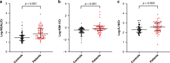
Patients with COVID-19 had significantly higher levels of urinary biomarkers (NGAL/Cr, KIM-1/Cr, and IL-18/Cr) than the healthy controls (p < 0.001 for all) (Fig. 1). A total of 27 patients (38%) showed at least one urinary biomarker positivity. Seventeen of them showed one biomarker positivity, eight patients had two biomarkers positive, and two patients had all the biomarkers positive. The most common positive urinary biomarker was KIM-1 (n = 21, 29.6%), followed by NGAL (n = 14, 19.7%) and IL-18 (n = 4, 5.6%).
Fig. 1. Comparison of urinary biomarkers between patients and healthy controls.
This figure shows the comparisons of logarithmically transformed urinary biomarkers [neutrophil gelatinase-associated lipocalin/creatinine (NGAL/Cr), kidney injury molecule-1/creatinine (KIM-1/Cr), and interleukin-18/creatinine (IL-18/Cr)] between healthy controls (n = 75) and patients with COVID-19 (n = 71). Original levels [median (Q1–Q3)], not logarithmically, for urinary biomarkers in healthy controls and patients with COVID-19, a NGAL/Cr levels: 29.8 (15.6–52.4) vs 71.3 (27.3–262.9) ng/mg, b KIM-1/Cr: 0.41 (0.16–0.72) vs 0.72 (0.34–1.86) ng/mg, c IL-18/Cr: 202.2 (110.7–371.2) vs 374.1 (151.0–948.9) pg/mg. Cr creatinine, AKI acute kidney injury.
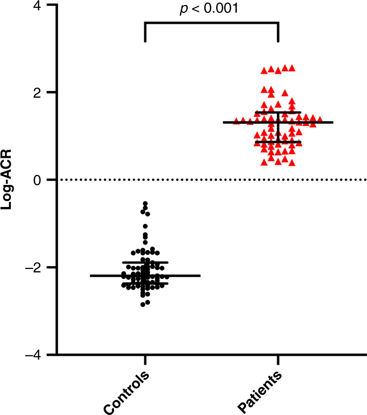
Patients with COVID-19 also had significantly higher urine ACR compared to the healthy controls [20.3 (7.3–34.8) vs 0.007 (0.003–0.013), p < 0.001] (Fig. 2). Eighteen out of 65 patients (26%), but none of the controls, had albuminuria. Thirteen of them had microalbuminuria, the remaining 5 had macroalbuminuria. Ten patients had microscopic hematuria. There was no difference in ACR levels between the patients with or without fever. In addition, there was no association between fever and ACR level.
Fig. 2. Comparison of ACR between patients and healthy controls.

This figure shows the comparisons of logarithmically transformed urinary albumin creatinine ratio (ACR) between healthy controls (n = 75) and patients with COVID-19 (n = 71). Patients with COVID-19 also had significantly higher urine ACR compared to the healthy controls [20.3 (7.3–34.8) vs 0.007 (0.003–0.013), p < 0.001]. ACR urinary albumin–creatinine ratio.
AKI and subclinical AKI
Based on the KDIGO criteria, 12 patients (16.9%) developed AKI; 11 patients were classified as AKI stage 1, and the remaining one as AKI stage 2. In 11 patients, AKI was present on admission and in 1 developed on the third day of the hospitalization. None of the patients required kidney replacement therapy. None of the healthy controls met AKI criteria.
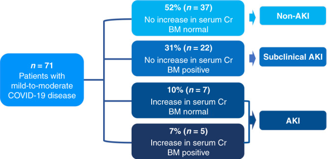
Twenty-two patients (31%) had subclinical AKI with elevated urinary biomarkers without an increase in sCr (Fig. 3). The levels of urinary biomarkers did not differ between the subclinical AKI and AKI groups. Patients with subclinical AKI had higher serum cystatin-C (0.95 (0.79–1.22) vs 0.78 (0.71–0.97), p = 0.036) and higher urine ACR (25.0 (14.5–119) vs 10.7 (5.9–24.3), p = 0.006) than non-AKI patients. Ten patients in the subclinical AKI group (46%), 3 in the AKI group (25%), and 5 in the non-AKI group (15%) showed albuminuria (p = 0.061).
Fig. 3. Distribution of the patients with AKI, subclinical AKI, and non-AKI.

Patients with COVID-19 (n = 71) were classified as acute kidney injury (AKI), subclinical AKI, and non-AKI groups regarding increase in serum creatinine (Cr) and biomarker (BM) positivity.
Table 2 shows the comparisons of the three groups (AKI, subclinical AKI, and non-AKI groups) in terms of clinical features and laboratory findings at the presentation. AKI group had significantly higher neutrophil (NEU) count and more patients with neutrophilia than both subclinical AKI and non-AKI groups (p < 0.05 for all). However, none of the clinical or laboratory findings differed between the subclinical AKI and non-AKI groups.
Table 2.
Comparisons of clinical features and initial laboratory findings between the AKI, subclinical AKI, and non-AKI groups.
| AKI group (n = 12) | Subclinical AKI group (n = 22) | Non-AKI group (n = 37) | pa | |
|---|---|---|---|---|
| Age (years) | 8.9 ± 4.8 | 10.2 ± 6.4 | 9.1 ± 6.5 | 0.75 |
| Sex (female), n (%) | 5 (42) | 12 (55) | 21 (57) | 0.66 |
| BMI-SDS | 0.53 ± 1.49 | 0.08 ± 1.26 | 0.14 ± 1.38 | 0.62 |
| Systolic BP-SDS | 0.46 ± 1.17 | 0.50 ± 1.00 | 0.32 ± 0.86 | 0.86 |
| Diastolic BP-SDS | 0.00 ± 0.58 | 0.48 ± 0.90 | 0.40 ± 0.79 | 0.53 |
| Severity of pulmonary disease, grade 2/grades 3–4, n | 12/0 | 19/3 | 33/4 | 0.43 |
| Laboratory findings | ||||
| Hemoglobin, g/dL | 12.7 ± 1.5 | 12.5 ± 2.2 | 12.5 ± 2.0 | 0.95 |
| Neutrophil, ×103/μL*,** | 9.7 ± 5.3 | 5.8 ± 5.2 | 4.6 ± 3.3 | 0.003 |
| Neutropenia, n (%) | 0 | 2 (10) | 2 (5) | 0.53 |
| Neutrophilia, n (%)*,** | 6 (50) | 5 (23) | 5 (14) | 0.037 |
| Lymphocyte, ×103/μL | 2.7 ± 1.4 | 2.6 ± 2.1 | 3.0 ± 1.8 | 0.67 |
| Lymphopenia, n (%) | 3 (25) | 9 (41) | 8 (22) | 0.24 |
| Platelet, ×103/μL | 340 ± 118 | 253 ± 82 | 260 ± 116 | 0.055 |
| CRP, mg/L | 41.7 ± 52.8 | 28.1 ± 47.7 | 16.5 ± 26.6 | 0.16 |
| Procalcitonin, ng/mL | 0.33 ± 0.53 | 0.41 ± 0.92 | 0.34 ± 1.16 | 0.97 |
| Ferritin, ng/mL | 100.1 ± 73.6 | 88.8 ± 131.4 | 74 ± 95 | 0.78 |
| D-dimer, μg/mL | 0.81 ± 0.49 | 2.00 ± 4.00 | 1.69 ± 2.71 | 0.65 |
| LDH, IU/L | 279 ± 135 | 269 ± 115 | 274 ± 69 | 0.96 |
| CK, IU/L | 76 ± 40 | 118 ± 108 | 157 ± 133 | 0.21 |
| Albumin, g/dL | 4.4 ± 0.3 | 4.2 ± 0.4 | 4.3 ± 0.34 | 0.47 |
| Blood pH | 7.39 ± 0.05 | 7.39 ± 0.04 | 7.40 ± 0.05 | 0.74 |
| HCO3, mEq/L | 23.7 ± 2.9 | 23.6 ± 3.4 | 23.1 ± 2.2 | 0.70 |
| Uric acid, mg/dL | 4.8 ± 2.1 | 3.4 ± 1.5 | 3.9 ± 1.2 | 0.12 |
| Creatinine, mg/dL | 0.60 ± 0.36 | 0.46 ± 0.18 | 0.44 ± 0.20 | 0.13 |
| Change in creatinine, % | 65 ± 17 | 15 ± 22 | 14 ± 17 | <0.001 |
| eGFR, mL/min/1.73 m2 (Schwartz) | 109 ± 27 | 130 ± 38 | 128 ± 28 | 0.13 |
Normal ranges for laboratory values: hemoglobin: 10–14 g/dL (1–3 months), 9.5–13.5 g/dL (3–6 months), 11–13.5 g/dL (6–24 months), 10–14 g/dL (2–5 years), 11.4–15.5 g/dL (5–8 years), 11.6–15.5 g/dL (8–12 years), 11.8–16 g/dL (12–18 years); neutrophil: 1–9 × 10³/μL (1–3 months), 1–8.5 × 10³/μL (3–24 months), 1.5–8.5 × 10³/μL (2–8 years), 1.5–8 × 10³/μL (8–15 years), 1.8–8 × 10³/μL (15–18 years); lymphocyte: 2.5–16.5 × 10³/μL (1–3 months), 4–13.5 × 10³/μL (3–6 months), 4–10.5 × 10³/μL (6–24 months), 2–9.5 × 10³/μL (2–5 years), 1.5–7 × 10³/μL (5–12 years), 1.2–6.2 × 10³/μL (12–18 years); platelet: 150–400 × 103/μL; CRP: < 5 mg/L; procalcitonin: <0.5 ng/dL; ferritin: 7–142 ng/mL; D-dimer: <0.5 μg/mL; LDH: 110–430 IU/L; CK < 171 IU/L; albumin: 3.8–5.4 g/dL; blood pH: 7.38–7.45; HCO3: 21–26 mEq/L; uric acid: 2–6 mg/dL.
AKI acute kidney injury, SD standard deviation, BMI body mass index, SDS standard deviation score, BP blood pressure, NLR neutrophil lymphocyte ratio, CRP C-reactive protein, LDH lactate dehydrogenase, CK creatine kinase, HCO3 bicarbonate, eGFR estimated glomerular filtration rate.
*p < 0.05 between the AKI and subclinical AKI groups; **p < 0.05 between the AKI and non-AKI groups.
aData presented mean ± SD and ANOVA with Bonferroni correction was used for comparisons of the three groups.
Bold values denote statistical significance at the p < 0.05.
Binary logistic regression analysis revealed that NEU count is an independent risk factor for AKI (p = 0.014, 95% CI 1.036–1.372). None of urinary biomarkers showed a significant association with NEU or inflammatory markers (CRP or PCT).
Ten of the 12 children with AKI were examined at a median of 4.3 months (range, 3.0–6.1 months) after discharge. All of these 10 patients had normal sCr level at follow-up, and the mean sCr level decreased from 0.60 ± 0.36 mg/dL (maximum creatinine during hospitalization) to 0.40 ± 0.25 mg/dL (p = 0.005).
Discussion
This study provides evidence for the development of AKI and subclinical AKI in children with mild-to-moderate COVID-19. In this cohort of children with less severe disease and a low number of concomitant complications, the incidence of AKI was 16.9% and the incidence of subclinical AKI was 31%. The fact that patients with AKI have higher indicators of inflammation than subclinical and non-AKI patients draw attention to the relationship between kidney injury and inflammation in this patient population.
AKI is a common condition in patients with COVID-19; however, its incidence varies by age, severity of illness, comorbidities, countries, regions, and AKI definitions.6 Studies in adults have reported higher incidence rates of AKI of up to 76%, particularly in patients with multiple comorbidities and those admitted to the ICU.10 Pediatric studies at the beginning of the pandemic did not report abnormal kidney function in children with COVID-19,28 but further studies demonstrated AKI ranging from 1.5 to 44% in children.8,9,28–30 Most of the pediatric studies included critically ill patients. The main risk factors for developing AKI were admission to the pediatric ICU, MIS-C, diarrhea, or vomiting as the initial symptoms or the presence of comorbidities. Based on our hypothesis, we excluded patients admitted to the ICU to avoid secondary factors associated with AKI. In this population with a relatively less severe COVID-19, the incidence of AKI according to KDIGO criteria was 16.9%. This finding suggests that children with the less severe disease may develop AKI due to COVID-19.
In addition to patients with AKI, almost one-third had subclinical kidney injury with biomarker positivity. The ADQI workgroup recommends using KDIGO criteria for a standardized definition of AKI in COVID-19 and also states the importance of using new biomarkers to diagnose and determine the prognosis of AKI in COVID-19.10 To date, few studies in adults but no pediatric studies have evaluated new biomarkers in COVID-19.4,15–19,29 Several urinary biomarkers such as NGAL, KIM-1, alpha-1 microglobulin, and tissue inhibitor of metalloproteinases-2 have emerged in adult studies as the useful diagnostic tool to detect AKI earlier and more sensitively than sCr. Our study revealed that urinary biomarkers including NGAL, KIM-1, and IL-18 were significantly higher in patients with COVID-19 than in healthy controls. Our study also showed that a significant number of patients had subclinical AKI due to COVID-19. This finding suggests that using biomarker positivity combined with traditional markers may allow early recognition of AKI and contribute toward an improved outcome in COVID-19 patients.
Relatively little is known about the pathogenesis of AKI in COVID-19. The latest evidence reveals that two main mechanisms are responsible for the development of AKI in COVID-19.31,32 The first is the direct cytopathic effects of the virus in the kidneys, which lead to endothelial dysfunction, complement activation, and inflammation in the tubules and glomerulus. Another is that complications of the disease, such as hypovolemia, hypoxia, hemodynamic changes, or secondary infections, contribute to lesions in the kidneys.9,31,33 In our cohort, patients presenting with gastrointestinal symptoms that may cause hypovolemia were less frequent, only two patients had secondary bacterial infection, and none had severe hypoxia, hemodynamic changes, or hyperinflammatory status. The direct effect of kidney tropism seems to be more prominent mechanism in the development of AKI in this cohort of children with mild/moderate COVID-19 and a low number of concomitant complications. It is also important to note that patients with AKI had significantly higher NEU counts than both subclinical AKI and non-AKI patients. Logistic regression for prediction AKI showed that NEU was the only independent factor for AKI. Taken together, these findings suggest that the systemic pro-inflammatory response in early phase of COVID-19 may play a key role in developing kidney injury.
Until now, several adult and pediatric studies showed the clinical importance of albuminuria for patient with AKI in various clinical settings.34–36 Recent studies on COVID-19 widely focuses more on proteinuria, which showed that the frequency of proteinuria reaches up to 44% in COVID-19 adult patients, on hospital admission.2 In our cohort, urinary albumin levels were significantly higher in patients with COVID-19 compared with healthy controls. In addition, almost half of the patients with subclinical AKI had albuminuria. Although it is known that febrile episodes can cause transient proteinuria,37 we could not show any difference in ACR levels between the patients with or without fever. On the other hand, unlike other infections that cause transient proteinuria, it is considered that COVID-19 acts directly on the glomeruli and tubules through multiple mechanisms.3 Unfortunately, we do not have short-term follow-up of ACR levels after recovery from COVID -19 to determine whether albuminuria is a permanent or transient sign of infection. Therefore, further studies are needed to evaluate albuminuria in a short-term follow-up period to conclude the presence of transient or permanent albuminuria.
The present study evaluates AKI and subclinical AKI in a relatively homogenous group of children with mild-to-moderate COVID-19 clinical course. Subclinical AKI is defined in this pediatric cohort by the assessment of three urinary biomarkers. Our study has several limitations. The first limitation is that our cohort included not only PCR-positive confirmed cases but also patients with a history of contact and characteristic radiological findings for COVID-19. The most definitive diagnosis of COVID-19 is usually made with a RT-PCR test, but the sensitivity reported in clinical practice ranges from 42 to 83%, depending on symptom duration, viral load, and quality of the test sample. Therefore, for symptomatic patients with positive contact history but not positive PCR test, the diagnosis was often made on the basis of radiological diagnostic criteria on CT. We attempted to standardize the radiologic evaluation according to the consensus reports to minimize errors in radiological evaluation. The second limitation of the study is that our cohort did not include a control group of patients with infections other than COVID-19 to assess the direct association of COVID-19 and AKI. The third, urine output criteria were not used for AKI definition since urine output was not accurately followed in non-critically ill patients. In addition, most patients were most likely treated with intravenous (IV) fluid at the time of hospitalization; however, no accurate data were available on the IV fluid. The last one is the cross-sectional design of our study, which did not allow us to assess the long-term data on urinary biomarkers in children with subclinical or clinical AKI.
In conclusion, this study reveals that even children with a mild or moderate disease course are at increased risk for AKI; however, most patients show early stages of AKI. In this cohort, neutrophil count show association with AKI, which may reflect the role of the inflammation cascade in the development of AKI. Use of biomarkers enable diagnosis of significant number of patients with subclinical AKI in patients without elevation in sCr.
Further studies evaluating patients with various disease severity are needed to improve our knowledge about the frequency and clinical consequences of AKI and subclinical AKI in children with COVID-19.
Supplementary information
Author contributions
S.S. and N.C. designed the project, provided funding acquisition, contributed to analysis and interpretation of the results, drafting the initial manuscript, editing the manuscript, and subsequent critical revisions. R.Y.C., A.A., E.K.Y., A.A.K.S., D.A., G.A., K.C.D., and D.K. completed the acquisition of clinical and laboratory data. H.C., S.C., and L.S. contributed to the review of the article. All authors read and approved the final version of the manuscript and agree to be accountable for all aspects of the work.
Funding
This work was supported by Scientific Research Projects Coordination Unit of Istanbul University-Cerrahpasa (Project number: 34892).
Data availability
The datasets generated during and/or analyzed during the current study are available from the corresponding author on reasonable request.
Competing interests
The authors declare no competing interests.
Ethics approval and consent to participate
The study protocol and this consent procedure were approved by the local Ethics Committee (number: 57696, April 2020). The procedure was conducted with the written and informed consent of the parents or guardians of the minors and in accordance with all applicable ethical and legal rules concerning medical research involving human studies in the Declaration of Helsinki ethical statement.
Footnotes
Publisher’s note Springer Nature remains neutral with regard to jurisdictional claims in published maps and institutional affiliations.
Supplementary information
The online version contains supplementary material available at 10.1038/s41390-022-02124-6.
References
- 1.Gupta A, et al. Extrapulmonary manifestations of Covid-19. Nat. Med. 2020;26:1017–1032. doi: 10.1038/s41591-020-0968-3. [DOI] [PubMed] [Google Scholar]
- 2.Cheng YC, et al. Kidney disease is associated with in -hospital death of patients with Covid-19. Kidney Int. 2020;97:829–838. doi: 10.1016/j.kint.2020.03.005. [DOI] [PMC free article] [PubMed] [Google Scholar]
- 3.Perico L, et al. Immunity, endothelial injury and complement-induced coagulopathy in Covid-19. Nat. Rev. Nephrol. 2021;17:46–64. doi: 10.1038/s41581-020-00357-4. [DOI] [PMC free article] [PubMed] [Google Scholar]
- 4.Sun DQ, et al. Subclinical acute kidney injury in Covid-19 patients: a retrospective cohort study. Nephron. 2020;144:347–350. doi: 10.1159/000508502. [DOI] [PMC free article] [PubMed] [Google Scholar]
- 5.Lim, J. H. et al. Fatal outcomes of Covid-19 in patients with severe acute kidney injury. J. Clin. Med.9, 1718 (2020). [DOI] [PMC free article] [PubMed]
- 6.Hirsch JS, et al. Acute kidney injury in patients hospitalized with Covid-19. Kidney Int. 2020;98:209–218. doi: 10.1016/j.kint.2020.05.006. [DOI] [PMC free article] [PubMed] [Google Scholar]
- 7.Ali H, et al. Survival rate in acute kidney injury superimposed Covid-19 patients: a systematic review and meta-analysis. Ren. Fail. 2020;42:393–397. doi: 10.1080/0886022X.2020.1756323. [DOI] [PMC free article] [PubMed] [Google Scholar]
- 8.Bjornstad, E. C. et al. Preliminary assessment of acute kidney injury in critically ill children associated with Sars-Cov-2 infection: a multicenter cross-sectional analysis. Clin. J. Am. Soc. Nephrol.16, 446–448 (2020). [DOI] [PMC free article] [PubMed]
- 9.Stewart DJ, et al. Renal dysfunction in hospitalised children with Covid-19. Lancet Child Adolesc. Health. 2020;4:e28–e29. doi: 10.1016/S2352-4642(20)30178-4. [DOI] [PMC free article] [PubMed] [Google Scholar]
- 10.Nadim MK, et al. Covid-19-associated acute kidney injury: Consensus Report of the 25th Acute Disease Quality Initiative (ADQI) Workgroup. Nat. Rev. Nephrol. 2020;16:747–764. doi: 10.1038/s41581-020-00356-5. [DOI] [PMC free article] [PubMed] [Google Scholar]
- 11.Kellum JA, Lameire N, Group KAGW. Diagnosis, evaluation, and management of acute kidney injury: a KDIGO Summary (Part 1) Crit. Care. 2013;17:204. doi: 10.1186/cc11454. [DOI] [PMC free article] [PubMed] [Google Scholar]
- 12.Ostermann M, Joannidis M. Acute kidney injury 2016: diagnosis and diagnostic workup. Crit. Care. 2016;20:299. doi: 10.1186/s13054-016-1478-z. [DOI] [PMC free article] [PubMed] [Google Scholar]
- 13.Murray PT, et al. Potential use of biomarkers in acute kidney injury: report and summary of recommendations from the 10th Acute Dialysis Quality Initiative Consensus Conference. Kidney Int. 2014;85:513–521. doi: 10.1038/ki.2013.374. [DOI] [PMC free article] [PubMed] [Google Scholar]
- 14.Komaru Y, Doi K, Nangaku M. Urinary neutrophil gelatinase-associated lipocalin in critically ill patients with coronavirus disease 2019. Crit. Care Explor. 2020;2:e0181. doi: 10.1097/CCE.0000000000000181. [DOI] [PMC free article] [PubMed] [Google Scholar]
- 15.Luther, T. et al. Covid-19 patients in intensive care develop predominantly oliguric acute kidney injury. Acta Anaesthesiol. Scand.65, 364–372 (2020). [DOI] [PMC free article] [PubMed]
- 16.Kerget, B. et al. Evaluation of the relationship between Kim-1 and suPAR levels and clinical severity in Covid-19 patients: a different perspective on suPAR. J. Med. Virol. 93, 5568–5573 (2021). [DOI] [PMC free article] [PubMed]
- 17.He L, et al. Incorporation of urinary neutrophil gelatinase-associated lipocalin and computed tomography quantification to predict acute kidney injury and in-hospital death in Covid-19 patients. Kidney Dis. 2021;7:120–130. doi: 10.1159/000511403. [DOI] [PMC free article] [PubMed] [Google Scholar]
- 18.Ouahmi H, et al. Proteinuria as a biomarker for Covid-19 severity. Front. Physiol. 2021;12:611772. doi: 10.3389/fphys.2021.611772. [DOI] [PMC free article] [PubMed] [Google Scholar]
- 19.Huart J, et al. Proteinuria in Covid-19: prevalence, characterization and prognostic role. J. Nephrol. 2021;34:355–364. doi: 10.1007/s40620-020-00931-w. [DOI] [PMC free article] [PubMed] [Google Scholar]
- 20.Simpson S, et al. Radiological Society of North America Expert Consensus document on reporting chest CT findings related to Covid-19: endorsed by the Society of Thoracic Radiology, the American College of Radiology, and RSNA. Radiol. Cardiothorac. Imaging. 2020;2:e200152. doi: 10.1148/ryct.2020200152. [DOI] [PMC free article] [PubMed] [Google Scholar]
- 21.Prokop M, et al. Co-Rads: a categorical CT assessment scheme for patients suspected of having Covid-19-definition and evaluation. Radiology. 2020;296:E97–E104. doi: 10.1148/radiol.2020201473. [DOI] [PMC free article] [PubMed] [Google Scholar]
- 22.Neyzi O, et al. Reference values for weight, height, head circumference, and body mass index in Turkish children. J. Clin. Res. Pediatr. Endocrinol. 2015;7:280–293. doi: 10.4274/jcrpe.2183. [DOI] [PMC free article] [PubMed] [Google Scholar]
- 23.Flynn, J. T. et al. Clinical practice guideline for screening and management of high blood pressure in children and adolescents. Pediatrics140, e20171904 (2017). [DOI] [PubMed]
- 24.Schwartz GJ, et al. New equations to estimate GFR in children with CKD. J. Am. Soc. Nephrol. 2009;20:629–637. doi: 10.1681/ASN.2008030287. [DOI] [PMC free article] [PubMed] [Google Scholar]
- 25.Celkan TT. What does a hemogram say to us? Turk. Pediatr. Ars. 2020;55:103–116. doi: 10.14744/TurkPediatriArs.2019.76301. [DOI] [PMC free article] [PubMed] [Google Scholar]
- 26.Buttarello M, Plebani M. Automated blood cell counts: state of the art. Am. J. Clin. Pathol. 2008;130:104–116. doi: 10.1309/EK3C7CTDKNVPXVTN. [DOI] [PubMed] [Google Scholar]
- 27.Colantonio DA, et al. Closing the gaps in pediatric laboratory reference intervals: a caliper database of 40 biochemical markers in a healthy and multiethnic population of children. Clin. Chem. 2012;58:854–868. doi: 10.1373/clinchem.2011.177741. [DOI] [PubMed] [Google Scholar]
- 28.Qiu H, et al. Clinical and epidemiological features of 36 children with coronavirus disease 2019 (Covid-19) in Zhejiang, China: an observational cohort study. Lancet Infect. Dis. 2020;20:689–696. doi: 10.1016/S1473-3099(20)30198-5. [DOI] [PMC free article] [PubMed] [Google Scholar]
- 29.Husain-Syed F, et al. Acute kidney injury and urinary biomarkers in hospitalized patients with coronavirus disease-2019. Nephrol. Dial. Transpl. 2020;35:1271–1274. doi: 10.1093/ndt/gfaa162. [DOI] [PMC free article] [PubMed] [Google Scholar]
- 30.Basalely, A. et al. Acute kidney injury in pediatric patients hospitalized with acute Covid-19 and multisystem inflammatory syndrome in children associated with Covid-19. Kidney Int.100, 138–145 (2021). [DOI] [PMC free article] [PubMed]
- 31.Izzedine H, Jhaveri KD. Acute kidney injury in patients with Covid-19: an update on the pathophysiology. Nephrol. Dial. Transpl. 2021;36:224–226. doi: 10.1093/ndt/gfaa184. [DOI] [PMC free article] [PubMed] [Google Scholar]
- 32.Naicker S, et al. The novel coronavirus 2019 epidemic and kidneys. Kidney Int. 2020;97:824–828. doi: 10.1016/j.kint.2020.03.001. [DOI] [PMC free article] [PubMed] [Google Scholar]
- 33.Hardenberg JB, et al. Critical illness and systemic inflammation are key risk factors of severe acute kidney injury in patients with Covid-19. Kidney Int. Rep. 2021;6:905–915. doi: 10.1016/j.ekir.2021.01.011. [DOI] [PMC free article] [PubMed] [Google Scholar]
- 34.Zappitelli M, et al. The association of albumin/creatinine ratio with postoperative AKI in children undergoing cardiac surgery. Clin. J. Am. Soc. Nephrol. 2012;7:1761–1769. doi: 10.2215/CJN.12751211. [DOI] [PMC free article] [PubMed] [Google Scholar]
- 35.Ware LB, Johnson AC, Zager RA. Renal cortical albumin gene induction and urinary albumin excretion in response to acute kidney injury. Am. J. Physiol. Ren. Physiol. 2011;300:F628–F638. doi: 10.1152/ajprenal.00654.2010. [DOI] [PMC free article] [PubMed] [Google Scholar]
- 36.Greenberg JH, Parikh CR. Biomarkers for diagnosis and prognosis of AKI in children: one size does not fit all. Clin. J. Am. Soc. Nephrol. 2017;12:1551–1557. doi: 10.2215/CJN.12851216. [DOI] [PMC free article] [PubMed] [Google Scholar]
- 37.Marks MI, McLaine PN, Drummond KN. Proteinuria in children with febrile illnesses. Arch. Dis. Child. 1970;45:250–253. doi: 10.1136/adc.45.240.250. [DOI] [PMC free article] [PubMed] [Google Scholar]
Associated Data
This section collects any data citations, data availability statements, or supplementary materials included in this article.
Supplementary Materials
Data Availability Statement
The datasets generated during and/or analyzed during the current study are available from the corresponding author on reasonable request.


