Abstract
The loss of stratospheric ozone and the accompanying increase in solar UV flux have led to concerns regarding decreases in global microbial productivity. Central to understanding this process is determining the types and amounts of DNA damage in microbes caused by solar UV irradiation. While UV irradiation of dormant Bacillus subtilis endospores results mainly in formation of the “spore photoproduct” 5-thyminyl-5,6-dihydrothymine, genetic evidence indicates that an additional DNA photoproduct(s) may be formed in spores exposed to solar UV-B and UV-A radiation (Y. Xue and W. L. Nicholson, Appl. Environ. Microbiol. 62:2221–2227, 1996). We examined the occurrence of double-strand breaks, single-strand breaks, cyclobutane pyrimidine dimers, and apurinic-apyrimidinic sites in spore DNA under several UV irradiation conditions by using enzymatic probes and neutral or alkaline agarose gel electrophoresis. DNA from spores irradiated with artificial 254-nm UV-C radiation accumulated single-strand breaks, double-strand breaks, and cyclobutane pyrimidine dimers, while DNA from spores exposed to artificial UV-B radiation (wavelengths, 290 to 310 nm) accumulated only cyclobutane pyrimidine dimers. DNA from spores exposed to full-spectrum sunlight (UV-B and UV-A radiation) accumulated single-strand breaks, double-strand breaks, and cyclobutane pyrimidine dimers, whereas DNA from spores exposed to sunlight from which the UV-B component had been removed with a filter (“UV-A sunlight”) accumulated only single-strand breaks and double-strand breaks. Apurinic-apyrimidinic sites were not detected in spore DNA under any of the irradiation conditions used. Our data indicate that there is a complex spectrum of UV photoproducts in DNA of bacterial spores exposed to solar UV irradiation in the environment.
The bacterial endospore is a highly evolved structure which is capable of maintaining the bacterial genome in a protected, viable state for extended periods of time; there are reliable reports of recovering viable spores from environmental samples at least 102 to 104 years old (reviewed in references 7, 8, 25, and 38). Over the last 50 years, much research has been devoted to understanding the mechanisms responsible for spore resistance properties and spore longevity in the environment. On the basis of laboratory simulations of extreme environments, detailed molecular models have been constructed to describe spore resistance to germicidal treatments, such as wet and dry heat, UV radiation, desiccation, and oxidative damage (for reviews see references 24 and 38). To date, the best-understood spore resistance mechanism involves the resistance of Bacillus subtilis spores to 254-nm UV radiation (UV-C). B. subtilis spores are approximately 10 to 20 times more resistant to UV-C than vegetative B. subtilis cells are (35, 39). The UV-C resistance of spores has been determined to be due to two interlocking mechanisms. First, DNA in spores irradiated with UV-C radiation accumulates as the major photoproduct the unique thymine dimer 5-thyminyl-5,6-dihydrothymine, which is informally referred to as “spore photoproduct” (2, 42; reviewed in references 36 and 37). Second, spores possess at least two major DNA repair pathways for accurate repair of spore photoproduct during spore germination; these pathways are the general nucleotide excision repair system (encoded by genes designated uvr) and a spore photoproduct-specific enzyme called spore photoproduct lyase encoded in part by the splB gene (5, 12, 17, 18). The results of experiments performed with B. subtilis strains in which either nucleotide excision repair or spore photoproduct lyase was inactivated by mutation indicated that spore photoproduct lyase plays a more important role in determining spore resistance to UV-C radiation than nucleotide excision repair plays, as spores of splB mutants are more sensitive to UV-C radiation than spores of uvrB or uvrC mutants are (5, 12, 13, 44).
In studies parallel to the laboratory studies mentioned above, B. subtilis spores have proven to be a particularly fruitful system for field studies of the consequences of long-term cellular exposure to solar radiation due to (i) the well-developed information about their genetics and molecular biology, (ii) the fact that they are simple and easy to use and to transport to and from monitoring sites, (iii) the fact that they are stable during long-term storage both before and after exposure, and (iv) fact that their inactivation response is reproducible (14, 40, 43; reviewed in reference 21). The nucleotide excision repair and spore photoproduct lyase DNA repair pathways are also major determinants of spore resistance to solar radiation, as mutant B. subtilis spores that lack both repair systems are extremely sensitive not only to laboratory UV-C radiation (12) but also to the UV wavelengths present in sunlight (14–16, 26, 40, 43, 44).
How well does the current laboratory model describe spore UV resistance in the environment? Solar radiation reaching the Earth's surface is considerably more complex than artificially produced monochromatic 254-nm UV-C radiation and consists of a mixture of UV, visible, and infrared radiation; the UV portion spans approximately 290 to 400 nm (the so-called UV-B and UV-A portions of the UV spectrum) (41). In agreement with the current laboratory model, it has been well-documented that DNA in spores exposed either directly to solar radiation or in the laboratory to UV wavelengths present in sunlight accumulate spore photoproduct as a major photoproduct (40). In contrast to the laboratory model, however, in spores exposed to UV-B radiation or full-spectrum sunlight, there is a shift towards nucleotide excision repair when the relative contributions of nucleotide excision repair and spore photoproduct lyase to spore UV resistance are compared (44). These findings were interpreted to indicate that environmentally relevant UV wavelengths also induce non-spore photoproduct damage in spore DNA which was preferentially repaired by nucleotide excision repair (44). In addition, exposing spores of nucleotide excision repair- or spore photoproduct lyase-deficient mutant B. subtilis strains to UV-A sunlight consisting of wavelengths of ≥320 nm resulted in lethal damage which was in large part repaired by neither nucleotide excision repair nor spore photoproduct lyase (44). Collectively, the data indicate that exposing spores to solar radiation may produce a DNA photoproduct(s) in addition to the spore photoproduct. What is the nature of the putative photoproducts? As suggested previously (based on considerations discussed extensively in reference 44), cyclobutane type pyrimidine dimers, apurinic-apyrimidinic sites, and breaks in the phosphodiester backbone of the DNA are possible types of solar radiation-induced damage in spore DNA. In this paper we describe experiments designed to test this hypothesis.
MATERIALS AND METHODS
Bacterial strains and culture conditions.
The B. subtilis strain used in this study was WN356 [metC14 Δ(splAB)::ermC sul thyA1 thyB1 trpC2 uvrB42], which lacks nucleotide excision repair and spore photoproduct lyase activities and has been described previously (20). Spores were routinely prepared by growth and sporulation of strain WN 356 for 48 to 72 h at 37°C in nutrient broth sporulation medium (29). Suspensions of sporulated cultures were treated with lysozyme (final concentration, 10 mg/ml) to remove vegetative cells and were purified further by repeated washing in various buffers and centrifugation, followed by heat shock (80°C, 10 min), as described previously (22). The resulting spore preparations were ascertained by phase-contrast microscopy to be ≥99.9% free of vegetative cells.
Artificial UV and solar exposure.
To assay for DNA damage, suspensions of purified spores (5.2 × 10¹⁰ CFU) were layered onto the bottom halves of sterile 10-cm-diameter polystyrene petri dishes and dried at 55°C. The resulting dried spore samples were subjected to artificial UV radiation by using either a UV-C lamp that produced predominantly 254-nm UV radiation or a UV-B lamp modified as described previously (44) to emit 290- to 320-nm UV-B radiation with peak emission at 302 nm (lamp models UVS-11 and UVM-57, respectively; UV Products, Inc., San Gabriel, Calif.). Dosimetry was performed by using a model UVX radiometer (UV Products) fitted with the appropriate UV-C or UV-B probe.
Spore samples were exposed to sunlight during the daily period when maximal solar intensity occurred; local noon was calculated for the longitude of Tucson, Ariz. (111°2′W), by using the Voyager II computer program (Carina Software, San Leandro, Calif.). For exposure to full-spectrum sunlight, samples were covered with a single layer of Saran Wrap (Dow Products), which transmits essentially all solar UV wavelengths (44). Spores were exposed to sunlight from which the UV-B portion of the spectrum had been removed (designated UV-A sunlight) by covering the samples with a 1.25-cm (0.5-in.)-thick glass plate previously determined to completely block transmission of UV wavelengths shorter than 325 nm (44).
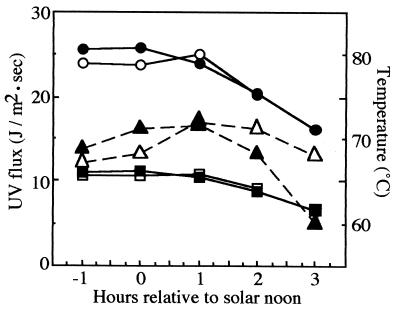
During solar exposures, ambient temperatures greater than 70°C were routinely recorded (see Fig. 3). In order to control for potential DNA damage caused by heat, spore samples shielded with a single layer of aluminum foil were exposed to solar radiation in parallel and treated identically. Solar dosimetry was performed by using a model UVX radiometer fitted with the appropriate UV-B and UV-A probes, and readings were obtained under the same shielding materials which covered the spore samples. Dose rate readings (joules per square meter per second) were taken at hourly intervals, and the average of two successive readings was used to estimate the total UV dose (in joules per square meter) received by a sample during the interval. In order to obtain the desired solar UV dose (especially for samples exposed to UV-A sunlight), it was often necessary to expose spores for several days. At the end of each daily exposure period, samples were transported to the laboratory and stored at room temperature in the dark until the following exposure period.
FIG. 3.
Solar UV dosimetry for the experiment performed on 21 and 22 July 1998. UV-A flux (circles) and UV-B flux (squares) were measured during exposure on 21 July (open symbols) and 22 July (solid symbols) with a model UVX radiometer as described in the text. The temperature (triangles) was measured with a surface contact thermometer. The total UV doses for the exposure period were 6.7 × 10⁵ J of UV-A radiation per m2 and 2.92 × 10⁵ J of UV-B radiation per m2.
DNA isolation, manipulation, and electrophoresis.
Exposed spore samples were resuspended in 10 ml of phosphate-buffered saline (10 mM potassium phosphate, 150 mM NaCl; pH 7.4), and spores were harvested from petri dishes with a sterile spatula. The resulting spore suspensions were collected by centrifugation, resuspended in decoating solution (8 M urea, 50 mM Tris base [pH 10], 1% sodium dodecyl sulfate, 50 mM dithiothreitol), and incubated at 60°C for 90 min to remove the protein coat. Decoated spores were washed, centrifuged, and resuspended three times with STE buffer (10 mM Tris-HCl [pH 8], 10 mM EDTA, 150 mM NaCl) and once with lysis buffer (50 mM NaCl, 100 mM EDTA). Spores were then lysed and chromosomal DNA was extracted and purified as previously described (1). To detect cyclobutane pyrimidine dimers, DNA was digested with phage T4 endonuclease V (Endo V) (Epicentre Technologies, Madison, Wis.), which cleaves the phosphodiester backbone 5′ to cyclobutane pyrimidine dimers (6). To detect apurinic-apyrimidinic sites, DNA was digested with endonuclease IV (Endo IV) (Epicentre Technologies) (10). Neutral agarose gel electrophoresis of DNA was performed by using standard techniques (28). In order to detect single-strand breaks generated in DNA either directly by UV treatment or as a result of Endo V or Endo IV cleavage at cyclobutane pyrimidine dimers or apurinic-apyrimidinic sites, DNA was denatured with 0.3 N (final concentration) NaOH and electrophoresed at 4°C through 0.8% alkaline agarose gels with buffer recirculation as described previously (28). Migration of DNA was determined relative to a set of molecular size standards whose sizes ranged from 0.5 to 12 kb (1-kb ladder; Life Technologies, Gaithersburg, Md.). After the gels were stained with ethidium bromide, digital photographic images of the gels were scanned and quantitated on a Macintosh computer using the public domain NIH Image program (developed at the U.S. National Institutes of Health and available on the Internet at http://rsb.info.nih.gov/nih-image).
RESULTS
In a control experiment, DNA was extracted from samples of unirradiated spores of strain WN356, prepared, and recovered by using techniques identical to those used in exposure experiments, and the DNA was separated on either 0.8% neutral or alkaline agarose gels along with high-molecular-weight markers (phage λ digested with HindIII). The chromosomal DNA migrated on neutral agarose as 23-kbp double-stranded fragments and on alkaline agarose as 23-kb single-stranded fragments (data not shown). Thus, DNA extracted from spores and purified was uniformly sheared to produce approximately 23-kbp double-stranded fragments, and no detectable additional single-strand breaks occurred during the purification procedure. Spores were then exposed to artificial UV-C radiation, artificial UV-B radiation, full-spectrum sunlight, or UV-A sunlight. Chromosomal DNA isolated from exposed spores was probed for double-strand breaks by neutral agarose gel electrophoresis and for single-strand breaks by denaturation in alkali, followed by alkaline agarose gel electrophoresis.
Artificial UV-C radiation.
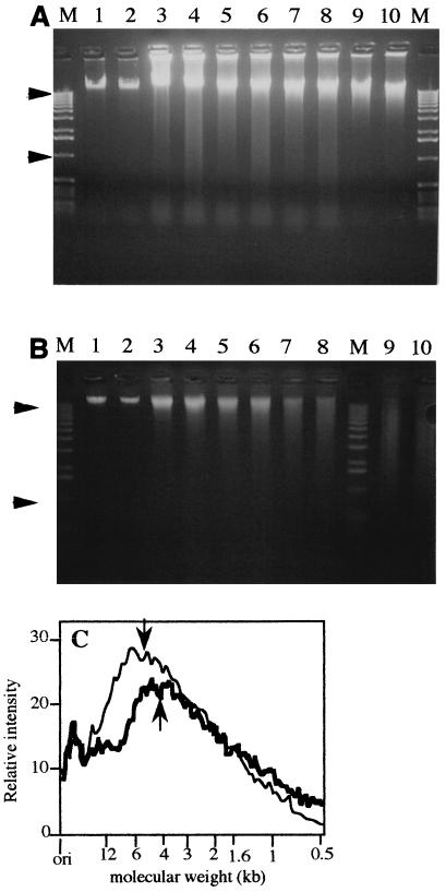
DNA isolated from spores exposed to UV-C radiation doses of 0, 2, 4, 8, and 16 kJ/m2 was electrophoresed through either a native 0.8% agarose gel or a denaturing 0.8% alkaline agarose gel (Fig. 1). UV-C treatment resulted in dose-dependent induction of double-strand DNA breaks in strain WN356 spores, which was manifested by DNA smears at progressively lower molecular sizes than 23 kbp on neutral agarose gels (Fig. 1A). In addition to double-strand breaks, some cross-linking of DNA was revealed by species that migrated more slowly than 23 kbp, particularly in spores exposed to 2 kJ of UV-C radiation per m2 (Fig. 1A). Prior digestion of the DNA with Endo V (Fig. 1A) or Endo IV (data not shown) did not appreciably change the electrophoretic patterns on neutral agarose gels.
FIG. 1.
Chromosomal DNA extracted from UV-C-irradiated spores of strain WN356 electrophoresed through a 0.8% neutral agarose gel (A) and a 0.8% alkaline agarose gel (B). Spores were irradiated with 0 (lanes 1 and 2), 2 (lanes 3 and 4), 4 (lanes 5 and 6), 8 (lanes 7 and 8), or 16 (lanes 9 and 10) kJ of UV-C radiation per m2, and isolated DNA was treated with Endo V before electrophoresis (lanes 2, 4, 6, 8, and 10). Lanes M contained molecular weight markers (1-kb ladder; the arrowheads indicate the positions of 12- and 1-kb markers). (C) Densitometric scan of lanes 9 (thin line) and 10 (thick line) from the alkaline agarose gel shown in panel B containing DNA extracted from spores irradiated with 16 kJ of UV-C radiation per m2 before (lane 9) and after (lane 10) digestion with Endo V. The average single-strand fragment lengths were 4.7 kb before Endo V digestion (down arrow) and 4.2 kb after Endo V digestion (up arrow). ori, origin of gel.
When the same DNA samples were denatured with alkali and electrophoresed through a 0.8% alkaline agarose gel, a clear UV-C dose-dependent increase in single-strand breaks was also detected (Fig. 1B). In addition to single-strand breaks, UV-C irradiation of spores induced the formation of a small amount of cyclobutane pyrimidine dimers in chromosomal DNA, as detected by T4 Endo V digestion followed by alkaline denaturation of digested DNA and alkaline agarose gel electrophoresis (Fig. 1B, lanes 7 through 10). In order to document the presence of cyclobutane pyrimidine dimers more clearly, a negative digital image of the ethidium bromide-stained gel containing DNA extracted from spores irradiated with 16 kJ of UV-C radiation per m2 (Fig. 1B, lanes 9 and 10) was subjected to densitometric scanning and a quantitation analysis (Fig. 1C). Plotting the scan versus migration of the molecular size standards revealed that at 16 kJ of UV-C radiation per m2 the average single-strand fragment length was reduced from 23 to 4.7 kb, which corresponded to approximately 1,300 single-strand breaks per B. subtilis chromosome (using 4.215 kbp as the circumference of the B. subtilis chromosome) (9). Digestion of the same DNA with Endo V before alkaline agarose gel electrophoresis resulted in a further reduction in the average single-strand fragment size from 4.7 to 4.2 kb, which was consistent with production of approximately 215 cyclobutane pyrimidine dimers per chromosome. Induction of apurinic-apyrimidinic sites in spore DNA by UV-C irradiation was not detected after digestion with Endo IV and alkaline agarose gel electrophoresis (data not shown).
Artificial UV-B radiation.
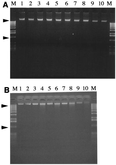
Spores were exposed to different doses of UV-B radiation (0, 20, 40, 80, and 160 kJ/m2) from a commercial UV-B lamp, and then chromosomal DNA was extracted and electrophoresed through a 0.8% neutral agarose gel (Fig. 2A) and a 0.8% alkaline agarose gel (Fig. 2B). Irradiation of spores with UV-B radiation resulted in dose-dependent formation of cyclobutane pyrimidine dimers, as detected by digestion with phage T4 Endo V prior to electrophoresis through alkaline agarose (Fig. 2B). Neither double-strand breaks (Fig. 2A) nor single-strand breaks (Fig. 2B) nor apurinic-apyrimidinic sites (data not shown) were detected in DNA from spores irradiated with artificial UV-B radiation up to the maximum dose used.
FIG. 2.
Chromosomal DNA extracted from spores of strain WN356 irradiated with artificial UV-B radiation was electrophoresed through a neutral 0.8% agarose gel (A) and a 0.8% alkaline agarose gel (B). Spores were irradiated with 0 (lanes 1 and 2), 20 (lanes 3 and 4), 40 (lanes 5 and 6), 80 (lanes 7 and 8), or 160 (lanes 9 and 10) kJ of UV-B radiation per m2, and isolated DNA was treated with Endo V before electrophoresis (lanes 2, 4, 6, 8, and 10). Lanes M contained molecular weight markers (1-kb ladder; the arrowheads indicate the positions of 12- and 1-kb markers).
Full-spectrum sunlight.
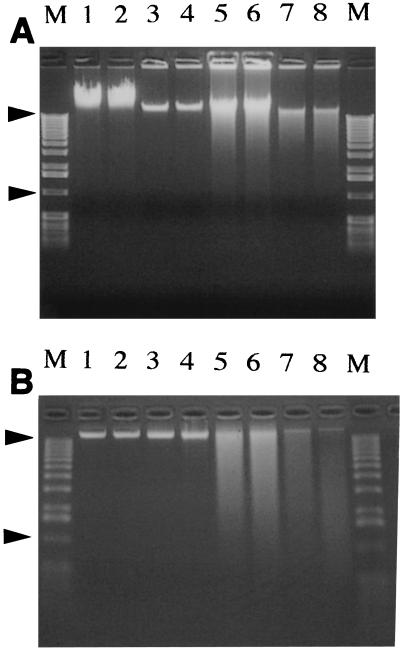
On 2 successive days (21 and 22 July 1998) spores covered with Saran Wrap were exposed to full-spectrum solar radiation. During the exposure period, the temperature and the UV-B and UV-A fluxes were recorded at hourly intervals (Fig. 3). During this experiment the spores were exposed to a total dose of 6.7 × 10⁵ J of UV-A radiation per m2 and 2.9 × 10⁵ J of UV-B radiation per m2. DNA was extracted from the sunlight-irradiated spores and electrophoresed through both a 0.8% neutral agarose gel (Fig. 4A) and a 0.8% alkaline agarose gel (Fig. 4B). Exposure of spores to full-spectrum sunlight resulted in the formation of single-strand breaks and cyclobutane pyrimidine dimers in the chromosomal DNA of the spores (Fig. 4B, lanes 3 and 4). Some double-strand breaks were detected (Fig. 4A, lanes 3 and 4), but no apurinic-apyrimidinic sites were present (data not shown). Despite the fact that the temperature exceeded 70°C twice during the experiment (Fig. 3), the DNA damage in spores exposed to full-spectrum sunlight was not caused by heat, as spores exposed in parallel to the same heat regimen but shielded from solar radiation exhibited no detectable DNA damage (Figs. 4A and B, lanes 7 and 8).
FIG. 4.
Chromosomal DNA extracted from spores of strain WN356 exposed to full-spectrum sunlight on 21 and 22 July 1998 was electrophoresed through a 0.8% neutral agarose gel (A) and a 0.8% alkaline agarose gel (B). Spores were not exposed to light (lanes 1 and 2), exposed to full-spectrum sunlight (6.7 × 10⁵ J of UV-A radiation per m2 plus 2.9 × 10⁵ J of UV-B radiation per m2) (lanes 3 and 4), exposed in parallel to only UV-A sunlight (2.68 × 10⁵ J/m2) (lanes 5 and 6), or exposed in parallel to only heat (lanes 7 and 8). Isolated DNA was treated with Endo V before electrophoresis (lanes 2, 4, 6, and 8). Lanes M contained molecular weight markers (1-kb ladder; the arrowheads indicate the positions of 12- and 1-kb markers).
UV-A sunlight.
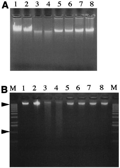
During the experiment on 21 and 22 July 1998 described above, a parallel set of spores was also exposed under 0.5-in.-thick plate glass to UV-A sunlight (total dose, 2.68 × 10⁵ J/m2). At this dose no significant spore DNA damage was detected by agarose gel electrophoresis (Fig. 4A and B, lanes 5 and 6). Therefore, in a separate experiment performed on clear days from 5 to 16 October 1998, spores were exposed under 1.25-cm-thick plate glass to a larger total dose of UV-A sunlight, 1.1 × 10⁶ J/m2. We found that the DNA extracted from spores exposed to UV-A sunlight in this experiment and electrophoresed through 0.8% neutral and 0.8% alkaline agarose gels contained double-strand breaks (Fig. 5A, lanes 5 and 6) and single-strand breaks (Fig. 5B, lanes 5 and 6) but virtually no cyclobutane pyrimidine dimers (Fig. 5B, lanes 5 and 6) or apurinic-apyrimidinic sites (data not shown). Again, damage to spore DNA was due to direct exposure to solar radiation and not to heat, as a parallel set of spores shielded from UV by aluminum foil but exposed to the same temperature regimen exhibited no detectable DNA damage (Fig. 5A and B, lanes 3 and 4).
FIG. 5.
Chromosomal DNA extracted from spores of strain WN356 exposed to UV-A sunlight on 5 to 16 October 1998 and to full-spectrum sunlight on 4 and 5 August 1998 was electrophoresed through a 0.8% neutral agarose gel (A) and a 0.8% alkaline agarose gel (B). Spores were not exposed to light (lanes 1 and 2), exposed in parallel to only heat (lanes 3 and 4), exposed in parallel to only UV-A sunlight (1.1 × 10⁶ J/m2) (lanes 5 and 6), or exposed to full-spectrum sunlight (8.23 × 10⁵ J of UV-A radiation per m2 plus 3.53 × 10⁵ J of UV-B radiation per m2) (lanes 7 and 8). Isolated DNA was treated with Endo V before electrophoresis (lanes 2, 4, 6, and 8). Lanes M contained molecular weight markers (1-kb ladder; the arrowheads indicate the positions of 12- and 1-kb markers).
DISCUSSION
Bacterial spores in the environment must maintain the integrity of their DNA for extended periods of time. Although spores are more resistant to UV radiation than vegetative cells are, vegetative cells can constantly repair their DNA in the environment. In contrast, spores are metabolically inactive and accumulate unrepaired DNA damage in their genomes during dormancy (21). Furthermore, upon germination, spores must rapidly repair the cumulative damage in their genomic DNA prior to gene expression (21, 35). UV radiation plays an important role in regulating levels of microorganisms in the environment (3, 11). The recent decreases in atmospheric ozone levels pose a serious threat to the ecological balance of bacterial populations in the environment (3, 11). While spore DNA photochemistry and repair have been well defined in the laboratory (21, 38), probing the types of adducts caused by solar UV radiation in spores should provide a better understanding of the resistance of spores in the environment and their ability to cope with exposure to solar UV radiation.
It is well established that spore photoproduct is the major UV photoproduct in spore DNA irradiated with UV-C radiation (2, 30) and full-spectrum sunlight (40) and that DNA repair processes are important determinants of spore survival when spores are exposed to laboratory UV-C, UV-B, or solar UV radiation (14, 40; reviewed in references 21, 38, and 44). In particular, spore photoproduct lyase and nucleotide excision repair have been identified as the two major DNA repair pathways which remove spore photoproduct from UV-C-irradiated spores during spore germination (17, 18), while there is evidence that recombinational repair also plays a role in spore resistance to UV-C radiation (19). Nucleotide excision repair and spore photoproduct lyase also make important contributions to B. subtilis spore resistance to solar radiation (14, 40, 43, 44). While it has been determined that spore photoproduct is also the major photoproduct in the DNA of spores exposed to sunlight, the observation that fewer spore photoproduct dimers were detected per lethal event at solar wavelengths suggested that (an)other DNA photoproduct(s) could also be formed in spores exposed to solar radiation (40). In support of this suggestion, a study of the relative efficiencies of nucleotide excision repair and spore photoproduct lyase for repairing DNA damage in spores exposed to sunlight revealed that some nucleotide excision repair-reparable DNA damage other than spore photoproduct appeared to occur in spore DNA exposed to solar radiation (44). In an attempt to elucidate the nature of additional spore DNA photoproducts, in the present study we probed for the presence of double-strand and single-strand breaks, cyclobutane pyrimidine dimers, and apurinic-apyrimidinic sites in UV-irradiated spores by using both neutral and alkaline agarose gel electrophoresis and treatment of spore DNA with the enzymes Endo IV and Endo V.
UV-C irradiation of spores resulted in dose-dependent production of detectable amounts of double-strand breaks, single-strand breaks (approximately 1,300 breaks per chromosome when the dose was 16 kJ/m2), and cyclobutane pyrimidine dimers (approximately 215 dimers per chromosome when the dose was 16 kJ/m2) (Fig. 1). It is important to note that the maximum dose used in this experiment (16 kJ/m2) probably converted nearly 40% of the total chromosomal thymine into spore photoproduct (30, 35), which corresponded to roughly 4.2 × 10⁵ spore photoproduct dimers per chromosome; therefore, in response to UV-C irradiation, the single-strand breaks and cyclobutane pyrimidine dimers produced in spore DNA accounted for approximately 0.3 and 0.16% of the spore photoproduct produced, respectively. Because strain WN356 lacks both nucleotide excision repair and spore photoproduct lyase, its spores are very sensitive to UV-C irradiation; the 90% lethal dose (LD90) is only 5 J/m2 (20). It has been calculated that for spores which lack nucleotide excision repair and spore photoproduct lyase one lethal hit by 254-nm UV-C irradiation corresponds to approximately 527 spore photoproduct dimers per chromosome (40) and that wild-type spores are 33-fold more resistant to UV-C irradiation than spores which lack nucleotide excision repair and spore photoproduct lyase (44). Therefore, the UV-C doses used in this experiment to detect cyclobutane pyrimidine dimers and single-strand breaks exceeded the LD90 of wild-type spores by more than a factor of 20, and it can reasonably be concluded that cyclobutane pyrimidine dimers and single-strand and double-strand breaks probably do not have major physiological consequences for spore survival in response to 254-nm UV-C irradiation.
However, we observed that spores irradiated with artificial UV-B radiation accumulated cyclobutane pyrimidine dimers at physiologically relevant doses. Cyclobutane pyrimidine dimers were detected after treatment with UV-B doses as low as 20 kJ/m2, which is less than the LD90 of UV-B radiation for wild-type spores (approximately 30 kJ/m2) (14, 27, 40). The observation that appreciable quantities of cyclobutane pyrimidine dimers are produced in spore DNA irradiated with UV-B radiation is consistent with the observation of Xue and Nicholson (44) that nucleotide excision repair is a more important repair pathway for spore survival in the presence of environmentally relevant UV-B radiation than in the presence of UV-C radiation.
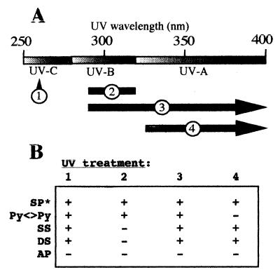
In order to understand the photochemistry of spore DNA in the environment compared to the artificial laboratory model, spores were irradiated with full-spectrum sunlight or sunlight containing only UV-A wavelengths and longer wavelengths. Full-spectrum solar radiation induced double-strand breaks, single-strand breaks, and cyclobutane pyrimidine dimers, whereas UV-A sunlight (wavelengths, >325 nm) induced both double-strand breaks and single-strand breaks but no detectable cyclobutane pyrimidine dimers in spore chromosomal DNA. These results imply that in spores exposed to solar radiation, it is the UV-B component which causes formation of cyclobutane pyrimidine dimers, whereas the UV-A portion of the solar spectrum is responsible for causing double-strand breaks and single-strand breaks. These results are summarized in Fig. 6. Therefore, in the solar environment, dormant spores must repair at least spore photoproduct, cyclobutane pyrimidine dimers, single-strand breaks, and double-strand breaks during germination.
FIG. 6.
(A) UV treatments used in this study. The following treatments were used: 1, artificial UV-C radiation (wavelength, 254 nm); 2, artificial UV-B radiation (290 to 310 nm); 3, full-spectrum sunlight (>290 nm); and 4, UV-A sunlight (>325 nm). (B) Summary of B. subtilis spore photochemistry in the presence of different artificial and environmental UV wavelengths. For UV treatments see above. SP*, spore photoproduct; Py<>Py, cyclobutyl pyrimidine dimers; SS, single-strand breaks; DS, double-strand breaks, AP, apurinic-apyrimidinic sites. +, damage detected; −, damage not detected. Spore photoproduct data are from reference 40.
No apurinic-apyrimidinic sites were detected in any of our DNA preparations, as revealed by digestion with Endo IV and alkaline agarose gel electrophoresis, even though in a control experiment apurinic-apyrimidinic sites were readily detected by Endo IV digestion of B. subtilis chromosomal DNA heated in vitro at 90°C for 30 min (data not shown). DNA is protected from depurination-depyrimidination by the major small acid-soluble proteins (α/β type SASP) and, to a lesser extent, by the relatively dehydrated state of the spore core (31–33). α/β-type SASP constitute 5 to 12% of total spore dry mass and bind spore DNA (reviewed in references 36 and 37), and SASP binding to DNA has been shown to protect the spore DNA from processes that may give rise to apurinic-apyrimidinic sites, such as acceleration of spontaneous base loss due to heat or oxidative damage (4, 31, 32, 34). Studies of wild-type B. subtilis spores and α− β− mutants have shown that α/β-type SASP are responsible for retarding the formation of apurinic-apyrimidinic sites during dry heat treatment in spore DNA (31, 32). In contrast, our control dried spores that were subjected to dry heat did not exhibit any type of damage, as detected by the assays utilized (Fig. 4 and 5). The difference in the results is perhaps explained by the fact that in the previous experiments, spores were subjected to 120°C dry heat (32), whereas the temperatures to which spores were exposed in our experiments were considerably lower (typically between 60 and 70°C) (Fig. 3). Our findings suggest that in environmental settings where temperatures do not generally exceed 70°C, heat does not contribute significantly to spore DNA damage. Under these conditions it is likely that other cellular components, such as spore proteins, may be the targets of damage that may lead to spore death.
ACKNOWLEDGMENTS
This work was supported by grant GM47461 from the National Institutes of Health and by grant USDA-HATCH-ARZT-136753 from the Arizona Agricultural Experimental Station to W.L.N.
REFERENCES
- 1.Cutting S M, Vander Horn P B. Genetic analysis. In: Harwood C R, Cutting S M, editors. Molecular biological methods for Bacillus. Sussex, England: John Wiley and Sons; 1990. pp. 27–74. [Google Scholar]
- 2.Donnellan J E, Jr, Setlow R B. Thymine photoproducts but not thymine dimers are found in ultraviolet irradiated bacterial spores. Science. 1965;149:308–310. doi: 10.1126/science.149.3681.308. [DOI] [PubMed] [Google Scholar]
- 3.Elasri M O, Miller R V. Study of the response of a biofilm community to UV radiation. Appl Environ Microbiol. 1999;65:2025–2031. doi: 10.1128/aem.65.5.2025-2031.1999. [DOI] [PMC free article] [PubMed] [Google Scholar]
- 4.Fairhead H, Setlow B, Setlow P. Prevention of DNA damage in spores and in vitro by small, acid-soluble proteins from Bacillus subtilis. J Bacteriol. 1993;175:1367–1374. doi: 10.1128/jb.175.5.1367-1374.1993. [DOI] [PMC free article] [PubMed] [Google Scholar]
- 5.Fajardo-Cavazos P, Salazar C, Nicholson W L. Molecular cloning and characterization of the Bacillus subtilis spore photoproduct lyase (spl) gene, which is involved in repair of UV radiation-induced DNA damage during spore germination. J Bacteriol. 1993;175:1735–1744. doi: 10.1128/jb.175.6.1735-1744.1993. [DOI] [PMC free article] [PubMed] [Google Scholar]
- 6.Friedberg E C, Walker G C, Siede W, editors. DNA repair and mutagenesis. Washington, D.C.: American Society for Microbiology; 1995. [Google Scholar]
- 7.Gest H, Mandelstam J. Longevity of microorganisms in natural environments. Microbiol Sci. 1987;4:69–71. [PubMed] [Google Scholar]
- 8.Kennedy M J, Reader S L, Swierczynski L M. Preservation records of microorganisms: evidence of the tenacity of life. Microbiology. 1994;140:2513–2519. doi: 10.1099/00221287-140-10-2513. [DOI] [PubMed] [Google Scholar]
- 9.Kunst F, Ogasawara N, Moszer I, Albertini A M, Alloni G, Azevedo V, Bertero M G, Bessieres P, Bolotin A, Borchert S, Borriss R, Boursier L, Brans A, Braun M, Brignell S C, Bron S, Brouillet S, Bruschi C V, Caldwell B, Capuano V, Carter N M, Choi S K, Codani J J, Connerton I F, Cummings N J, Daniel R A, Denizot F, Devine K M, et al. The complete genome sequence of the Gram-positive bacterium Bacillus subtilis. Nature. 1997;390:249–256. doi: 10.1038/36786. [DOI] [PubMed] [Google Scholar]
- 10.Ljungquist S. A new endonuclease from Escherichia coli acting at apurinic sites in DNA. J Biol Chem. 1976;262:2808–2814. [PubMed] [Google Scholar]
- 11.Miller R V, Wade J, Mitchell D, Elasri M. Bacterial responses to ultraviolet light. ASM News. 1999;65:535–541. [Google Scholar]
- 12.Munakata N. Genetic analysis of a mutant of Bacillus subtilis producing ultraviolet-sensitive spores. Mol Gen Genet. 1969;104:258–263. doi: 10.1007/BF02539290. [DOI] [PubMed] [Google Scholar]
- 13.Munakata N. Mapping of the genes controlling excision repair of pyrimidine photoproducts in Bacillus subtilis. Mol Gen Genet. 1977;156:49–54. [Google Scholar]
- 14.Munakata N. Killing and mutagenic action of sunlight upon Bacillus subtilis spores: a dosimetric system. Mutat Res. 1981;82:263–268. doi: 10.1016/0027-5107(81)90155-x. [DOI] [PubMed] [Google Scholar]
- 15.Munakata N. Genotoxic action of sunlight upon Bacillus subtilis spores: monitoring studies at Tokyo, Japan. J Radiat Res. 1989;30:338–351. doi: 10.1269/jrr.30.338. [DOI] [PubMed] [Google Scholar]
- 16.Munakata N. Biologically effective dose of solar ultraviolet radiation estimated by spore dosimetry in Tokyo since 1980. Photochem Photobiol. 1993;58:386–392. doi: 10.1111/j.1751-1097.1993.tb09579.x. [DOI] [PubMed] [Google Scholar]
- 17.Munakata N, Rupert C S. Genetically controlled removal of “spore photoproduct” from deoxyribonucleic acid of ultraviolet-irradiated Bacillus subtilis spores. J Bacteriol. 1972;111:192–198. doi: 10.1128/jb.111.1.192-198.1972. [DOI] [PMC free article] [PubMed] [Google Scholar]
- 18.Munakata N, Rupert C S. Dark repair of DNA containing “spore photoproduct” in Bacillus subtilis. Mol Gen Genet. 1974;130:239–250. doi: 10.1007/BF00268802. [DOI] [PubMed] [Google Scholar]
- 19.Munakata N, Rupert C S. Effects of DNA-polymerase-defective and recombination-deficient mutations on the ultraviolet sensitivity of Bacillus subtilis spores. Mutat Res. 1975;27:157–169. doi: 10.1016/0027-5107(75)90075-5. [DOI] [PubMed] [Google Scholar]
- 20.Nicholson W L, Chooback L, Fajardo-Cavazos P. Analysis of spore photoproduct lyase operon (splAB) structure and function using targeted deletion-insertion mutations spanning the Bacillus subtilis ptsHI and splAB operons. Mol Gen Genet. 1997;255:587–594. doi: 10.1007/s004380050532. [DOI] [PubMed] [Google Scholar]
- 21.Nicholson W L, Fajardo-Cavazos P. DNA repair and the ultraviolet radiation resistance of bacterial spores: from the laboratory to the environment. Recent Res Dev Microbiol. 1997;1:125–140. [Google Scholar]
- 22.Nicholson W L, Setlow P. Sporulation, germination, and outgrowth. In: Harwood C R, Cutting S M, editors. Molecular biological methods for Bacillus. Sussex, England: John Wiley and Sons; 1990. pp. 391–450. [Google Scholar]
- 23.Nicholson W L, Setlow B, Setlow P. Ultraviolet irradiation of DNA complexed with α/β-type small, acid-soluble proteins from spores of Bacillus or Clostridium species make spore photoproduct but not thymine dimers. Proc Natl Acad Sci USA. 1991;88:8288–8292. doi: 10.1073/pnas.88.19.8288. [DOI] [PMC free article] [PubMed] [Google Scholar]
- 24.Piggot P J, Moran C P Jr, Youngman P, editors. Regulation of bacterial differentiation. Washington, D.C.: American Society for Microbiology; 1994. [Google Scholar]
- 25.Potts M. Desiccation tolerance of prokaryotes. Microbiol Rev. 1994;58:755–805. doi: 10.1128/mr.58.4.755-805.1994. [DOI] [PMC free article] [PubMed] [Google Scholar]
- 26.Quintern L E, Puskeppeleit M, Rainer P, Weber S, el Nagger S, Eschweiler U, Horneck G. Continuous dosimetry of the biologically harmful UV radiation in Antarctica with the biofilm technique. J Photochem Photobiol B Biol. 1994;22:59–66. doi: 10.1016/1011-1344(93)06954-2. [DOI] [PubMed] [Google Scholar]
- 27.Riesenman, P. J., and W. L. Nicholson. Role of the spore coat layers in Bacillus subtilis spore resistance to hydrogen peroxide, artificial UV-C, UV-B, and solar UV radiation. Appl. Environ. Microbiol., in press. [DOI] [PMC free article] [PubMed]
- 28.Sambrook J, Fritsch E F, Maniatis T. Molecular cloning: a laboratory manual. 2nd ed. Cold Spring Harbor, N.Y: Cold Spring Harbor Laboratory Press; 1989. [Google Scholar]
- 29.Schaeffer P, Millet J, Aubert J-P. Catabolic repression of bacterial sporulation. Proc Natl Acad Sci USA. 1965;54:704–711. doi: 10.1073/pnas.54.3.704. [DOI] [PMC free article] [PubMed] [Google Scholar]
- 30.Setlow B, Setlow P. Thymine-containing dimers as well as spore photoproducts are found in ultraviolet-irradiated Bacillus subtilis spores that lack small, acid-soluble proteins. Proc Natl Acad Sci USA. 1987;84:421–424. doi: 10.1073/pnas.84.2.421. [DOI] [PMC free article] [PubMed] [Google Scholar]
- 31.Setlow B, Setlow P. Heat inactivation of Bacillus subtilis spores lacking small, acid-soluble proteins is accompanied by generation of abasic sites in spore DNA. J Bacteriol. 1994;176:2111–2113. doi: 10.1128/jb.176.7.2111-2113.1994. [DOI] [PMC free article] [PubMed] [Google Scholar]
- 32.Setlow B, Setlow P. Small, acid-soluble proteins bound to DNA protect Bacillus subtilis spores from killing by dry heat. App Environ Microbiol. 1995;61:2787–2790. doi: 10.1128/aem.61.7.2787-2790.1995. [DOI] [PMC free article] [PubMed] [Google Scholar]
- 33.Setlow B, Setlow P. Role of DNA repair in Bacillus subtilis spore resistance. J Bacteriol. 1996;178:3486–3495. doi: 10.1128/jb.178.12.3486-3495.1996. [DOI] [PMC free article] [PubMed] [Google Scholar]
- 34.Setlow B, Setlow P. Heat killing of Bacillus subtilis spores in water is not due to oxidative damage. App Environ Microbiol. 1998;64:4109–4112. doi: 10.1128/aem.64.10.4109-4112.1998. [DOI] [PMC free article] [PubMed] [Google Scholar]
- 35.Setlow P. Resistance of bacterial spores to ultraviolet light. Comments Mol Cell Biophys. 1988;5:253–264. [Google Scholar]
- 36.Setlow P. I will survive: protecting and repairing spore DNA. J Bacteriol. 1992;174:2737–2741. doi: 10.1128/jb.174.9.2737-2741.1992. [DOI] [PMC free article] [PubMed] [Google Scholar]
- 37.Setlow P. DNA in dormant spores of Bacillus species is in an A-like conformation. Mol Microbiol. 1992;6:563–567. doi: 10.1111/j.1365-2958.1992.tb01501.x. [DOI] [PubMed] [Google Scholar]
- 38.Setlow P. Mechanisms for the prevention of damage to DNA in spores of Bacillus species. Annu Rev Microbiol. 1995;49:29–54. doi: 10.1146/annurev.mi.49.100195.000333. [DOI] [PubMed] [Google Scholar]
- 39.Stuy J H. Studies on the mechanism of radiation inactivation of micro-organisms. III. Inactivation of germinating spores of Bacillus cereus. Biochim Biophys Acta. 1956;22:241–246. doi: 10.1016/0006-3002(56)90146-9. [DOI] [PubMed] [Google Scholar]
- 40.Tyrrell R M. Solar dosimetry with repair deficient bacterial spores: action spectra, photoproduct measurements and a comparison with other biological systems. Photochem Photobiol. 1978;27:571–579. doi: 10.1111/j.1751-1097.1978.tb07648.x. [DOI] [PubMed] [Google Scholar]
- 41.Urbach F, Gange R W, editors. The biological effects of ultraviolet A radiation. New York, N.Y: Praeger Publishers; 1986. [Google Scholar]
- 42.Varghese A J. 5-Thyminyl-5,6-dihydrothymine from DNA irradiated with ultraviolet light. Biochem Biophys Res Commun. 1970;38:484–490. doi: 10.1016/0006-291x(70)90739-4. [DOI] [PubMed] [Google Scholar]
- 43.Wang T-C V. A simple convenient biological dosimeter for monitoring solar UV-B radiation. Biochem Biophys Res Commun. 1991;177:48–53. doi: 10.1016/0006-291x(91)91946-a. [DOI] [PubMed] [Google Scholar]
- 44.Xue Y, Nicholson W L. The two major spore DNA repair pathways, nucleotide excision repair and spore photoproduct lyase, are sufficient for the resistance of Bacillus subtilis spores to artificial UV-C and UV-B but not to solar radiation. Appl Environ Microbiol. 1996;62:2221–2227. doi: 10.1128/aem.62.7.2221-2227.1996. [DOI] [PMC free article] [PubMed] [Google Scholar]


