Abstract
Cellulose is the most abundant natural polymer and deserves the special attention of the scientific community because it represents a sustainable source of carbon and plays an important role as a sustainable energent for replacing crude oil, coal, and natural gas in the future. Intense research and studies over the past few decades on cellulose structures have mainly focused on cellulose as a biomass for exploitation as an alternative energent or as a reinforcing material in polymer matrices. However, studies on cellulose structures have revealed more diverse potential applications by exploiting the functionalities of cellulose such as biomedical materials, biomimetic optical materials, bio-inspired mechanically adaptive materials, selective nanostructured membranes, and as a growth template for inorganic nanostructures. This article comprehensively reviews the potential of cellulose structures as a support, biotemplate, and growing vector in the formation of various complex hybrid hierarchical inorganic nanostructures with a wide scope of applications. We focus on the preparation of inorganic nanostructures by exploiting the unique properties and performances of cellulose structures. The advantages, physicochemical properties, and chemical modifications of the cellulose structures are comparatively discussed from the aspect of materials development and processing. Finally, the perspective and potential applications of cellulose-based bioinspired hierarchical functional nanomaterials in the future are outlined.
Keywords: cellulose, metallic and metal oxide nanostructures, metal sulfide nanostructures, mesoporous nanostructures, hierarchical nanostructures, heterogeneous catalysis
1. Introduction and Scope
With the aim to meet the global challenges that humanity is facing nowadays such as a lack of energy and fossil resources as well as increasing environmental issues, green and progressive strategies are expected to sustain the research impulse. In this context, cellulose, with its derivatives, will certainly play a significant role in this demanding task since it has an annual average output of 1011–1012 tons in nature and only 2 × 10⁶ tons are used by people [1,2]. Bio-based materials are attractive as the basis for sustainable development and innovative solutions to a wide range of technological challenges as well as environmental problems. Cellulose and cellulose-based materials are not only earth abundant and biocompatible, but also contain intrinsic structures for transformative device performance [3]. While many innovative structures and devices as well as applications have been published and demonstrated, substantial challenges still exist in the fundamental science, and the commercialization of cellulose-based materials is a necessity in solving the emerging problems and demands [3,4,5,6]. The great advantage of cellulose or cellulose-based products and materials is that they can be produced from various byproducts or waste products emerging during food production such as various straws, peelings, husks, bagasse, corn stalks, or cobs, etc., or during wood processing [7,8,9,10].
Cellulose is the most abundant natural polymer globally, and is present in a large number of living species, mostly plants and bacteria, but also in animals [11,12]. It is a biodegradable and biocompatible as well as renewable polymer and it is a green alternative to fossil-fuel carbon sources [13,14]. Cellulose has also attracted the special attention of the scientific community as a versatile and abundant “green” template [15]. The formation of functional nanomaterials via biomineralization or biotemplating is a topic that is attracting an enormous amount of interest from academia and the industry because it is promising for a more precise control over the positioning and connecting of different functional nanostructures into complex nano- and macro-devices [16,17].
The natural cellulose molecule is actually a long polysaccharide chain. It consists of a variable number of β-1,4 linked glucopyranose rings as repeating units [18]. Cellulosic polymer chains are, due to a vast number of hydroxyl groups, associated via hydrogen bonds forming bundles of fibrils (microfibrillar aggregates), where highly ordered regions (crystalline domains) alternate with disordered regions (amorphous domains) inside the cellulose fibrils, which constitute the walls of plant cells [19,20]. This highly ordered structure gives cellulose a very high elastic modulus and specific strength, making it an ideal reinforcing material for polymer matrices [21,22,23] in a variety of forms such as macroscopic fibers (hemp, jute, and kenaf), microfibrillated cellulose (MFC), cellulose nanofibrils (CNFs), or cellulose nanocrystals (CNCs) [13,24].
On the other hand, the scientific and research communities have experienced an immense rise in interest in the areas of nanoscience and nanotechnology during the last few decades. These are nowadays at the center of our technological progress due to their outstanding capability to manipulate matter on a nanometer scale. The capability to directly design, synthesize, and control systems at the same scale as nature is a huge scientific and technological challenge [25]. Over billions of years, nature has evolved nanoscale biological systems for the efficient production of energy and materials. By mimicking such systems, scientists aim to reach the goal of a sustainable society in the future [25]. Therefore, nanomaterials and nanotechnology have been regarded as a huge leap toward miniaturization and nanoscaling, generating various subfields with the aim to study these materials in detail. Nanotechnology as a scientific field is highly multidisciplinary; contributions from biologists, chemists, physicists, and engineers are indispensable to the advancement in the understanding the formation, application, and impact of new nanostructures and innovative nanotechnologies, which can potentially provide a very efficient approach to the production of chemicals, fuels, energy, and energy efficient materials [25,26,27,28].
1.1. Types and Forms of Cellulose
1.1.1. Cellulose Fibers and Fibrils (CF)
Native cellulose, defined as the cellulose I polymorph, does not exist as an isolated individual molecule, but is found as highly organized assemblies of individual cellulose chains, forming extended structures-fibers. The ultrastructure of cellulose fibers exhibits a hierarchical arrangement, starting from cellulose molecules to elementary fibrils and further to micro- as well as macrofibers. When cellulose molecules are synthesized as individual molecules, they immediately undergo spinning in a hierarchical order at the site of biosynthesis. Typically, up to 36 individual cellulose molecules are self-organized into larger units known as elementary fibrils (protofibrils) that are further packed into so-called microfibrils, and these are finally assembled into cellulose fibers with diameters of a few micrometers and lengths of up to a few thousands millimeters. Depending on the biosynthesis conditions, celluloses from different sources may occur in different packing that results in the different morphologies and properties of cellulose fibers [11,20]. For various purposes, cellulose needs to be isolated from its source, followed by purification and possible modification. Methods of extraction, purification, and modification affect the physical and chemical properties of cellulose such as the chain lengths and their distribution, the degree of crystallinity, the mechanical properties, thermal stability, solubility, the distribution of functional groups in the monomer units of the cellulose polymer chain, and the ratio of inter- and intra-molecular hydrogen bonds in the cellulose molecules. These physico-chemical properties play a key role in the determination of industrial and commercial applications of cellulose fibers [29,30,31].
1.1.2. Regenerated Cellulose (RC)
Regenerated cellulose, defined as a cellulose II polymorph, is obtained by the chemical regeneration process comprising the dissolution or swelling of native cellulose I fibers, followed by its reprecipitation in water. Suitable solvents for cellulose include cupric hydroxide in aqueous ammonia (cuoxam) or cupriethylenediamine (cuen), ammonia or amine/thiocyanate, hydrazine/thiocyanate, lithium chloride/N,N-dimethylacetamide (LiCl/DMAc), and N-methylmorpholine-N-oxide (NMMO)/water systems, and lately, ionic liquids (IL). Most of these solvents are limited on a pilot-scale except for NMMO, which is commercially used for the production of regenerated cellulose fibers called Lyocell [32]. Although viscose technology is still the most widely used process to manufacture regenerated cellulose fibers, this technology has many disadvantages related to environmental issues. A special case is mercerization (i.e., the swelling of the native cellulose in concentrated sodium hydroxide solution followed by the removal of the swelling agent). All of these processes finally produce fibers of the cellulose II polymorph. During the conversion (I to II), the conformation of hydroxyl groups is changed, causing a rearrangement in the entire hydrogen-bonded network while the dimensions of regenerated fibers remain mostly unchanged. Compared to cellulose I with a parallel up arrangement, the chains in cellulose II are in an antiparallel arrangement, producing a more stable fibrillary structure that is preferable for various practical applications [11,20]. One of the easiest ways to prepare regenerated cellulose is by dissolving raw cellulose in a NaOH/urea or thiourea aqueous solution [33]. An innovative approach toward regenerated cellulose is the dissolution of raw cellulose in various ionic liquids, representing a more sustainable strategy. Here, the choice of cation and anion is critical not only to the degree of the dissolution, but also to the ultimate sustainability of such processes. Studies have revealed that the interactions between the anion and cellulose play a key role in cellulose solvation, however, opinions on the cation role are still conflicting [34]. Oxidized regenerated cellulose (ORC), obtained by transforming its primary alcohol groups into carboxyl groups, is of special interest because of the various attractive medical applications and is among one of the most widespread hemostatics being used in almost any kind of surgery [2].
1.1.3. Microcrystalline Cellulose (MCC)
Microcrystalline cellulose is a pure, partially degraded native cellulose I, derived from high-quality wood pulps. It is prepared either by enzyme-mediated reactive extrusion, steam explosion, or acid hydrolysis with strong mineral acids (H2SO4, HCl, or HBr). During this treatment, a partial degradation of cellulose fibers (preferentially in amorphous regions) takes place, producing a stable, chemically inactive, and physiologically inert cellulose material with attractive binding properties. The average dimension of MCC particles is above 5 µm. MCC offers the opportunity for a variety of applications (e.g., in the pharmaceutical industry as a tablet binder, in food applications as a texturizing agent, fat replacer, emulsifier, and bulking agent as well as an additive in paper and composite applications) [11]. MCC is one of the most useful and attractive fillers because of its high dilution potential, excellent compactibility at low pressures, and superior disintegration properties. Its chemical inertness and compatibility with a large number of drugs make MCC a highly attractive pharmaceutical agent [29]. Recently, various alternative sources of MCC have been studied. Although all microcrystalline cellulose is made of the same biopolymer, different raw materials can be used to obtain MCC tailored to specific needs [35].
1.1.4. Microfibrillated Cellulose (MFC)
Microfibrillated (MFC) and/or nanofibrillated cellulose (NFC) consist of long, entangled, and flexible cellulose (nano)fibers composed of alternating crystalline and amorphous phases. The difference between MFC and NFC is in the average width, which is much smaller in NFC (10–50 nm) than in MFC (10–500 nm). An aqueous MFC or NFC suspension consists of interconnected hydrophilic cellulose I (nano)fibers, inducing a gelation of the suspension at low mass contents of only a few percent. These can be isolated using different mechanical treatments, which usually involve a refining step followed by a high-pressure homogenization step, although cryocrushing and grinding methods have also been applied. Mechanical treatments produce a network of interconnected cellulose microfibrils with diameters from 10 to 500 nm and aspect ratios from 50 to 100. MFC and NFC are generally produced from wood pulp, though other cellulose sources originating from agricultural byproducts are also important such as wheat straw, sugar beet pulp, oil palm kernel, potato pulp, or sugar cane bagasse, etc. [13,36]. NFC production can be commercially competitive through the choice of less energy intensive processes and by using low-cost raw materials [37,38]. Additionally, to enhance NFC production and reduce energy consumption, various physical, chemical, and biological pretreatments have been applied. NFC is frequently produced through alkali and enzymatic pretreatment followed by grinding or by using TEMPO-mediated oxidation, followed by homogenization. These pretreatments substantially improve the production yield and properties of NFC. Therefore, NFC production is a combination of several operations, and by changing their sequence, different types of NFC can be isolated [39].
1.1.5. Bacterial Nanocellulose (BNC)
Cellulose fibers are secreted extracellularly by several bacterial species including Gram-negative bacteria such as Acetobacter, Alcaligenes, Azotobacter, Rhizobium, Salmonella, Pseudomonas, and Gram-positive bacteria such as Sarcina ventriculi. Bacterial cellulose (BNC) is produced most efficiently by the bacteria of the Acetobacter genus such as Acetobacter G. xylinus, Acetobacter hansenii, or Aacetobacter pasteurianus species during cultivation in an aqueous culture media containing carbon and nitrogen sources in a time of a few days. The resulting cellulosic structure is in the form of a pellicle of randomly assembled ribbon shaped fibrils with a width of less than 100 nm, which are further composed of bundles of much finer nanofibrils (2 to 4 nm in diameter). These bundles are relatively straight and dimensionally uniform, and they further form a continuous three-dimensional network. Compared to plant cellulose, BNC is chemically a cellulose of high purity without the presence of hemicellulose and lignin. It shows higher hydrophilicity and water holding capacity, and in general, a higher tensile strength, resulting from a higher degree of polymerization and ultrafine network architecture [20,40]. The microbial process is an environmentally-friendly and effective method to produce high-yield BNC from various sources. However, the physiochemical characteristics (crystallinity and morphology) as well as yield of BNC are significantly influenced by the culture medium used [39].
1.1.6. Cellulose Nanocrystals (CNCs)
CNCs, also called cellulose whiskers or nanocrystalline cellulose (NCC), are predominantly isolated from cellulosic fibers by a simple process involving the acid hydrolysis of the biomass using mostly concentrated H2SO4 or other strong acids. CNCs, isolated by using (60–64%) H2SO4, possess a higher colloidal stability than CNCs produced by other acids. Severe environmental pollution concerns due to strong acid hydrolysis has encouraged the use of recyclable acids (maleic acid, formic acid, citric acid), thus ensuring a more environmentally-friendly process design. Additionally, multiple acid blends (strong and weak acid) have also been employed, resulting in highly crystalline CNCs with a better surface chemistry due to the strong and weak acid contribution. The production of CNCs can also be achieved by using oxidizing agents, ionic liquids, and subcritical water. CNCs produced by oxidation comparatively possess higher colloidal stability, crystallinity, and uniform nanoscale dimension [39]. During CNC processing, disordered (amorphous) regions of cellulose are selectively degraded, while leaving the more acid resistant crystalline regions mostly intact to produce rod-shaped cellulose nanocrystalline particles [41,42,43]. Their dimensions depend on the cellulose source and type of the isolation process used. Consequently, their widths and lengths can vary from 5 to 20 nm and from 100 nm to 1–2 μm, respectively [44]. In polar solvents, CNCs do not flocculate due to electrostatic repulsion forces originating from their negative charges on the surface, which results in stable suspensions for several months. One of the most interesting features originates from the ability of these suspensions to self-organize into stable, chiral nematic phases (characteristic for liquid crystals) that give the CNC suspension unique optical properties when a critical CNC concentration is reached [13]. The CNCs isolated by phosphoric acid (PCNCs) showed excellent flame-retardant properties due to the ability of the phosphate groups to enhance char formation, thus making PCNCs a self-extinguishing material. The incorporation of PCNCs in the nanocomposite foam also substantially improved the mechanical properties. Furthermore, PCNCs are also promising biomedical materials that can be used as a bone-scaffolding material [13,45,46]. Aside from weak acids, ionic liquids (ILs) are also used to isolate CNCs as well as other types of nanocellulose. ILs can be used alone or in combination with mineral acids, enzymatic treatment, or with physical treatment. The most suitable ILs are [BMIm]HSO4 and [BMIm]Cl [13,47,48]. The production of nanocellulose using ionic liquids has many benefits including the usage of atmospheric pressure, small amounts of solvents, the regeneration of ionic liquids, and working with an odorless and relatively safe solvent. On the other hand, this method also has disadvantages that include the relatively high costs of ionic liquids and the unsatisfactory efficiency of the extraction process [49].
1.2. Cellulose Gels as Supports or Templates for Nanostructured Materials
1.2.1. Cellulose Hydrogels
Cellulose hydrogels are physically or chemically cross-linked three dimensional hydrophilic cellulose networks capable of absorbing large amounts of water (or biological fluids) and swelling. The hydrogel stability is ensured either by physical interactions (chain entanglements, van der Waals forces, hydrogen bonds, crystallite association, and/or ionic interactions) or chemical cross-links (covalent bonding) [50]. Aqueous CNC or MFC suspensions are generally characterized by their gel like properties due to the presence of long interconnected hydrophilic cellulose microfibrils and to intense interaction between the cellulose fibers or nanocrystals. The properties of bacterial cellulose (BC) hydrogels are unique and quite different from those based on plant cellulose because of its ultrafine network structure, high hydrophilicity, and high purity as well as culturing without using any cross linker. Cellulose-based hydrogels can be prepared from wastepaper or various cellulose-containing agricultural wastes. Hydrogels have been extensively studied for a wide variety of applications because their properties and chemical compositions can be easily manipulated. Frequently studied applications of hydrogels are biomedical applications such as drug delivery, wound dressings, and tissue engineering. Additionally, studies have reported evaluating cellulose-based hydrogels as water reservoirs and controlled release fertilizer carriers in horticulture and agriculture [51]. Lately, more technical applications of hydrogels have also been investigated. Cellulose-based hydrogels can be applied as various sensors [52,53,54], heat harvesting [55], fire prevention, and firefighting [56], or in cancer detection or therapy [57,58]. Moreover, cellulose-based composite hydrogels have been studied for the massive uranium extraction from seawater to cope with the severe shortage of onshore uranium reserves [59].
1.2.2. Cellulose Aerogels
Aerogels, on the other hand, are ultra-light weight and highly porous materials that are formed by the replacement of a liquid solvent in a gel by air. In this process, the volume of the gel body or the network structure is maintained, or in other words, the shrinkage phenomena are significantly reduced or eliminated. The interest in these materials originates from their strongly attractive characteristics including low density (typically between 0.004 and 0.500 g cm−3), high porosity (typically greater than 80%) as well as high specific surface area, low thermal conductivity, excellent shock absorption, and low dielectric permittivity [50,60].
Cellulose aerogels are prepared from aqueous cellulose suspensions—hydrogels. Subsequent replacement of water with air using freeze-drying (FD) or CO2 supercritical drying produces a porous material with the structure displaying entangled cellulose I nanofibers or nanocrystals. In general, the properties of the resulting material depend on several parameters including the initial cellulose nanofiber concentration, drying technique, speed of freezing the suspension before drying, and the chemical or enzymatic pre-treatment of the starting cellulose material. One of the main advantages of using cellulose nanofibers to form the porous material originates from the high reactivity of free hydroxyl groups on their surface. Consequently, the chemical functionalization of cellulosic aerogels represents an attractive approach to tailor the properties of these structures and further broaden their scope of applications. Thus far, two routes have been reported to obtain functionalized porous structures: (1) Preparing the aerogel and modifying it afterward, or (2) derivatizing the nanofibers prior to the formation of the aerogel [13]. Both approaches have been used for the formation of inorganic nanoparticles as well as their organized or assembled structures by applying cellulose as a template. The sources to produce cellulose aerogels are diverse, but cellulose-originated wastes, especially biomass and textile waste, are of primary interest since they are appropriate for the development of eco-friendly production technologies with the use of renewable feedstocks. Aerogels prepared from cellulose-rich waste are biodegradable, easy to functionalize for different uses, and are inexpensive due to the abundance of raw materials [61]. Cellulose aerogels have a wide area of potential applications such as particle filters, particle trappers, catalyst supports, and heat insulators [62,63] and have been evaluated for their capability to remove various pollutants from an aqueous medium [61,64,65]. Additionally, the application of cellulose aerogels for oil absorption applications has also been investigated [66]. Furthermore, cellulose composite aerogels with inorganic nanostructures have also studied been as antibacterial materials for their application in medicine [67,68]. Moreover, a polydopamine-filled cellulose aerogel was developed and studied as a device for seawater and wastewater purification [69]. Another type includes composite aerogels (i.e., cellulose aerogels combined with other materials such as Ag nanowires), which are used to produce materials with a unique combination of mechanical, electrical, magnetic, and thermal properties for application in EMI shielding, electrical switches, and solar–thermal energy conversion [70,71].
The successful design and formation of inorganic nanostructures require effective and convenient methods for their fabrication. In this context, the synthesis of nanomaterials from biopolymers as the templates is a promising approach, which enables the design of complex hierarchical material systems or nanodevices. Celluloses and nanocelluloses are particularly attractive as bio-templating materials because of their highly interactive surface due to a large number of reactive hydroxyl groups that can efficiently nucleate inorganic phases around the rod-like cellulose structures. Cellulose fibers and cellulose nanostructures are supposed to direct the formation, growth, and patterning of inorganic materials to produce various types of nanostructures (nanoparticles, nanowires, or nanotubes) with unique combinations of optical, electrical, and catalytic properties [13]. High open porosity, large surface area, and high mechanical strength make any form of cellulose almost an ideal candidate for the supporting medium in the synthesis of nanostructures.
Recent progress reported in the scientific literature suggests that a synergetic use of the cellulose (nano)fibers together with suitable functionalization pathways will constitute a breakthrough in the formation of templated metallic- and metal oxide hierarchical nanostructures in the near future. Therefore, this review focuses on the utilization of cellulose and nanocellulose materials as templates or building blocks for the formation of nanomaterials and hierarchical functional materials. It provides a review of the applications of any form of cellulose as a template, supporting medium, or growing vector for the formation of inorganic nanostructures. With respect to the presence of cellulose in the final product, these applications can be divided into two groups: cellulose-supported structures and pure inorganic cellulose-templated structures. The first group comprises the publications reporting on the nanostructures where cellulose remains a constitutive component of the final nanocomposite structure, whereas the second group constitutes the publications dealing with the formation of metallic- or metal oxide nanostructures followed by subsequent elimination of the cellulose by calcination, resulting in pure inorganic structures. A special subgroup represents the inorganic nanostructures supported by carbon fibers formed from cellulose fibers by calcination in an inert atmosphere. These two main groups can be further divided based on a type of inorganic material into: metallic nanostructures, metal oxide nanoparticles, metal sulfide and other inorganic compound nanostructures as well as complex hybrid structures containing various metallic and/or metal oxide nanoparticles. For this reason, the review section is divided into five chapters: (2) Cellulose supported metallic nanoparticles; (3) cellulose supported metal oxide nanostructures; (4) cellulose templated pure metal oxide nanostructures; (5) cellulose supported/templated nanostructures of metal sulfides, hydroxyapatites, and other inorganic compounds; and (6) cellulose supported/templated hybrid nanostructures. Each chapter is further organized with respect to the size of the cellulose structures used as a support/template: macrostructured cellulose (cellulose fiber and fibril—CF, regenerated cellulose—RC); microstructured cellulose (microcrystalline cellulose—MCC, microfibrillated or nanofibrillated cellulose—MFC); and nanostructured cellulose (bacterial nanocellulose—BNC, cellulose nanofibril—CNF, cellulose nanocrystal—CNC). The number of publications reporting on cellulose supported or templated inorganic nanostructures in the last 20 years numbers in the hundreds or even thousands. Many research groups have reported on the outstanding capability of any form of cellulose to stabilize and support the growing inorganic nanostructures, thus forming hierarchical nanostructures with exceptional combinations of properties that are practically impossible to prepare in any other way. A large number of publications have suggested that a review is necessary to identify and monitor the trends of where this research area is heading and to list the achievements produced. There have been about ten different review articles published in the last ten years dealing with cellulose and inorganic materials, however, they are limited either by the type of cellulose or by the type of inorganic nanostructures or by the application area of these hybrid nanomaterials. Therefore, this publication offers a review over a wide range of cellulose structures (from cellulose fibers to cellulose nanocrystals) and a wide range of cellulose supported or templated inorganic nanostructures (metals, metal oxides, metal sulfides, hybrid inorganic nanostructures) as well as a wide range of the various application areas of these hierarchical nanomaterials.
2. Cellulose Supported Metallic Nanostructures
Various procedures of the formation of metallic nanostructures on the surface of cellulose have been reported. Authors have frequently used nanocellulose as a metallic nanoparticle support, but other forms of cellulose, even native cellulose fibers, are also useful for this purpose. With respect to the type of metal, mostly noble and particles of some transition metals are deposited on the cellulose surface.
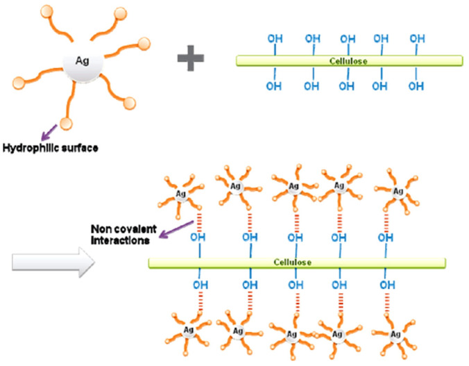
The most abundant metal studied is Ag since it has strong antibacterial activity and together with cellulose structures, it represents almost ideal material for wound dressings or other biomedical applications. There is a substantial number of publications on this topic [72]. Aside from cellulose–Ag, cellulose–Au hybrid materials have also been extensively studied [73]. Cellulose–Au hybrid materials are very attractive due to their chemical inertness and high resistance toward oxidation, making them interesting materials for biomedical applications. Additionally, Ag and Au have been studied as antiviral agents and their hybrid materials with cellulose can be applied in disinfectant wipes or as anti-viral layers in facemasks [74]. This potential application is highly important in the time of the COVID-19 pandemic. Cellulose structures decorated with Pt or Pd nanostructures are lightweight, flexible, environmentally-friendly membranes with strong catalytic activity. Many of the cellulose supported metallic nanostructures have shown strong catalytic activities for numerous chemical reactions [75]. Additionally, these lightweight hierarchical cellulose metallic nanostructures have potential applications in electronics, electro-optics, and chemical sensing. The ether oxygen and the hydroxyl functional groups of the cellulose anchor metal ions tightly on the cellulose fibers via ion–dipole interactions. They also stabilize metallic nanoparticles by the strong bonding interaction with the particle surface, thus enabling the formation of hierarchical metallic nanostructures that are otherwise almost impossible to prepare. Due to the strong interaction between the forming nanoparticles and the cellulose surface, the agglomeration and Ostwald ripening of these nanoparticles are generally prevented. Many authors have reported that cellulose itself possesses reducing properties and the capability to reduce the salts of noble metals to the metallic state, but in most cases, the authors have used reducing agents to form metallic particles on the surface of cellulose.
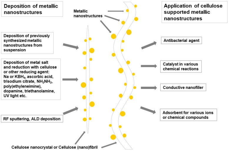
The presentation of the versatility of cellulose substrates, the formation processes of the metallic particles, and the potential application of these hierarchical structures is shown in Scheme 1.
Scheme 1.
Schematic representation of the cellulose/metallic particle nanocomposites, synthetic routes, and their most frequent applications.
2.1. Cellulose Supported Noble Metallic Nanostructures
Cellulose in any form has been applied as a support or template in the formation of various metallic nanostructures including noble (Ag, Au, Pt, Pd) as well as other transition (Co, Ni, Cu) metals [25]. Theoretically, by using strong reducing agents such as H2 or NaBH4, any metallic particles, even metallic Fe, can be formed on the cellulose support. On the other hand, noble metals can be reduced to the metallic state by cellulose itself or by using mild biobased reducing agents such as leaf extracts [76].
Various methodologies to synthesize gold and silver nanoparticles on the surface of the cellulose fibers have been explored and compared. Treatment of the cellulose fibers with an alkaline solution of HAuCl4 or AgNO3 led to the growth and deposition of Au and Ag nanoparticles on their surface. When the cellulose fibers were treated with lecithin or surface modified with thiol, a high temperature was not essential for the growth of nanostructures on the cellulose. With such treatment and modification, uniform metallic nanoparticles were obtained in relatively high yields (~43 wt.% of Au on the cellulose fibers modified with thiol) at room temperature. Reduction with borohydride resulted in substantially lower loading (~22 wt.%) and a wide size distribution of Au and Ag nanostructures on the cellulose fibers. These hierarchical structures were studied as catalysts in the reduction of 4-nitrophenol into 4-aminophenol. Thiol modified cellulose–gold nanoparticle composites quantitatively reduced 4-nitrophenol into 4-aminophenol in 90 min in the presence of NaBH4 [77].
Silver nanoparticles (AgNPs) synthesized with four different processes were studied as a disinfectant for cellulose-based wipes. These four techniques are: (1) cotton yarn as a reducing agent with trisodium citrate; (2) cotton fabric as a reducing agent with trisodium citrate; (3) an aqueous solution of PVA in the presence of glucose as a reducing agent; and (4) the photochemical reaction of polyacrylic acid in a AgNO3 solution. The prepared AgNPs with particle sizes between 10 and 70 nm were deposited on cellulose fabrics to be used as wipes for the disinfection of surfaces. The assessment of active solutions of silver nanoparticles for the antiviral and antimicrobial activities was evaluated. The results showed a significant effect of the AgNP preparation process on their disinfectant performance. The evaluation of antimicrobial and antiviral activities proved their effectiveness against fungi (A. niger and C. albicans), coronavirus (MERS-CoV) as well as bacteria (E. coli and P. mirabilis as Gram-negative bacteria, S. aureus and B. subtilis as Gram-positive bacteria). The AgNPs prepared by using cotton fabric as a reducing agent (2) showed the highest antiviral activity with 51.7% viral inhibition at 0.0625 μL with moderate cytotoxicity activity. Disinfectant cellulose-based wipes treated with antimicrobial and antiviral silver nanoparticles were successfully prepared to be applied for the prevention of the contamination and transmission of several pathogenic viruses (coronavirus) and microbes to humans in critical areas such as hospitals and health care centers [78].
Flexible and highly-porous cellulose sponges (CS)-based on the microfibrillated cellulose were prepared by cross-linking with γ-glycidoxypropyltrimethoxysilane (GPTMS) and polydopamine (PDA) and were applied as a support for the synthesis of Pd nanostructures (Pd NPs) (Figure 1). Catechol moieties of polydopamine formed chelates with Pd ions, enabling the nucleation and thus governing the growth of Pd nanostructures on the PDA modified cellulose fibers. The resulting spherical Pd NPs with a narrow particle size distribution between 2 and 6 nm were homogeneously dispersed on the surface of the cellulose fibers. The formed Pd NPs supported by CS exhibited a high catalytic activity in the Suzuki and Heck cross-coupling reactions with almost no leaching of Pd as well as good recyclability. The reported nanostructures represent an innovative approach toward the formation of highly effective, non-leaching, and recyclable metallic heterogeneous catalytic systems [79].
Figure 1.
The schematic of the formation of Pd NPs supported by cellulose sponges. Reprinted with permission from [79]. Copyright 2017 American Chemical Society.
Microcrystalline cellulose was applied as a cellulose support in the microwave-assisted synthesis of Ag nanostructures from the AgNO3 precursor using ascorbic acid (AAc) as a reducing agent to prepare cellulose–Ag nanocomposites. The properties of the nanocomposites were studied as a function of the microwave heating time and AAc concentration, and the average particle site of the Ag nanostructures varied between 50 and 250 nm. The results confirmed that the AAc concentration played an important role in the nanostructure formation, and a homogeneously decorated cellulose surface by Ag nanostructures was obtained. Since such a microwave-assisted method can be carried out without any seed or surfactant, it represents a fast and convenient approach to a cost-effective and large-scale production of cellulose–Ag nanocomposites [80].
A novel method was developed to synthesize and deposit silver nano particles (AgNPs) on the surface of bacterial cellulose (BNC) by applying polyvinyl alcohol (PVA) to form a nano porous matrix and to reduce the cytotoxicity of the deposited silver nanoparticles in comparison with those synthesized by the chemical reduction. Polyvinyl alcohol was used to reduce the absorbed silver ions (Ag+) on the surface of the BNC to the metallic silver nanoparticles (Ag0). The size and size distribution were controlled by adjusting the molar ratio of PVA:AgNO3. At the optimized reaction conditions, well dispersed and regular spherical AgNPS were formed with particle sizes ranging from 15 to 35 nm. The AgNPs displayed an optical absorption band around 420 nm. The cytotoxicity of these AgNPs were determined by using the MTT assay and the IC50 values to demonstrate the effect of the AgNP preparation method on the cytotoxicity of the formed AgNPs on the A549 viable cells. The results showed that PVA and BNC significantly reduced the concentration of Ag+ ions on the contact surface with A549 cells, thus significantly reducing the cytotoxicity of the so-prepared hybrid AgNPs–PVA–BNC materials [81].
An innovative cellulose–palladium nanocatalytical system (cell-OOCPhPPh2-Pd) has been developed using a convenient synthesis approach and simple precursors. Microcrystalline cellulose (MCC) was first tosylated and further reacted with 2-(diphenylphosphino) benzoic acid to produce microcrystalline cellulose–phosphinite. This modified MCC was then reacted with Pd(OCH3)2 to produce Pd doped MCC–phosphinite. Pd nanostructures have sizes from 1 to 5 nm and showed high activity in C–H activation as well as in three other types of cross-coupling reactions (Suzuki–Miyaura, Heck, and Sonogashira). All types of transformations were carried out with a single type of nanocatalyst and the yields were higher than 60%. The reaction products contained low Pd residues and the studied catalyst was readily recycled by simple filtration without significant loss in the catalytic activity, even after several runs. The above features suggest that this catalyst is highly promising for applications in the pharmaceutical industry [82].
Innovative composites of bacterial cellulose and Au nanoparticles were formed by a single-step biotemplated process in an aqueous suspension by applying polyethyleneimine (PEI) as a reducing agent. The thickness of the Au shell in the Au–BC nanocomposites was controlled by adding various halides, while the PEI, adsorbed to the surface of the BC nanofibers via hydrogen bonding, functioned both as a reducing agent and as a linker for the Au nanostructures. The average particle size of the grown Au nanoparticles was between 5 and 20 nm. Subsequently, horseradish peroxidase (HRP) was embedded into the fibrous structure of the Au–BC nanocomposites without any reduction in its bioactivity. Thus, the prepared HRP biosensor demonstrated a highly-sensitive detection of H2O2 with a detection limit close to 1 μM. The obtained Au–BC nanocomposites offer a promising support for the immobilization of other enzymes and enable biosensor fabrication for a wide range of applications in bioelectrocatalysis and bioelectroanalysis [83].
On many occasions, the authors used surfactants as stabilizers or agents for positioning particles on the cellulose surface. For example, tunicate CNCs were modified on their surface with metallic nanoparticles using the cationic surfactant cetyltrimethylammonium chloride (CTAB), which served as a stabilizer of the metallic nanoparticles and as a distributing agent for the nanoparticles on the surface of the CNCs (Figure 2). This synthetic path enabled the effective formation of Pt, Au, Cu, and Ag nanoparticles on the surface of the CNCs. With respect to the particle size distribution, the nanoparticles were polydisperse, which was ascribed to the competition of two processes, nucleation and particle growth, which governed their formation. The nanoparticles’ average size (5–20 nm) and the thickness of the metallic layer on the CNCs were both controlled by the pH of the salt solution, the reduction time, and the concentration of CTAB. The applied CTAB stabilized the metallic nanoparticles and increased their interaction with the cellulosic substrate, resulting in the enhanced coverage of the surface of the CNCs with metallic nanostructures [16].
Figure 2.
The synthesis mechanism for the formation of Ag nanoparticles on the surface of the CNCs. Reprinted with permission from [16]. Copyright 2010 American Chemical Society.
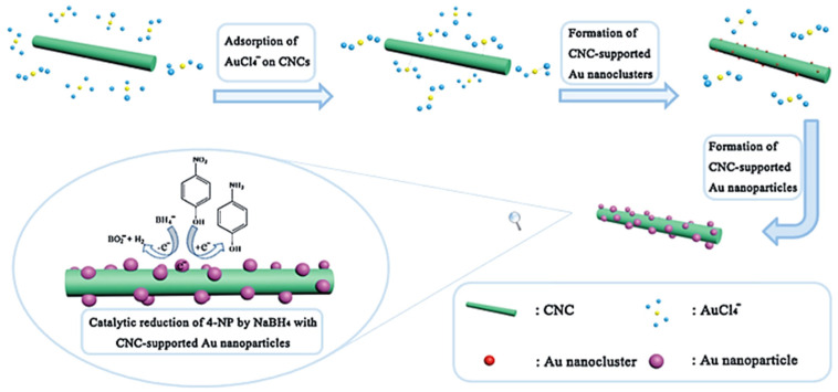
CNCs were used as a support to synthesize gold nanoparticles (Au NPs) by heating the aqueous mixture of polyethylene glycol, CNCs, and HAuCl4 at 80 °C for 1 h. In this way, extreme conditions and toxic chemicals as well as a complicated procedure were avoided (Figure 3). The prepared CNC-supported Au NPs demonstrated high catalytic activity for the reduction of 4-nitrophenol using sodium borohydride. The maximum turnover frequency and the apparent rate constant reached 641 h−1 and 1.47 × 10⁻² s−1, respectively. Due to the large concentration of highly dispersed Au NPs (average size 2–15 nm) that was deposited onto the CNC surface, a high catalytic performance was observed. The reported procedure proved to be environmentally-friendly, low-cost, and simple and has a large potential in medical and industrial applications [84].
Figure 3.
The schematic illustration of the formation process of CNC-supported Au NPs and their catalytic activity in the reduction of nitrophenol. Reprinted from [84]. Copyright 2015, with permission from Elsevier Ltd.
2.2. Cellulose Supported Non-Precious Metallic Nanostructures
Aside from noble metal nanoparticles, semi- and non-precious metallic nanoparticles have also been successfully formed on the surface of various cellulose structures. A green synthetic approach to prepare cobalt/cellulose nanocomposites with antibacterial and magnetic properties has been developed by the in situ reduction of cobalt nitrate on the substrate surface using hydrogen gas or NaBH4 as reducing agents. Spherical, cellulose-stabilized cobalt metallic nanoclusters with an average diameter of 7 nm were formed with cubic cobalt (α-cobalt) as the main component when hydrogen gas was used as a reducing agent. The cellulose-stabilized, metallic cobalt nanoclusters were homogenously distributed on the surface of the cellulose fibers. The cobalt/cellulose nanocomposites were contaminated with almost insignificant traces of boron. In contrast, the in situ reduction of cobalt ions on the cellulose surface with sodium borohydride leads to amorphous cobalt/cellulose composites with a significant contamination of boron. All of the cobalt/cellulose nanocomposites showed significant antibacterial action against the tested bacterial isolates [85].
CNCs were used as a support in the growth of copper nanoparticles (Cu NPs) by the reduction of CuSO4·5H2O, representing an environmentally-friendly, low-cost, and simple preparation method. Aqueous NaOH, ascorbic acid, and hydrazine were used as the pH controller, antioxidant, and reducing agent, respectively. The product was high purity spherical Cu NPs with the average size of ~3 nm. Compared to the unsupported Cu NPs, the reported Cu NPs–NCC demonstrated superior catalytic activity and high sustainability for the reduction of methylene blue in an aqueous solution of NaBH4 at RT. The prepared Cu NPs–NCC quantitatively reduced the methylene blue in the reaction time of 12 min with the rate constant of 0.7421 min−1 and the correlation coefficient (R2) of 0.9922 [86].
CNCs were applied as a support in the formation of metallic iron nanoparticles (Fe NPs, 3–10 nm or 15–40 nm, depending on the quantity of CNCs) by an environmentally-friendly single-step procedure. Hydroxyl groups on the surface of the CNCs served as anchor points for the stabilization of Fe NPs and as a reducing agent in the growth of Fe NPs. Additionally, CNCs acted as corrosion inhibitors for metallic Fe NPs and increased their catalytic activity since they retained the metallic state even after 5 days of air exposure. The CNC-stabilized Fe NPs exhibited a catalytic activity toward the methylene blue degradation. It was demonstrated to be a promising and nontoxic nanocatalyst for the hydrogenation reaction of 4-nitrophenol into 4-aminophenol. Additionally, the autonomous motion of the CNC-supported Fe NPs was observed in the presence of H2O2 and their trajectories were controlled externally by the pH gradient and magnetic field. It was demonstrated that their speed and controlled trajectory could be remotely tuned, making these nanomotors potential innovative nanomachines in drug delivery, imaging applications, and as sensors [87].
The number of publications on metallic nanostructures supported and templated by cellulose structures is vast and not all of them can be included within this review. Therefore, the additional information on publications dealing with the cellulose-metallic nanostructures is summarized in Table 1. Most commonly, the cellulose is decorated or coated with noble metal (Au and Ag) nanostructures and the most widely used reducing agent is NaBH4, or simply cellulose itself, while frequent applications of these hierarchical structures are in catalysis, medicine (as antibacterial agents), and electronics (as conductors) (Table 1). The most common method of deposition is the adsorption of metallic ions to the cellulose surface and their subsequent reduction to metallic particles. Alternative approaches include the separate formation of metallic nanoparticles and their subsequent deposition to cellulose fibers, or physical methods such as atomic layer deposition or plasma sputtering.
Table 1.
The list of publications on cellulose supported metallic nanostructures. The list is organized from large cellulose structures (CF and RC) via microstructured celluloses (MCC and MFC) to nanostructured celluloses (BNC and CNCs).
| Inorg. Particle (Size—nm) | Cellulose Used | Precursor (Synthetic Path) | Application | Reference |
|---|---|---|---|---|
| Precious Metallic Cellulose Supported Nanostructures | ||||
| Au NPs (~2 nm) |
CF | Bis(ethylenediamine) Au(III)Cl3, (CH3)2Au(III) acetylacet.,(red.by NaBH4) | Catalyst (glucose oxidation) | [88] |
| Ag NPs (8–20 nm) |
RC | AgNO3 (hydrothermal reduction) | Catalyst (reduction of 4-nitrophenol) | [89] |
| Au NPs (<5 nm) |
MFC | HAuCl4 (reduction by NaBH4) | Catalyst (reduction of 4-nitrophenol) | [90] |
| Au–Ag NPs | MFC | Conductive nanofiller | [91] | |
| Au–Pd NPs (4–9 nm) | MFC | HAuCl4, [Pd(NH3)4]·Cl2 (reduction by NaBH4) | Catalyst (reduction of 4-nitrophenol) | [92] |
| Au/Ag NPs | BNC | HAuCl4, AgNO3 (reduc. by poly(ethyleneimine) | - | [93] |
| Au NPs | BNC | HAuCl4 (reduction by cellulose) | Conductive nanofiller | [94] |
| AuNR@AgNCs | BNC | HAuCl4, NaBH4, AgNO3, CTAC | SERS–detection of TNT | [95] |
| Au NPs (2–10 nm) |
CNCs | HAuCl4, trisodium citrate, oleylamine, mercaptocation | Chiral photonic materials | [96] |
| Au NPs (20–30 nm) |
CNCs | HAuCl4, NaOH (reduction by CNCs) | Photothermal nanocomposite materials | [97] |
| Au NPs (30.5 nm) | CNCs | HAuCl4 (reduction by cellulose) | Catalyst (reduction of 4-nitrophenol) | [98] |
| Au NPs (4.5–7.1 nm) | CNCs | HAuCl4 (reduction by NaBH4) | Biosensor (for 2-mercaptoethanol) | [99] |
| Au NPs (2–3 nm) |
CNCs | HAuCl4 (reduction by—HS groups on the CNC surface) | Catalyst (alkyne–aldehyde–amine-coupling) | [100] |
| Au NPs (2–4 nm) |
CNCs | HAuCl4 (reduced with no and by NaBH4) | Catalyst (reduction of 4-nitrophenol) | [101] |
| Au NPs (~3 nm) |
CNCs | HAuCl4 (reduction by NaBH4) | Catalyst (reduction of 4-nitrophenol) | [102] |
| Au, Ag NPs | CNCs | HAuCl4, AgNO3, ascorbic acid | Catalyst (reduction of 4-nitrophenol, 4-aminophenol) | [103] |
| Au NPs(30–80 nm) | CNF | HAuCl4, Na3Cit·2H2O | Substrate–SERS spectroscopy | [104] |
| Au NPs (30–80 nm) |
CNCs | HAuCl4, K2CO3, NaOH | Seeds for Au coating of CNCs with tunable optical properties | [105] |
| Au NPs (35 nm) |
CNCs | HAuCl4 (reduction by trisodium citrate) | Sorption and detection of Au nanoparticles in H2O | [106] |
| Pt NPs (1–5 nm) |
CF | H2PtCl6, Na rhodizonate | Catalyst (reduction of 4-nitrophenol, methyl orange) | [107] |
| Pt NPs | CNF | H2PtCl6, (reduction by NaBH4) | Catalyst (reduction of 4-nitrophenol) | [108] |
| Pt NPs (5–30 nm) |
CNCs | H2PtCl6 (reduction by CNCs) | — | [109] |
| Pt NPs (11–101 nm) | CNCs | H2PtCl6 (reduction by wood nanomaterial) | Catalyst (reduction of 4-nitrophenol) | [110] |
| Pt NPs (~2 nm); |
CNCs | H2PtCl6 (reduction by CNCs) | Electrocatalyst (oxygen reduction) | [111] |
| Pd NPs (~20 nm) |
BNC | PdCl2 (reduction by KBH4) | Catalyst (Heck reaction) | [112] |
| Pd NPs (~20 nm) |
BNC | K2PdCl4 (reduction by NaBH4) | Catalyst (Suzuki–Miyaura reaction) | [113] |
| Pd NPs (3.6 nm) |
CNCs | PdCl2 (reduction by H2) | Catalyst (hydrogenation of phenol; Heck Coupling) | [114] |
| Pd NPs (1–7 nm) |
CNCs | PdCl2 (reduction by CNCs) | Catalyst (red. of methylene blue and 4-nitrophenol) | [115] |
| Pd-Cu NPs | BNC | PdCl2 and CuCl2 (reduction by KBH4) | Catalyst (water denitrification) | [116] |
| Ru NPs (~8 nm) |
MFC | RuCl3 (reduction by NaBH4) | Catalyst (aerobic oxidation of benzyl alcohol) | [117] |
| Ag-Au NPs (8–10 nm) |
CF | AgNO3, HAuCl4, NaOH, urea | Antibacterial agent | [118] |
| Ag NPs | CF | RF sputtering | Antibacterial agent | [119] |
| Ag NPs (3–15 nm) |
MCC | AgNO3, UV light reduction | Catalyst (reduction of p-nitrophen. to p-aminophen.) | [120] |
| Ag NPs (~6 nm) |
MFC | AgNO3 (reduction by NaBH4) | Catalyst (reduction of rhodamine B) | [121] |
| Ag nanowires | MFC | Previously formed Ag nanowires | Conductor in transparent nanopaper | [122] |
| Ag NPs (3–4 nm) |
MFC | AgNO3, UV light triggered reduction by MFC | Aerogels | [123] |
| Ag nanowires | MFC | Previously formed Ag nanowires | Conductive nanofiller | [124] |
| Ag NPs (~4 nm) |
MFC | AgNO3 (reduction by NaBH4) | Catalyst (aza–Michael reaction) | [117] |
| Ag NPs (~6 nm) |
MFC | AgNO3 (reduction by NaBH4) | Antibacterial agent | [125] |
| Ag nanowires | MFC | - | Conductive nanofiller | [126] |
| Ag NPs | BNC | AgNO3 (reduction by sodium citrate) | SERS substrate-pesticides detection | [127] |
| Ag NPs (var. sizes) | CF, MCC, CNCs | AgNO3, (reduction by cellulose) | - | [128] |
| Ag NPs (<10 nm) | BNC | AgNO3 (reduction by NaBH4) | Antibacterial agent | [129] |
| Ag NPs (17 nm) |
BNC | AgNO3 (reduction by cellulose) | Antibacterial agent | [130] |
| Ag NPs (~30 nm) | BNC | AgNO3 (red. by NH2NH2, NH2OH, ascorbic acid) | Antibacterial agent | [131] |
| Ag NPs (8–15 nm) |
BNC | AgNO3 (reduced by triethanolamine) |
Antibacterial agent | [132] |
| Ag NPs (~16 nm) | BNC | AgNO3, (reduction by BNC) | Antibacterial agent | [133] |
| Ag NPs (5– 50 nm) |
CNFs | AgNO3 (reduction by Na citrate) | Flusilazole adsorption and analysis | [134] |
| Ag NPs | CNFs | AgNO3 (reduction by Na citrate) | SERS probe for carbendiazim | [135] |
| Ag NPs (10–50 nm) |
CNCs | AgNO3 (reduction by CNC) | Electrocatalyst (reduction of oxygen) | [136] |
| Ag NPs (<10 nm) | CNCs | AgNO3 (reduction by NaBH4) | DNA biosensor | [137] |
| Ag NPs (10–15 nm) | CNCs | AgNO3 (reduction by NaBH4) | Antibacterial agent | [138] |
| Ag NPs (1 nm–10 μm) | CNCs | AgNO3 (reduction by CNCs) | Antibacterial agent | [139] |
| Ag NPs (20–45 nm) | CNCs | AgNO3 (reduction by CNCs) | Antibacterial agent | [140] |
| Ag NPs (2–3 nm) |
CNCs | AgNO3 (reduction by NaBH4) | — | [141] |
| Ag NPs (~10 nm) | CNCs | AgNO3 (reduction by dopamine) |
Catalyst (reduction of 4-nitrophenol) | [142] |
| Ag NPs (~7 nm) |
CNCs | AgNO3 (reduction with dopamine hydrochloride) |
Antibacterial agent | [143] |
| Ag NPs (10–80 nm) | CNCs | AgNO3 (reduction by NaBH4) | — | [144] |
| Ag NPs (1–2 nm) |
CNCs | Ag wire and AgNO3 (reduction by CNCs) | Catalyst (hydrogenation of aldehydes, nitrophenol, alkenes and alkynes) | [145] |
| Ag NPs (10–50 nm) | CNCs | AgNO3 (reduction by NaBH4) | Plasmonic activators for shape memory polymers | [146] |
| Semi- and non-precious cellulose supported metallic nanostructures | ||||
| Cu NPs | CF | CuSO4, poly(ethylenimine) | - | [147] |
| Cu NPs (~5 nm) |
MFC | CuCl2 (reduction by ascorbic acid) | Catalyst for the reduction of 4-nitrophenol | [148] |
| Cu NPs | CNCs | Cu(OCOCH3)2, NaOH (reduction by ascorb. acid) | Conductive nanofiller | [149] |
| Cu NPs (50 nm) |
CNCs | CuSO4, (reduction by ascorbic acid and NaBH4) | Catalyst for C–N coupling reactions | [150] |
| Cu NPs (10–20 nm) |
CNCs | CuSO4 (reduction by hydrazine) | Catalyst for oxidation of sulfides and alcohols | [151] |
| Fe NPs (200–300 nm) | CF | FeSO4 (reduction by NaBH4) | Adsorbent for Cd(II) ions | [152] |
3. Cellulose Supported Metal Oxide Nanostructures
Cellulose structures were used as supports and templates for the formation of various metal oxide nanostructures. In contrast to metallic nanostructures that are mainly limited to noble or semi-noble metals, practically any type of metal oxide can be deposited on the cellulose surface with TiO2, Fe3O4, and ZnO being the most abundant. For this purpose, various cellulose types have been applied, spanning from natural cellulose fibers to cellulose nanocrystals in the original or chemically modified form.
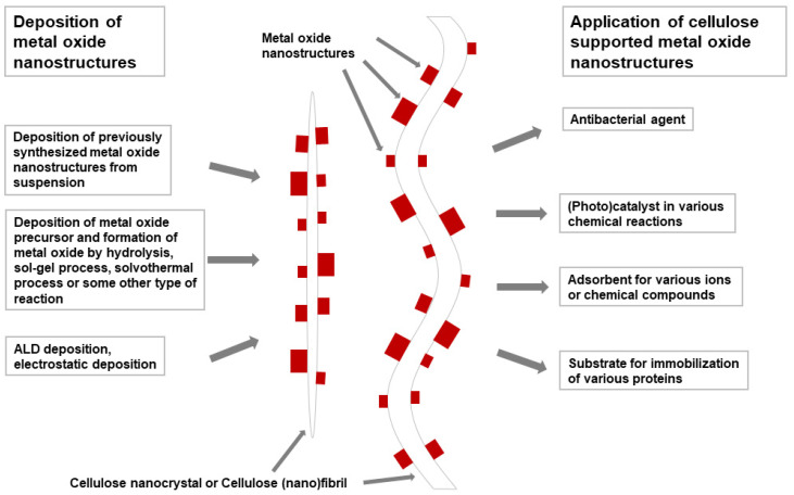
Various metal oxide nanostructures were deposited or formed on the cellulose surface that serves as the support and template in the formation of nanostructures. By using cellulose templates, various hierarchically organized nanostructured metal oxide particles were synthesized. The most widely used metallic oxides have been TiO2, ZnO, Fe3O4, and Fe2O3. Using cellulose as a support enables the formation of lightweight, highly flexible, porous, mechanically resistant, and environmentally-friendly structures accompanied with interesting catalytic, electronic, optical, magnetic, and antibacterial properties. The principal application of such structures have been found in heterogeneous catalysis and in the removal of toxic heavy metal ions (Pb2+, As5+, Mn2+, and Cr3+) from water. In particular, for the latter application, cellulose structures are particularly suitable since a comparison of the heavy metal ion absorption capacities of various inorganic sorbents and organically-modified cellulose structures is significantly in favor of the latter ones [153]. Cellulose gels with their specific structure, high specific surface, and porosity as well as a high concentration of functional groups are very suitable for the removal of various pollutants from water [153]. The incorporation of various inorganic compounds into cellulose gels enhances the pollutant removal capability and efficiency, and introduces a specific affinity toward certain pollutants. Aquatic pollutants that can be removed in this way comprise various metal ions, organic dyes, and some anions. Additionally, the ZnO/cellulose hierarchical nanostructures show a high applicative potential in medicine as wound protective materials due to the antibacterial properties of nano ZnO. Moreover, CuO, Cu2O, ZnO, and TiO2 were studied as antiviral agents and their hybrid materials with cellulose are potentially applicable in disinfectant wipes or as anti-viral layers in facemasks [74]. For example, CuO showed an intense antiviral activity and such hybrid nanomaterials were applied as an antiviral protection layer in respiratory facemasks. The practical tests demonstrated that CuO impregnated masks safely reduced the risk of influenza virus environmental contamination without altering the filtration capacities of the masks and that the manufacture of the mask with CuO layers would not add any significant costs to the price of such masks [154]. These applications are highly important nowadays, leading to the development of masks with improved protection against COVID-19. On the other hand, Fe3O4/cellulose structures are lightweight, flexible, magnetic materials that offer a wide range of potential applications. Cellulose can stabilize not only the metallic nanoparticles, but also the metal oxide nanoparticles by the strong bonding interaction of metal oxide nanoparticles with the cellulose surface, enabling the formation of hierarchical metallic oxide nanostructures that are otherwise practically impossible to prepare. The presentation of the versatility of cellulose substrates, the formation processes of metal oxide nanostructures, and the potential applications of these hierarchical structures is given in Scheme 2.
Scheme 2.
The schematic representation of the cellulose/metal oxide particle nanocomposites, the synthetic routes, and the most frequent applications of such materials.
3.1. Cellulose Supported TiO2
The most frequently used metallic oxide for the decoration of the cellulose surface is TiO2, since it offers various functionalities (photocatalytic activity, energy conversion, heavy metal absorption, etc.) and a broad range of potential applications including self-cleaning and dynamically responsive materials. Rutile TiO2 nanoparticles (average particle size 100–250 nm) have been prepared via in situ synthesis using cellulose fibers as a support to form cellulose fiber/nano-TiO2 composites. Additionally, CF/TiO2 was also prepared by the electrostatic deposition of commercial TiO2. Subsequently, composite membrane beds were prepared using a wet-laid procedure. After this, the dynamic Pb2+ adsorption was studied by passing the feed solution through the prepared beds using a single-pass flow mode. The impacts of parameters such as the bed stacking pattern, flow rate, and bed height on the bed breakthrough performance were studied. The results demonstrated that the CF/in situ-TiO2 bed had more favorable characteristics than the others, since its Pb2+ loading capacity was 9-times higher than that of the CF/self-assembled TiO2 bed and 13-times higher than that of the pure CF bed before 10% breakthrough occurred. Moreover, this bed showed high selectivity for Pb2+ toward Ca2+, and was easily regenerated for repeated use by rinsing with 0.1 M HCl solution. The excellent performance of the in situ-TiO2/CF bed was ascribed to its faster adsorption kinetics and larger fiber adsorption capacity compared to other types of beds with equal bed structures [155].
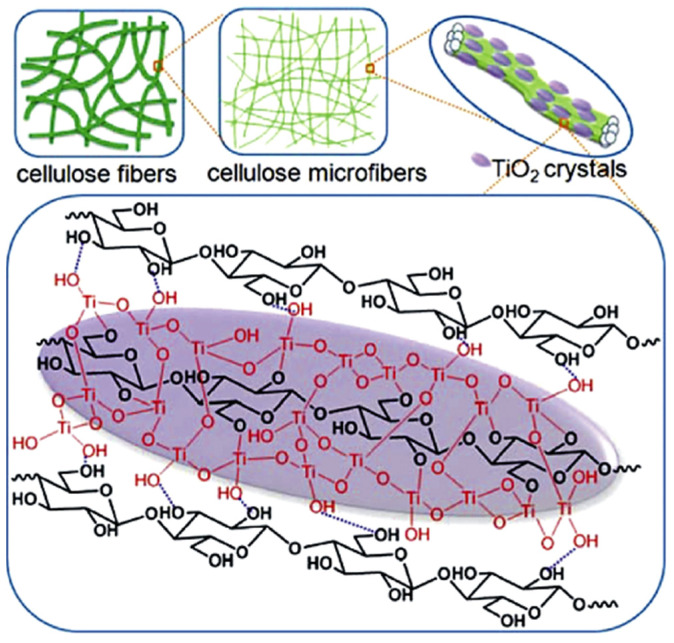
TiO2 nanocrystals (TiO2 NCs) were formed in situ on the surface of the cellulose fibers (CFs) by a simple hydrolysis of TiOSO4 to prepare the cellulose/TiO2 nanocomposites. Initially, cellulose was applied as a scaffold for the TiO2 NC immobilization. However, the results showed that CF also functioned as a chemical template, directing the crystal growth of TiO2 (Figure 4). Consequently, uniformly dispersed spindle rutile TiO2 crystals (length ~180 nm and width ~50 nm) were formed on the CF surface, which exhibited a tendency to the adsorption of heavy metal ions such as Pb2+. The CF/TiO2 composites demonstrated an improved high selectivity for lead (Pb2+) removal, high adsorption capacity, and good regenerability. Nonwoven filter beds were easily prepared using the prepared composite fibers that were further used in the dynamic filtration experiment. The CF/TiO2 beds showed a 12-fold increase in the filtered bed volume before the breakthrough occurred when compared to that of the pure CF beds. This study represents a green route toward the preparation of highly efficient and low-cost nanosorbents for water decontamination using CF as the support and template [156].
Figure 4.
The schematic illustration of the in situ directional growth of spindle TiO2 along the cellulose microfibers. Reprinted from [156]. Copyright 2015, with permission from Elsevier Ltd.
The TiO2 nanostructures (particle size 2–10 nm) were formed by in situ precipitation in the micronanoporous structure of the regenerated cellulose matrix using a sol–gel method to prepare the titania/cellulose composite films. The titania nanoparticles were formed inside the cellulose matrix, which provided reaction nanocavities for the formation of TiO2 nanostructures and hydroxyl groups for their interactive adsorption. The composite films showed high photocatalytic activity in the degradation reaction of phenol using low intensity UV light. The nano TiO2/regenerated cellulose films with interesting mechanical properties proved to be an efficient and reusable catalyst in the photodegradation of organic pollutants such as phenol [157].
A thin TiO2 film was formed on lightweight native nanocellulose aerogels using a chemical vapor deposition method (CVD), producing a new type of photo switching functional material between water-repelling and water-absorbing states. Uniform titania coatings with an average thickness of ~7 nm were prepared on cellulose fibers of the aerogel. As shown by weighing, the TiO2-coated aerogel samples practically did not absorb water during exposure, which was also proven by a high surface contact angle for water of 140°. After UV exposure, they intensively absorbed water (16-times their own weight) and showed a very low surface contact angle that is characteristic of superabsorbents. The recovery of the original properties was observed after storage in the dark for 2 weeks. Additionally, the TiO2-coated nanocellulose aerogels demonstrated a photocatalytic activity in the photooxidative decomposition of methylene blue, which, combined with the highly porous structure, provides a high potential in water purification applications [158].
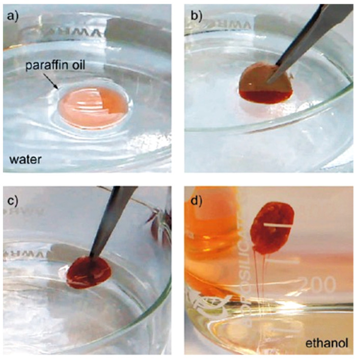
Cellulosic aerogels have been modified with TiO2 nanoparticles using a chemical vapor deposition process, introducing oleophobic functionality in such systems. It was demonstrated that functionalization of the native cellulose nanofibrils of the aerogel with an oleophilic and hydrophobic titania coating produced a water floating and selectively oil-absorbing material. These surface modified aerogels enabled the removal of organic contaminants from the water surface due to the low density combined with the capability to absorb large amounts of nonpolar liquids and oils (Figure 5), and moreover, they were recyclable and reusable after washing or alternatively, they could be incinerated together with the absorbed liquid [159].
Figure 5.
Oil spill removal from water. Paraffin oil (colored for clarity) floating on water (a); oil being absorbed into the aerogel (b); all of the floating oil has been absorbed (c); oil-filled aerogel can be washed simply by immersing it in a solvent such as ethanol. The oil is removed as shown by the red streaks (d). Reprinted with permission from [159]. Copyright 2010 American Chemical Society.
3.2. Cellulose Supported Fe3O4 and Other Iron Oxides
Cellulose structures are often decorated with Fe3O4 to introduce magnetic properties on the cellulose surface for various applications including magnetic resonance imaging for enzyme immobilization, clinical diagnosis, hyperthermia anticancer treatment, and magnetic drug targeting. Iron oxides such as magnetite (Fe3O4) and maghemite (γ-Fe2O3) have been proposed for these technological applications because of their high magnetic transition temperatures, high saturation magnetization, and their biocompatibility. In particular, water-dispersible polymer-based magnetic micrometer-sized spheres have attracted high attention for immobilizing various biopolymers such as enzymes and other drugs for diagnostic applications, controlled drug delivery, cell labeling, and separation purposes.
Fe3O4 nanoparticles (particle size 5–12 nm) were precipitated on the surface of the regenerated cellulose fibers to prepare nano-magnetic cellulose (MCGT) with excellent magnetic properties during the process of cellulose regeneration. Subsequently, a highly efficient adsorbent was formed by grafting MCGT with glycidyl methacrylate and tetraethylenepentamine and the final product was evaluated for the removal of Ag(I), Cu(II), and Hg(II). The measured adsorption capacities were 1.2, 1.5, and 2 mmol g−1 for the Ag(I), Cu(II), and Hg(II), respectively. The kinetic studies revealed that adsorption followed the pseudo-second-order kinetic model, whereby the adsorption equilibrium was reached within 3–5 min. The thermodynamic parameters revealed the spontaneous and exothermic adsorption of ions. Grafted MCGT was easily separated and regenerated from the adsorption medium by using an external magnetic field while thiourea was applied for the removal of adsorbents. It was tested for a real industrial wastewater treatment, demonstrating a high removal efficiency of pollutants, making the prepared MCGT a promising substrate for these purposes [160].
Fe3O4 spherical nanoparticles were formed on the bacterial cellulose (BC) and new BC/Fe3O4 nanocomposites (BC/Fe3O4) were prepared during BC biosynthesis from the G. xylinum fermentation by using an innovative pH controlling method. The fermentation produced Fe3O4 nanoparticles with the average diameter of 15 nm being uniformly distributed in BC. The BC/Fe3O4 nanocomposites with a 33 wt.% of Fe content showed the saturated magnetization (σs) of 41 emu g−1 and the coercivity of 27 Oe. The adsorption and elution characteristics of the BC/Fe3O4 nanocomposites for the Cr3+, Pb2+, and Mn2+ ions were studied. Their adsorption capacities were found to differ significantly at the same concentration of an ion and they were ranked in the order Cr3+ < Mn2+ < Pb2+, whereas the sequence of the elution capacity followed the order Cr3+ < Pb2+ < Mn2+. The BC/Fe3O4 nanocomposites were recyclable by applying magnetic field separation after the elution of the adsorbed heavy metal ions [161].
Never dried bacterial cellulose (BC) nanofibrils were surface functionalized with aminated magnetite nanoparticles (AMH). Particles were formed by the solvothermal reaction of FeCl3, 1,6-hexanediamine, and BC pellicles in ethylene glycol medium at 200 °C with a 6 h reaction time. Cellulose accelerated the growth of Fe3O4 nanostructures and facilitated their deposition on the cellulose nanofibers. The AMH attached on the cellulose nanofibril surface significantly improved the mechanical and thermal properties as well as increased the amine concentration of the BC pellicles. The BC–AMH pellicles exhibited a high adsorption capacity toward the As5+ ions due to their high AMH contents. Almost 90 mg of As5+ per gram of the BC–AMH composite was achieved. The high mechanical and chemical stability, large surface area as well as the high content of surface amine groups in the BC–AMH pellicles show that these structures have potential in applications such as enzyme immobilization, drug delivery, and catalysis [162].
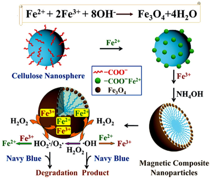
The Fe3O4 nanostructures were deposited on the surface of the carboxyl cellulosic nanospheres by using a simple co-precipitation process to form magnetic cellulose nanocrystal particles (MNPs) (Figure 6). The morphology and structure of the Fe3O4 nanoparticles (size ~50 nm) were controlled by cellulosic nanospheres that functioned as a template for their stabilization. The MNPs were efficiently recycled and reused by using an external magnetic field. The spherical MNPs showed a superparamagnetic behavior accompanied by a sensitive magnetic response under an external magnetic field, and functioned as highly efficient nanocatalysts in the Fenton-like system for the rapid removal of Navy blue textile dye from an aqueous solution. By using MNPs that were prepared with H2O2 at the feed ratio of 1:2, 90.6% of Navy blue was removed in the first min and 98.0% in 5 min time, while 75.0% of the removal efficiency was preserved in the fifth cycle by optimizing the reaction conditions. Because of the low-cost, large surface area, good dispersibility, strong catalytic activity, and facile preparation as well as the ability of magnetic recycling and reuse, the reported MNPs showed potential application as highly effective heterogeneous nanocatalysts [163].
Figure 6.
The formation and catalytic mechanism of MNPs. Magnetic iron oxides were anchored on the cellulose nanospheres by using a co-precipitation process; the rapid removal of textile dye Navy blue was achieved in the Fenton-like system. Reprinted from [163]. Copyright 2015, with permission from Elsevier Ltd.
3.3. Cellulose Supported ZnO
A frequently used metal oxide for cellulose decoration is definitely ZnO due to its wide range of outstanding physical and chemical properties. Optical and electrical properties make it attractive in various optical, electrical, and electro-optical applications, whereas chemical properties make it highly promising in a large number of catalytical and accelerating applications. Finally, the antibacterial properties of ZnO show that it has high potential in various medical and other protective applications.
The ZnO nanostructures were formed on regenerated cotton cellulose by a simple one-pot wet chemical method using the Zn–amine complex to prepare nano ZnO–cellulose hybrid materials. The Zn–amine complex was prepared from ZnNO3 as a precursor and triethanolamine without thermal treatment. The hybrid material contained around 40 wt.% of ZnO nanoparticles (size ~250 nm) with a uniform distribution of nano ZnO through the cross section of the material. The ZnO–cellulose hybrid film was formed by the initial adsorption of Zn2+ ions to the surface of the hydrophilic cellulose fibrils, followed by the alkaline hydrolysis of the Zn–amine complex and the growth of ZnO nanostructures. Possible applications of the reported nano ZnO–cellulose hybrids are in optoelectronics as flexible display devices and biomedical or strain sensors [164].
The ZnO nanoparticles were formed simultaneously with the ZnO/cellulose nanocrystal (ZnO/CNC) nanohybrids by the hydrolysis of commercial microcrystalline cellulose (MCC) with citric/hydrochloric acid, followed by precipitation using an aqueous solution of zinc nitrate. The prepared carboxylated CNCs functioned as the stabilizing and supporting agent for the growing ZnO nanoparticles, anchoring them to the surface. ZnO nanostructures with hexagonal wurtzite structure with an average diameter of 42.6 nm were deposited onto the surface of the CNCs. The prepared ZnO/CNC nanohybrids demonstrated a high antibacterial activity and improved the thermal stability and photocatalytic activity for methylene blue (MB) as 93% of MB was decomposed after the UV light exposure within 100 min [165]. Therefore, the reported ZnO/CNC hybrids are promising photocatalytic or biomedical materials.
Spherical nanocrystalline ZnO particles with an average size of 100 nm have been formed through a simple in situ polyol method by using an amidoxime modified bacterial cellulose (Am–BC) as a template. The hydroxyl and amidoxime groups of the Am–BC functioned as reactive sites for the growth of zinc oxide NPs. The Am–BC nanoreactor as a template produced ZnO NPs in a much higher yield compared to those based on the unmodified BC. Moreover, Am–BC prevented the aggregation of ZnO NPs, resulting in well-dispersed and regular ZnO NPs assembled in situ into the Am–BC network, which showed excellent photocatalytic properties. The reported synthetic process represents a simple and effective approach for the formation of other metallic nanostructures [166].
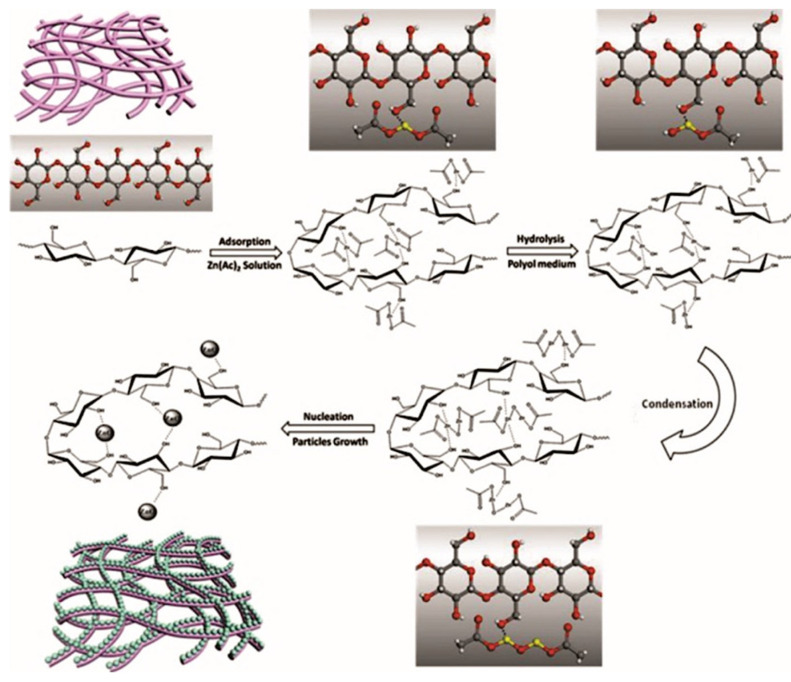
The BC was applied as a bio-template in the precipitation of crystalline ZnO nanoparticles by a simple polyol method (Figure 7). Their particle size and morphology were to a large extent affected by the hydrolysis time and concentration of the Zn(OOCCH3)2 precursor. Under the optimal reaction conditions (hydrolysis time: 10 min, concentration of Zn2+:0.5 wt.%), nano ZnO with an average size of 100 nm was formed. The BET surface areas of the pure BC fibers and the ZnO/BC composites were found to be 55.36 and 101.34 m2 g−1, respectively, indicating these structures as promising photocatalytic materials. The obtained ZnO/BC composites showed a relatively high photocatalytic activity with a maximal decolorization efficiency of 70% at 0.5 wt.% of Zn2+ [167].
Figure 7.
The schematic illustration of the formation of the ZnO nanoparticles impregnated into the BC [167]. Copyright 2013, with permission from Elsevier Ltd.
By using an ultrasonic-assisted in situ synthesis, nano ZnO particles were formed and incorporated simultaneously into bacterial cellulose (BC) pellicles. The BC pellicles were initially dispersed in a zinc acetate solution, followed by the addition of Zn2+-absorbed BC pellicles to the ammonium hydroxide solution accompanied by simultaneous ultrasonic treatment. The influences of the immersion time of the BC pellicles in the zinc acetate solution as well as the ultrasonic exposure time on the weight percent and particle size of the deposited nano ZnO were studied. The average particle size of the ZnO formed within the BC pellicles was in the range of 54–63 nm. The ZnO particles deposited inside the BC showed an unusual morphology, which was explained by the growth, with their direction being perpendicular to the alignment of the BC nanofibrils. The prepared BC-nano ZnO sheets showed a high antibacterial activity against Gram-negative and Gram-positive bacteria. The reported BC sheets decorated with nano ZnO are promising materials for effective antibacterial wound dressings [168].
Composites of regenerated bacterial cellulose (RBC) with nano ZnO (ZnO-RBC) were prepared for enhanced biomedical applications by using previously synthesized ZnO NPs. Various concentrations of ZnO NPs were admixed to the solution of RBC dissolved in N-methylmorpholine-N-oxide, and RBC nanocomposite films containing 1 and 2 wt.% of ZnO were cast from the solutions by using an applicator. The prepared ZnO–RBC nanocomposites showed substantially improved mechanical, thermal, and biological properties. Moreover, the testing of the ZnO–RBC nanocomposites revealed significantly enhanced antibacterial properties and materials that were biocompatible with excellent cell adhesion capabilities. The potential applications of the studied ZnO–RBC nanocomposites are in bioelectroanalysis and other biomedical fields [169].
Flower-like zinc oxide (ZnO) nanorod clusters were grown on the surface of spherical cellulose nanocrystals (SCNs with cellulose II crystal structure) applied as a growth substrate by a simple chemical precipitation process. In strongly alkaline (pH = 11) conditions, flower-like ZnO nanorod cluster nanohybrids were grown at a temperature of 100 °C in 2 h of reaction time. On the other hand, rod-like ZnO particles were formed at a temperature of 90 °C in weakly alkaline (pH = 9.3 and 10.5) conditions. A possible growth mechanism for the flower-like ZnO on SCNs is that by increasing the NH3 concentration (pH = 11), a larger number of hydroxyl groups favors the formation of Zn(OH)42− rather than Zn(NH3)42+. This restricts the continuous growth of ZnO nanorods, resulting in the formation of new ZnO nuclei on sites of the SCN surface, and finally, in the growth of flower-like ZnO nanorod clusters. The flower-like nanohybrids exhibited enhanced antibacterial activity against E. coli and S. aureus, and higher photocatalytic activity compared to the rod-like nanohybrids and commercial ZnO [170].
3.4. Cellulose Supported Miscellaneous Inorganic Oxides
Additionally, other metallic oxides have been successfully formed on the cellulose surface for various purposes. For example, Cu2O was formed by an innovative synthetic approach that exploited the dual properties of the thiol-grafted cellulose paper and was developed for promoting copper-catalyzed [3+2]-cycloadditions of organic azides with alkynes (Click reaction) and for adsorbing/eliminating of the residual copper species from the reaction solution. The cellulose paper was functionalized with thiol (-SH) groups by grafting the cellulose with thioglycolic acid. The -SH grafted cellulose (Cel–SH) had a dual function, that is, promoting the Cu-catalyzed Huisgen synthesis of 1,4-disubstituted triazoles through the reduction of CuSO4·5H2O to Cu2O as well as adsorbing and eliminating copper byproducts from the reaction medium. The thiol-modified cellulose paper reduced CuII to catalytically active CuI and acted as an effective adsorbent for copper, thereby simplifying the synthetic process and resulting in the reaction mixture being almost free of copper residues after a single filtration (Table 2). Highly complex organic molecules were synthesized to demonstrate the robustness of the reported cellulose-supported catalytic system as well as the related synthetic approach [171].
Table 2.
The adsorption properties of Cel–SH for the copper-catalyzed (cellulose supported Cu2O) Huisgen 1,3-dipolar cycloaddition of azide with the terminal alkyne forming 1-(benzyl-1H-1,2,3-triazol-4-yl)methanol [171]. Copyright 2016 John Wiley & Sons Inc. Reproduced with permission.
| Entry [a] | SH Loading | Yield [b] | Cu Adsorbed |
|---|---|---|---|
| (mol%) | [%] | [%] | |
| 1 | 0 | <5 | 0 |
| 2 | 3.2 | 41 | 4 |
| 3 | 8 | 43 | 58 |
| 4 | 16 | 87 | 94 |
| 5 | 24 | 88 | 97 |
| 6 | 32 | 91 | 97.5 |
[a] Reaction conditions: benzyl azide (1 mmol), propargyl alcohol (1.5 mmol), CuSO4·5H2O and Cell–SH were stirred in 5 mL of t-BuOH/H2O (1:1) for 14 h at the specified temperature. [b] Yield of the isolated product.
Magnetic CoFe2O4 nanoparticles were deposited into the cellulose structure to prepare lightweight and flexible magnetic cellulose aerogels by a simple method. Their porosities varied from 52 to 78%, while their density and internal specific surface area were between 0.25–0.39 g cm−3 and 300–320 m2 g−1, respectively. The concentration of the incorporated CoFe2O4 nanoparticles increased by increasing the precursor concentration, however, the particle size practically did not change. The deposited CoFe2O4 nanoparticles showed a superparamagnetic behavior as well as interesting mechanical properties and changed the microstructure of the cellulose aerogels. Because of the cellulose sustainability and availability in large quantities, the reported process with a simple concept is suitable for the industrial-scale up and is also applicable for many other types of nanoparticles [172].
The lightweight porous magnetic aerogels were prepared from the bacterial cellulose (BC) aerogels as templates, which were compacted into a stiff magnetic nanopaper. Non-agglomerated ferromagnetic CoFe2O4 nanostructures, with diameters ranging from 40 to 120 nm, were formed from FeSO4 and CoCl2 precursors in a mixture of NaOH and KNO3 on BC nanofibrils functioning as templates. The reported dry magnetic aerogels were flexible, porous (98%), lightweight, and they were actuated by a small magnet to absorb water and release it during the compression. Due to their high porosity, large surface area, and flexibility, these aerogels are promising materials for electronic actuators and microfluidic device applications [173].
An alternative method for the treatment of brain aneurysms was developed by applying magnetized bacterial nanocellulose (BNC) as a platform for both magnetic and cell attraction as well as for cell proliferation (Figure 8). The preparation of magnetic bacterial nanocellulose (MBNC) was carried out via the reaction of Fe II and Fe III precursors, precipitating superparamagnetic iron oxide nanoparticles, which were afterward coated with polyethylene glycol to enhance their biocompatibility. The biocompatibility and cytotoxicity testing of MBNC showed suitable cell viability and minimal cytotoxicity. The study of cellular magnetic attraction and the cell migration tests revealed the viability of MBNC as a multifunctional coating of stents for the treatment of brain aneurism [174].
Figure 8.
The schematic presentation of the concept of endovascular treatment at the neck orifice in cerebral aneurisms (a). The process consists of arterial media reconstruction by using a stent covered with the magnetic bacterial nanocellulose (MBNC) (b) designed to attract cells to the aneurysm region (c). With time, new tissue grows over the stent, closing the neck of the aneurysm (d) and ultimately, the removal of the stent (e). The photographic image of the Nitinol stent covered with MBNC revealed their good adhesion to the stent [174]. Copyright 2017 John Wiley & Sons Inc. Reproduced with permission.
Cellulose nanocrystal (CNC) films and electrospun composite CNC fibers with magnetic properties were prepared. Magnetism was introduced into the CNC by CoFe2O4 nanoparticles immobilized on the CNC nanofibrils. The CNCs (ca. 150 nm length) and the aqueous dispersion of the cobalt–iron oxide nanoparticles (10–80 nm diameter) were applied as precursors for the preparation of composite fibers as well as composite films. The CNC–CoFe2O4 composite fibers showed ferromagnetic properties, having a saturation magnetization of ~20 emu g−1. The potential application of precursor dispersions was demonstrated, and possible applications of the studied CNC-based magneto-responsive materials in the biomedical and magneto-optical components were highlighted such as magnetic shielding, packaging, separation applications, cell treatment, and drug delivery [175].
The number of publications on cellulose supported/templated inorganic oxide nanostructures is also extensive and therefore not all of them can be included in the text of the review. Consequently, the information on the inorganic oxides supported by the cellulose structures is collected in Table 3. The most commonly used method of metal oxide deposition on the cellulose is the adsorption of metallic ions via hydroxyl groups, followed by the formation of oxide particles by a hydrothermal or solvothermal reaction, or by a sol–gel process. Alternative methods of deposition involve a separate formation of nanoparticles, followed by their deposition onto the cellulose surface or commercial powders as well as various physical methods such as laser vaporization or ultrasonication, etc. The types of cellulose substrate range from natural cellulose fibers (CF) via microcrystalline cellulose (MCC) or microfibrillated cellulose (MFC) to bacterial nanocellulose (BNC) and cellulose nanocrystals (CNCs). The most common applications of these structures range from various (photo)catalytic systems to antibacterial agents in medicine and as adsorbents for drinking and wastewater purification.
Table 3.
The list of publications on the cellulose supported metal oxide nanostructures. The list is organized from large cellulose structures (CF and RC) via microstructured celluloses (MCC and MFC) to nanostructured celluloses (BNC and CNCs).
| Inorg. Particle (Size—nm) | Cellulose Used | Precursor (Synthetic Path) | Application | Reference |
|---|---|---|---|---|
| Cellulose Supported TiO2 Nanostructures | ||||
| TiO2 NPs (5–100 nm) | CF | Ti(OBu)4, HCl (hydrothermal reaction) | Photocatalyst for CO2 reduction | [176] |
| TiO2 NPs (40–250 nm) | CF | Commercial TiO2 (hydrothermal treatment) | Photocatalyst, antibacterial agent | [177] |
| TiO2 NPs (~4 nm) | CF | TiCl4 (hydrothermal reaction—ultrasonication) | Photocatalyst in self-cleaning fabric | [178] |
| TiO2 NPs (10–20 nm) |
CF | Ti(IV) isopropoxide (hydrothermal reaction) | Photocatalyst and antibacterial agent | [179] |
| TiO2 NPs | CF | TiOSO4, urea | Adsorbent for phosphate removal | [180] |
| TiO2 NPs | MCC | TiCl4, EtOH (sol–gel process) | Adsorbent for Pb2+, Cd2+, Zn2+ ions | [181] |
| TiO2 NPs (300–900 nm) | MCC | TiCl3, NH3 (hydrothermal reaction) | Photocatalyst (H2 production) | [182] |
| TiO2 nanorods (l.: 26 nm; w.: 16 nm) | MFC | Commercial TiO2 NPs | — | [183] |
| TiO2 NPs | MFC | Commercial TiO2 NPs | Antibacterial agent | [184] |
| TiO2 NPs-anatase | BNC | TiO2 powder, laser vaporization | Photocatalyst, purification of drinking water | [185] |
| TiO2 NPs (5–15 nm) |
BNC | Ti(IV) n-butoxide, sol–gel process | Detector for phosphopeptides | [186] |
| TiO2 NPs | BNC | Ti(IV) n-butoxide Solvothermal process |
Photocatalyst (methyl orange degradation) | [187] |
| TiO2 NPs | CNF | Commercial TiO2 | Photoanodes | [188] |
| TiO2 NPs | BNC | Ti(IV) isopropoxide, Sol-Gel process |
Antibacterial agent | [189] |
| TiO2 NPs | BNC | Commercial TiO2 | Adsorbent for water contaminants | [190] |
| Black TiO2 NPs | BNC | Ti[OCH(CH3)2]4, microwave-assist. sonochem. process | Interfacial solar evaporator |
[191] |
| TiO2 NPs | CNCs | TiOSO4 (hydrolysis with sulfuric acid) | UV absorber in skin care products | [192] |
| TiO2 NPs (50–100 nm) |
CNCs | Commercial nano TiO2 | Drug release system | [193] |
| Cellulose supported iron oxide nanostructures | ||||
| Fe3O4 NPs | CF | FeSO4, NH3, H2O2, PEG, previously formed Fe3O4 | Magnetic fibers | [194] |
| Fe2O3 NPs (50–200 nm) | CF | FeSO4, NaOH (ultrasonication) | Magnetic paper | [195] |
| Fe3O4 NPs | CF | FeCl3, FeCl2, NaOH | Cancer treatment with 5-Fluorouracil | [196] |
| Fe2O3 NPs | CF | (NH4)2Fe(SO4)2, NaH2PO2, | Encapsulation and delivery-biologically active compounds | [197] |
| Fe2O3 nanoplatelet (48 nm) | RC | FeCl2, FeCl3 (reaction with NaOH) | — | [198] |
| Fe3O4 NPs | CF | FeCl2, FeCl3 (reaction with NaOH) | Immobilization of prenyltransferase NovQ | [199] |
| Fe3O4 NPs | MCC | FeSO4, FeCl3, NH3 | Biotechnology, water purification | [200] |
| Fe3O4 NPs (~3 nm) |
MCC | FeCl3, FeCl2, NH3 | Antibacterial and contrasting agent | [201] |
| Fe3O4 NPs (~9 nm) |
BNC | Fe(III) acetylacetonate, (microwave sol–gel process) |
Magnetic paper | [202] |
| Fe3O4 NPs | BNC | Fe2(SO4)3, FeSO4, reaction with NaOH | Absorbing agent (removal of Cr(VI) ions) | [203] |
| Fe3O4 NPs | CNFs | Fe(NO3)3, FeSO4, NaOH, citric acid, nano TiO2 | Recoverable catalyst-H2 photogeneration | [204] |
| Fe3O4 NPs (~10 nm) |
CNCs | FeCl2, FeCl3, NH3 (precipitation) | Substrate for protein separation | [205] |
| Fe3O4 NPs | CNCs | FeCl2 and FeCl3 reaction with ammonia | Targeted delivery of doxorubicin | [206] |
| Fe3O4 NPs | CNCs | FeCl2, FeCl3 (reaction with ammonia) | Semiconductor in antistatic paper | [207] |
| Cellulose supported ZnO nanostructures | ||||
| ZnO nanorods | CF | (Zn(CH3COO)2, hexamethylenetetramine | Antibacterial agent in packaging | [208] |
| ZnO nanowires | CF | Previously synthesized ZnO NWs, Zn(CH3COO)2 (solvothermal hydrolysis) | Piezoelectric accelerometer | [209] |
| ZnO NPs (3–30 nm) |
CF | Zn(CH3COO)2, reaction with NaOH | Antibacterial agent | [210] |
| ZnO nanorods | CF | Zn(NO3)2 (hydrothermal growth) | Semi-conductor | [211] |
| ZnO rods | CF | Zn(NO3)2, (CH2)6N4, (hydrothermal synthesis) | Antibacterial rubber | [212] |
| ZnO NPs | CF | Zn(NO3)2, urea (hydrothermal process) | Photocatalyst (methyl orange degradation) | [213] |
| ZnO nanorods | BNC | Zn(NO3)2, (CH2)6N4, (hydrothermal synthesis) | — | [214] |
| ZnO NPs | BNC | Zn(NO3)2, solution plasma process | Antibacterial agent | [215] |
| ZnO NPs (100–200 nm) | BNC | Zn(OOCCH3)2, solvothermal process | Antibacterial agent | [216] |
| ZnO NPs | BNC | Zn(NO3)2, NaOH | Antibacterial dressing for burn wounds | [217] |
| ZnO NPs | CNCs | Commercial nano ZnO | Semiconductor in flexible electronic applications | [218] |
| ZnO nanorod clusters | CNCs | Zn(CH3COO)2, reaction with NaOH | Antibacterial agent | [219] |
| ZnO NPs (10–50 nm) | CNCs | ZnCl2, NaOH | Catalyst in degrad. of tetracycline | [220] |
| Cellulose supported miscellaneous oxide nanostructures | ||||
| BiVO4 | CF | Bi(NO3)3·5H2O, citric acid | Photocatalyst for methyl orange | [221] |
| Ti3(PO4)4 | CF | Waste Ti metal, H2SO4, Na3PO4 | Photocatalyst for crystal and methyl violet | [222] |
| MnO2 (150 nm) | MCC | MnSO4, KMnO4 | Adsorbent of Pb2+, H2O purification | [223] |
| In2O3–10 wt.% SnO2 | MFC | RF sputtering | Semiconductive nanofiller | [128] |
| Ag2O NPs (2–20 nm) |
MFC | AgNO3, NH3, NaOH | Adsorbent for Iˉ ions | [224] |
| H3PW12O40 | BNC | Commercial PTA | Photochromic agent | [225] |
| Mn3O4 (300 nm) | BNC | KMnO4, reduction by carboxymethyl cellulose | Electrode material | [226] |
| ZnO-CdS NPs | BNC | Zn(NO3)2, CdSO4 |
Photocatalyst (degr. of methyl orange) | [227] |
| BaTiO3 | BNC | Ti(OC4H9-n)4, H4Ba6(O)(OCH2CH2OCH3)14, hydrolysis | Piezoelectric paper | [228] |
| BaTiO3 | CNCs | Commercial BaTiO3 | Piezoelectric energy harvester | [229] |
| La2CuO4 | BNC | La(NO3)3/Cu(NO3)2 hydrothermal process |
Catalyst-methanol steam reforming | [230] |
| CoFe2O4 | BNC | FeCl3 and CoCl2, reaction with NaOH |
Magnetic paper | [231] |
| V2O5 | BNC | Vanadium(V) oxytriisopropoxide hydrolysis with HCl | Semiconductive nanofiller | [232] |
| CuO NPs | BNC | Previously prepared CuO NPs | Antimicrobial agent | [233] |
| CuO NPs | BNC | Cu(OOCCH3)2, CH3COOH, ethylene glycol | Antibacterial agent | [234] |
| CuO NPs (~7 nm) | CNCs | CuSO4 (reduction by NaBH4) | Catalyst (reduction of 4-nitrophenol) | [235] |
| FeMnOx
(50–100 nm) |
CF | KMnO4, FeSO4, NaOH, cetyltrimethyl ammonium Br | Adsorbent for As(III) and As(V) removal | [236] |
| CeO2 NPs (10–40 nm) |
RC | Ce(NO3)3, NaOH | UV shielding | [237] |
4. Cellulose Templated Pure Metal Oxide Nanostructures
The sol–gel mineralization of cellulose nanofibers is a flexible way to produce inorganic frameworks of a controlled size, structure, and porosity. For example, hollow nano-objects have raised interest in applications such as sensing, encapsulation, and drug-release [238]. A porous inorganic structure template by the cellulose structures can be prepared by the formation of metallic oxides on the surface of the cellulose and subsequent calcination. In some cases, the solvothermal removal of the cellulose template is also used [239]. Various porous structures of a wide range of pure metal oxides have been reported to be prepared by chemical or physical methods using different cellulose structures as templates. Porosity significantly increases the specific surface of these materials, giving these materials a high potential in catalysis, photovoltaics, sensorics, electrochemical energy storage, etc. [15]. The tunable porosity of inorganic oxide thin films is a key factor for their successful applications in photovoltaics, sensing, and photocatalysis. Direct cellulose templating of inorganic oxides has been carried out with nanocellulose aerogels, microfibrillated cellulose, bacterial cellulose, nanocrystalline cellulose, filter paper, and green leaves. It is expected that the pore size is a function of the shape and dimensions of the applied cellulose template. The resulting materials were sponge-like inorganic oxide monoliths, freestanding films, or powders. Additionally, titania powders biotemplated with cotton fibers were further processed to a paste that was afterward coated on conducting substrates and applied in a solar-cell assembly.
Cellulose in all of its structures and variations proved to be a highly suitable sacrificial biotemplate for the synthesis and formation of porous and mesoporous hierarchical structures or aerogels of the whole variety of inorganic oxides. All types of cellulose have been applied as biotemplates for the porous structures of inorganic oxides. The list includes TiO2, ZnO, Fe2O3, CuO, Mn3O4, Nb2O5, Co3O4, SnO2, and more complex mixed oxides such as indium tin oxide, Cu0.5Co0.5Fe2O4, or BaFe12O19/CoFe2O4. Unfortunately, there are very few reviews based solely on cellulose-templated porous pure inorganic materials [240]. The most frequently studied cellulose template porous material is TiO2, which is a very promising catalyst in the photoelectochemical water splitting of cells [241]. Porous materials are also important in the sorption of gases such as CO2, which is highly important for the reduction in the CO2 concentration in the environment [242]. Recently, the last area of application is gaining significant importance since the number of electrically driven vehicles is rapidly increasing [243]. Cellulose, as an environmentally-friendly shape persistent biotemplate, offers the possibility of the formation of new shapes and forms of inorganic oxides with tunable porosity through mostly simple and environmentally-friendly procedures followed by calcination. Cellulose is an ideal organic support and sacrificial template since it contains a lot of oxygen, and upon calcination in air, it completely degrades, leaving pure inorganic compounds with high porosity. If the calcination is performed in an inert atmosphere, carbon-supported or carbon-doped materials are formed [240]. Aside from a high specific surface, the density of the prepared material was significantly reduced, and in many cases, special structures were formed such as hollow 1-D fibers, microrods, nanowires, or nanotubes. The capability of the cellulose nanocrystals to organize into lyotropic liquid crystalline structures was exploited in the formation of nematic mesoporous silica films that were further applied as templates for inorganic oxides organized into photonic crystals with periodic order and tunable optical properties [244]. In addition to classical chemical synthesis, other innovative processes have also been applied such as the sonochemistry process, microwave-assisted ionic liquid process, evaporation-induced self-assembly, and atomic layer deposition method. In Scheme 3, the versatility of cellulose substrates, the formation processes of metal oxide porous nanostructures, and the potential applications of these structures are presented. The versatility of the cellulose substrates, the formation processes of carbon fibers supported metal oxide nanostructures, and the potential applications of these hierarchical structures is presented in Scheme 4.
Scheme 3.
The schematic representation of the cellulose/metal oxide nanocomposites and porous metal oxide structures, synthetic routes, and the most frequent applications of these materials.
Scheme 4.
The schematic representation of the carbon fiber supported metal oxide nanostructures, synthetic routes, and most frequent applications of such materials.
4.1. Cellulose Templated Porous TiO2
This section addresses the formation of (nano)porous TiO2 structures using any type of cellulose as a sacrificial template. The formation of nanotube aerogels with a framework composed of inorganic hollow nanotubes was reported. A preparation route of TiO2, Al2O3, and ZnO nanotube aerogels by applying biological aerogel templates such as microfibrillated cellulose (MFC) or cellulose nanocrystals (CNC) (Figure 9) using the atomic layer deposition method (ALD) was demonstrated. Freeze-drying or supercritical-drying was used for the preparation of aerogel templates as freestanding objects or films on substrates from nanocellulose hydrogels. Supercritical-drying produced nanocellulose aerogels without major interfibrillar aggregation, even in thick films. Uniform oxide layers were readily formed by ALD onto the fibrils, producing organic–inorganic core-shell nanofibers. The cellulose core was removed by calcination at 450 °C, forming inorganic self-supporting aerogels containing hollow nanotubular networks. They were also dispersed in ethanol by grinding to prepare a hollow nanotube slurry, which was cast to form a porous inorganic film. The TiO2 nanotube network was tested as a fast response humidity sensor [238].
Figure 9.
(a) The schematic presentation of the inorganic hollow nanotube preparation process. First the nanocellulose hydrogel is dried to obtain the aerogel, which is then coated with inorganic oxides using the ALD method to form composite organic/inorganic nanofibers. Finally, the composite was calcined to inorganic hollow nanotubes. (b) The SEM images demonstrating the effect of different drying methods: freeze-drying by freezing the nanocellulose hydrogel in liquid nitrogen, followed by the sublimation of ice in vacuum, leads to aerogels with sheet-like aggregates; freeze-drying in liquid propane leads to fibrillar aerogels with suppressed aggregation, given the sample is sufficiently thin; supercritical-drying leads to fibrillar aerogels without aggregates, even in thick samples; drying at ambient conditions leads to the collapse of the structure. (c) Photographs of the aerogel samples: (1) Liquid propane freeze-dried aerogel of 2 mm thickness; (2) supercritically-dried sample with ca. 12 mm in diameter and 10 mm in height; (3) atmospherically-dried sample that completely collapsed. Wet dimensions were the same as in the supercritically-dried sample; (4) Supercritically-dried aerogel after ALD showed a slight yellow color on the surface. Reprinted with permission from [238]. Copyright 2011 American Chemical Society.
Cellulose nanocrystals (CNCs) were applied as a shape-persistent template allowing for the straightforward formation of mesoporous TiO2 films (Figure 10). The formed TiO2 structures were high porosity anatase films with narrow pore size distributions and well-defined pores. By varying the template to the TiO2 ratio, the pore size, pore anisotropy, surface area, and diameter of the titania nanoparticles in the films were controlled. Then, by applying a post-treatment at high humidity followed by slow template removal, the pore widening was achieved and this procedure was useful for the multilayer deposition to prepare thick films. Homogeneous and transparent films were prepared by the spin- or dip-coating on various substrates such as glass, silicon, and transparent conducting oxide (TCO). The formed mesoporous TiO2 films exhibited high activity in the degradation of 4-chlorophenol as well as in the photocatalytic conversion of nitrogen oxide and they were successfully applied as anodes in dye-sensitized solar cells. The results revealed that CNC-templating offered the potential to supersede conventional processes toward porous TiO2 and that this process showed a potential for application in the preparation of mesoporous films with other inorganic oxides [15].
Figure 10.
The synthesis approach to the NCC-templated mesoporous TiO2 film. Reprinted with permission from [15]. Copyright 2014 American Chemical Society.
Mo-doped titania thin films with high transparency were formed on an ITO glass substrate by spin-coating the titania composite precursor, which was prepared by mixing titanium(IV) ethoxide and HCl, followed by the addition of CNCs and molybdenum(V) chloride. Subsequently, the prepared composite film was calcined at 400 °C for 1 h to obtain an ITO supported titania porous film. Due to the presence of CNCs as a sacrificial template, deposited titania films, composed of TiO2 crystals with average particle sizes of 10 nm, contained slit-like pore channels with a pore width ranging from 5.7 to 7.8 nm and a surface area of 150 m2 g−1. The photocatalytic properties of the formed films were studied for the degradation of trichloro ethylene (TCE) relative to the non-templated solid titania film as well as to the undoped CNC-templated porous titania film. This evaluation showed that both the CNC-templating and the Mo-doping significantly increased the photocatalytic activity of the TiO2 films for TCE degradation. Additionally, the Mo-doped TiO2 films exhibited very strong visible-light absorption and are expected to have high potential as sustainable coatings for photocatalysis and solar cells. The reported synthesis method of TiO2 with the application of CNCs as a sacrificial template was demonstrated to be a low-cost, simple, and energy saving process to fabricate highly transparent and mesoporous thin photocatalytic films, offering a significant enhancement in the photocatalytic performance with a two-fold faster degradation of TCE [245].
Recently, a new class of mesoporous materials with a chiral nematic pore structure templated by the lyotropic liquid-crystalline phase of cellulose nanocrystals (CNCs) was developed. The condensation of Si(OMe)4 as a silica precursor in the presence of CNCs produced a composite material in which CNC particles with a nematic chiral organization were embedded in the SiO2 matrix. The helical pitch in the mesoporous SiO2 was tuned by varying the ratio of the CNC to Si(OMe)4, thus changing the reflected light wavelength in the range from the near IR to the UV. After the removal of the CNC with 12 M HCl at 85 °C followed by a short oxidizing treatment with H2SO4/H2O2 (5:1), porous SiO2 was obtained as a freestanding film with a long-range chiral nematic structure. The mesoporous anatase TiO2 was synthesized from TiCl4 by calcination at 600 °C by using a nematic chiral mesoporous SiO2 film, acting as a hard template, which was subsequently removed by etching with 2 M NaOH to prepare a free-standing structure of the mesoporous TiO2 with a specific surface area between 150 and 230 m2 g−1. The structure of the chiral nematic SiO2, originating from the lyotropic liquid crystalline ordering of CNC, was preserved in the final product, producing a chiral nematic mesoporous TiO2 film, reflecting the circularly polarized light. This study is the original approach toward organizing a crystalline metal oxide into a chiral nematic structure. The reported synthetic method is highly attractive for preparing new catalytic and photonic materials [246].
4.2. Cellulose Templated Porous Iron Oxides
Cotton cellulose was applied as a sacrificial template in the in situ formation of porous and hollow CoFe2O4/BaFe12O19 microrods. The cellulose template was removed by calcination at 250 °C for 2 h and additionally at 900 °C for 3 h. The resulting composites possessed the porous hollow microrod morphology with soft ferrite (CoFe2O4) and hard ferrite (BaFe12O19) being uniformly deposited along the microrods. Due to the exchange–coupled interaction and their specific morphological effects, the microwave absorption characteristics of the microrods were substantially enhanced, as shown by electromagnetic studies. The microwave absorption bandwidth with the reflection loss less than −10 dB was expanded to 8.1 GHz, while at the same time, the weight content of the ferrite component in the composites was reduced to 60 wt.%. Therefore, the application of cellulose as a template proved to be an efficient approach to simultaneously reduce the mass of conventional ferrites and enhance the microwave absorption properties. The reported simple formation process of hollow porous CoFe2O4/BaFe12O19 microrods also increases the opportunities to develop a variety of soft/hard ferrite composite materials for other potential applications such as sensors, permanent magnets, and ultra-high-density data storage [247].
A simple method for the synthesis of crystalline and highly porous α-Fe2O3 thin films with various nanomorphologies has been developed by using the CNCs as a biotemplate. FeCl3, FeCl2, and Fe(NO3)3 were used as precursors in the CNC-templated synthesis. It was demonstrated that after the post-synthetic humidity treatment at 100 °C, the crystallization of Fe2O3 proceeded via the formation of β-FeOOH of a highly anisotropic shape. The porosity and homogeneity of the Fe2O3 films were preserved despite the complex crystallization behavior of Fe2O3 due to the shape persistence of CNCs. The calcination procedure of the CNC/Fe2O3 precursor composites showed a strong impact on the morphology and porosity of the Fe2O3 films. The calcination was performed at temperatures between 300 and 600 °C, depending on the type of precursor. It was demonstrated that a great variety of Fe2O3 networks could be synthesized and the morphology of their films could be tuned by simple templating with CNCs. The reported synthesis approach is advantageous for the formation of porous homogeneous Fe2O3 films on various kinds of substrates [248].
4.3. Miscellaneous Cellulose Templated Porous Metallic Oxides
Natural cellulosic fibers (kapok fibers) were applied as sacrificial templates in the formation of hollow SnO2 nanotubes by applying the surface sol–gel process followed by calcination at 550 °C for 5 h. The prepared nanostructures, consisting of SnO2 nanoparticles being smaller than 12 nm in size, were hollow nanotubes with a diameter between 50 and 100 nm, while the thickness of the tube wall was estimated to be from 10 to 20 nm. The resulting SnO2 nanotubes showed a high BET surface area of 185.15 m2 g−1 and very high aspect ratios. The coulombic efficiencies larger than 97.8% and stable discharge capacities of ~600 mA h g−1 were measured by using the current density of 100 mA g−1. The characteristics of the nanostructured SnO2 particles embedded in the nanotube resulted in high electronic conductivity and high capacity of the electrode. The hollow structure provided enough void space so that the SnO2 nanoparticles changed a volume without a collapse, causing excellent cycling stability of the electrode. This preparation approach is low-cost, facile, and is suitable for the scale-up production of nanotubular materials that are applicable in gas sensors or in lithium-ion batteries [249].
Cellulose was applied as a template in the preparation of octahedral, face-raised Co3O4 crystals to be applied as a catalyst in the selective oxidation of alcohols. Initially, the CoCl2 precursor and cellulose were mixed in solution, dried, and calcined at 500 °C for various times of 2–6 h. The edge length of the prepared Co3O4 structures was about 200 nm and they formed microtubes with a width of 2–3 μm and a length of ca. 10 μm. The formation of the Co3O4 octahedral structure and assembly process were facilitated by the thermal decomposition and carbonization of cellulose (Figure 11). The face-raised Co3O4 octahedral nanocrystals showed higher saturation magnetization and remanence due to smaller grain sizes with higher uniformity compared to the normal Co3O4 octahedra. Additionally, the Co3O4 octahedra grown by the cellulose-assisted process exhibited both the higher selectivity and higher activity in the catalytic oxidation of alcohols compared to the normal Co3O4 octahedra. For example, the reported Co3O4 nanocrystals showed a very high catalytic activity after eight cycles, giving them a high potential in heterocatalytical applications [250].
Figure 11.
The schematic illustration describing a time-dependent growth of Co3O4. Reprinted with permission from [250]. Copyright 2014 American Chemical Society.
The cellulose/CuO nanocomposites were prepared by an ionic liquid microwave-assisted process from the microcrystalline cellulose solution, CuCl2 precursor, and BmimBF4 medium. The reported procedure combined three highly important green chemistry principles: sustainable resources, green solvents, and an environmentally-friendly method. With longer microwave-heating time, the crystallinity of CuO increased and the CuO shape changed from nanosheets to bundles composed of irregular nanosheets, and finally, to regular nanoparticles. The ratio of the cellulose solution to ionic liquid significantly affected the microstructure and morphology of CuO in the nanocomposites. The pure CuO structures with various morphologies and particle sizes were obtained by the calcination of different precursors (cellulose/CuO nanocomposites) at 800 °C for 3 h. The reported synthetic approach opens a novel path toward the formation of inorganic oxides and to the application of cellulose with a high-added value [251].
Pure ZnO nanoparticles with a wurtzite structure have been prepared by using bacterial cellulose (BC) embedded with zinc acetate as a template and by subsequent calcination. The morphology and average particle size of ZnO nanoparticles were tuned by varying the calcination temperature and the concentration of zinc acetate in an aqueous solution. The presence of BC strongly influenced the size, morphology, and size distribution of the formed ZnO. Highly crystalline ZnO nanostructures with a narrow particle size distribution from 20 to 50 nm and high photocatalytic activity were prepared by using BC as a template containing 1 wt.% zinc acetate and by calcination at 600 °C for 2 h. The reported process provided an efficient, facile as well as environmentally-friendly approach toward the formation of the highly defined and homogeneously dispersed ZnO nanostructures. The reported process can be applied for the preparation of other nanoscaled metal oxide particles. The only disadvantage of this approach is the rather high calcination temperature, which can cause sintering of the ZnO nanoparticles [252].
Inspired by the unique structural and surface characteristics of cellulose nanocrystals (CNCs), Kong et al. used an evaporation-induced self-assembly process to prepare niobium oxide/CNC composite films (Figure 12) [253]. After the calcination of the CNCs in air at 600 °C, mesoporous T-Nb2O5 films were obtained that were composed of interconnected nanocrystals (size ~50 nm). By varying the NbCl5/CNC mass ratio, it was possible to control the crystallite dimensions (20–50 nm) and the surface area of the Nb2O5 nanostructures. When studied as the electrode materials, these T-Nb2O5 nanocrystals showed very high Li+ intercalation pseudo capacitance, wide temperature adaptability, and excellent cycle performance. The superior charge storage and kinetics benefits of the prepared T-Nb2O5 nanocrystals enabled an innovative approach toward the design of materials providing high energy density and high power density at the same time for electrochemical energy storage applications.
Figure 12.
The schematic presentation of the Nb2O5 nanostructures prepared by an evaporation-induced self-assembly process directed by CNCs (a), the SEM micrographs of the CN film (b), the CN/NbCl5 composite film (c), and the mesoporous film of Nb2O5 after calcination (d). Reprinted from [253]. Copyright 2014, with permission from Elsevier B.V.
Europium (Eu3+) doped, chiral nematic, mesoporous luminescent films of ZrO2 (EDCNMZrO2) with tunable optical properties were prepared by a hard template process using silica templated by cellulose nanocrystals (CNCs). Chiral nematic SiO2 films were formed by replicating the chiral nematic structure of CNC using an evaporation-induced self-assembly (EISA) approach, followed by calcination at 600 °C for 6 h, and subsequent silica etching. The obtained EDCNMZrO2 showed selective suppression of the spontaneous emission of Eu3+ ions as well as the possibility of modulating their luminescence lifetime. This study is a showcase of how the capability of CNC to organize into the liquid crystal nematic phase can be exploited as an indirect template to prepare the inorganic (ZrO2) photonic material, which has potential application in novel smart optical nanodevices [254].
4.4. Inorganic Nanostructures Supported by Carbon Fibers Formed from Cellulose
A special case represents the carbon nanofibers formed from various cellulose structures by pyrolysis in an inert atmosphere. Such structures are thermally stable and electrically conductive. When bacterial nanocellulose is used, templated carbon networks are obtained. Depending on the raw cellulose material, a whole variety of porous carbon nanostructures can be formed. For example, when wood was first delignified, the resulting cellulose hierarchical structure was exposed to cellulose enzymes to partially degrade its structure to form micro- and nanopores, followed by carbonation at 1000 °C for 3 h in a N2 atmosphere, a highly porous hierarchical carbon structure was obtained. Such structures with a high specific surface have been applied as electrodes in supercapacitors with high areal/volumetric energy density and excellent stability [255]. Porous carbon hierarchical structures based on wood cellulose are frequently studied as electrodes in supercapacitors or in batteries [256,257]. Various cellulose raw materials including agro waste can be applied to form porous hierarchical carbon structures [258]. Carbonized cellulose structures have a wide range of potential applications. For example, regenerated cellulose (RC) fibers with low crystallinity have been used as a template for the formation of inorganic hollow structural materials by a simple pyrolysis procedure. The RC–CuO composite fibers were composed of cellulose fibers and a high concentration of CuO NPs inside the fibers. The obtained heterogeneous system allowed for the specific thermal decomposition mechanism of the cellulose template during the pyrolysis process, involving the migration of the CuO NPs from the center to the fibers’ edge (Figure 13). At the end, Cu@C and CuO hollow microfibers with a controllable morphology and size were formed in an argon and air atmosphere, respectively. Aside from the copper-based hollow structures, other metallic hollow structural materials such as the Ni@C as well as the hollow structures of many inorganic oxides (NiO, Fe2O3, Mn2O3, Fe3O4@C, MnO@C) have been prepared through this process. The reported approach to create hollow fibers was demonstrated to be scaled-up easily and the application of elaborated processes as well as expensive starting materials was avoided. The potential applications of the prepared hollow inorganic fibrous materials are in microfluidics, as microreactors, and in catalysis [259].
Figure 13.
(a) The SEM and (b) TEM images of the CuO–RC filaments. (c,d) SEM images of CuO-and (e,f) SEM images of the Cu@C hollow fibers. (g) The XRD patterns of the CuO–RC fibers, CuO, and Cu–C hollow fibers. (h) The formation of CuO and Cu–C hollow fibers by the fast pyrolysis of CuO–RC filaments. CuO and Cu–C hollow fibers were obtained by the pyrolysis of CuO–RC filaments at 600 °C for 1 h in an air and argon atmosphere, respectively [259]. Copyright 2016 John Wiley & Sons Inc. Reproduced with permission.
Nitrogen-doped carbon networks were formed by carbonizing the polyaniline (PANI) coated BNC at 850 °C for 2 h in a flow of N2 where the bacterial nanocellulose (BNC) was applied as the precursor and template. The obtained carbon networks functioned as conductive networks for the integration of active electrode materials providing the fast electron transfer and as a support for the preparation of high capacitance electrode materials. MnO2 was formed on the surface of the activated carbon network by the redox reaction using KMnO4 as a precursor. The prepared asymmetric supercapacitors, based on activated carbon/carbon-MnO2, showed a maximum power density of 227 kW kg−1 and a high energy density of 63 W h kg−1 in an aqueous solution of 1.0 M Na2SO4. Their energy density was higher than most values reported for the activated carbon//MnO2 asymmetric supercapacitors. Additionally, the resulting supercapacitor also showed a formidable cycling performance with a 92% specific capacitance retention after 5000 cycles. The reported approach represents an environmentally-friendly and low-cost preparation process of electrode materials with potential application in high-performance energy storage devices [260].
The thermal reduction of metallic nanoparticles in the presence of cellulose is questionable due to its rather low thermal stability, but the thermal reduction of metals in an inert atmosphere accompanied with carbonization of the cellulose is possible. For example, metallic Ni nanoparticles deposited on bacterial cellulose-derived carbon fibers (Ni-NPs/BCCF) were synthesized using a simple impregnation-pyrolysis method as efficient electrocatalysts for a two electron oxygen reduction reaction (2e− ORR). NiCl2 was first deposited on BC by its immersion in an aqueous solution of the precursor (NiCl2-conc. = 0.1 mol L−1) for 24 h and subsequently freeze-dried. The resulting material was pyrolyzed first at 360 °C for 2 h and then at 700 °C for an additional 2h both in an Ar atmosphere. After cooling down to RT, the solid product was ground in a mortar, washed several times with deionized water and ethanol, and dried for further use. In 0.1 mol L−1 KOH, the Ni-NPs/BCCF-20.7 sample showed a high electrocatalytic performance toward 2e− ORR, exhibiting a H2O2 selectivity of ~90% and an onset potential of 0.75 V vs. the reversible hydrogen electrode (RHE). By applying this catalyst, a high Faradaic efficiency (93.9% ± 4.2% at 0.5 V vs. RHE in bulk ORR electrolysis) and a high H2O2 yield rate (162.7 ± 13.7 mmol gcat−1 h−1 at 0.2 V vs. RHE) were achieved. This work provides new insights into the design and development of high efficiency 2e− ORR electrocatalysts for H2O2 production [261].
The number of publications on cellulose-templated or cellulose-based carbon fiber supported inorganic structures is quite numerous and they are summarized in Table 4. Table 4 itself is divided into two parts: the first part is devoted to the publications on cellulose-templated porous purely inorganic structures, while the second part comprises the publications on inorganic nanostructures supported by cellulose-based carbon (nano)fibers. The preparation procedures in both cases were similar; that is, the formation of inorganic structures templated by cellulose, followed by thermal treatment of the structure. The difference was only in the type of atmosphere used during the thermal processing. In the first case, the thermal treatment was performed in air, causing the complete degradation of the cellulose structure and leaving predominantly porous inorganic structures, while in the second case, thermal treatment was carried out in an inert atmosphere, causing the carbonization of cellulose and producing carbon fiber supported inorganic structures. The resulting structures were predominantly tested for (photo)catalytical systems, anodes in Li batteries, or electrodes in various sensors.
Table 4.
The list of publications on cellulose-templated porous metal oxide structures or metal oxide nanostructures supported by carbon fibers formed from cellulose structures. The list is organized from large cellulose structures (CF and RC) via the microstructured celluloses (MCC and MFC) to the nanostructured celluloses (BNC and CNCs).
| Inorg. Particle (Size—nm) | Cellulose Used | Precursor (Synthetic Path) | Application | Reference |
|---|---|---|---|---|
| Cellulose Templated Inorganic Oxides | ||||
| TiO2 | MCC | titanium isopropoxide, HNO3, H3PO4, NaOH, urea | Sorbent microextr. of organic compounds | [262] |
| TiO2 nanofibers | CNF | titanium isopropoxide, NH3 | Photocatalyst—degr. of methylene blue | [263] |
| TiO2 network | CNCs | Ti(N(CH3)2)4, atomic layer deposition | Photoelectrochemical water splitting | [250] |
| TiO2 | CNCs | titanium(IV) ethoxide |
Sorbent for microextraction | [264] |
| TiO2 | CNCs | Ti-n-butoxide | Photocatalyst–(degradation of methylene blue) | [265] |
| TiO2 | CNCs | tetra-n-butyl titanium | Photocatalyst–degradation of Rhodamine B | [266] |
| α-Fe2O3 | CF | FeCl3, calcination | Catalyst (oxidation of alcohols with H2O2) | [267] |
| α-Fe2O3 | CF | FeCl3, calcination | Magnetic materials, catalyst | [268] |
| α-Fe2O3 | CF | FeCl3, calcination | – | [269] |
| Fe2O3 | RC | FeCl2, NaOH | – | [270] |
| Fe3O4 NPs (10 nm) | BNC | Fe(NO3)3, urea, hydrothermal reaction | Electrode in Li batteries | [271] |
| Fe3O4 NPs (10 nm) | BNC | Fe(NO3)3, urea, hydrothermal reaction | Flexible electrodes in Li batteries | [272] |
| Fe2O3 NPs | BNC | Fe(NO3)3, pyrolysis | Anode in Li-ion batteries | [273] |
| ZnO | CF | ZnCl2, NaOH, ethylenediamine | Photocatalyst (degradation of methyl orange) | [274] |
| ZnO | CF | Zn(NO3)2, urea, calcination | Photocatalyst (degradation of methyl orange) | [275] |
| ZnO (500–1000 nm) | MCC | Zn(NO3)2, NH3, microwaves, hydrothermal reaction | Photocatalyst (degr. of methylene blue and rhodamine B) | [276] |
| In2O3–SnO2 nanotubular (ITO) | CF | In(III) acetylacetonate, Sn(IV) isopropoxide | Active multilayers in photoanode | [277] |
| Co3O4 NPs (50–100 nm) | RC | CoCl2, calcination | Electrode in Li batteries | [278] |
| MnO2 NPs | MCC | Mn(CH3COO)2, CO(NH2)2, KOH, (CH2)6N4, NaOH, calcin | – | [279] |
| Co0.5Cu0.5Fe2O4 | CF | Cu(II) acetate, Co(II) acetate, Fe(NO3)3, sol-gel process, calcination | – | [280] |
| V2O5, V2O3, Fe3O4, WO3 (200–1000 nm) | CF | Vanadium(V) oxychloride, tungsten(VI) chloride, iron(III) chloride | – | [281] |
| CoFe2O4 NPs (5–10 nm) | BNC | Fe(NO3)3, Co(NO3)2, NH4OH | Catalyst in fuel cells | [282] |
| MnO2 NPs | BNC | KMnO4, K2SO4, reduction by C | Electrode in supercapacitors | [283] |
| MnFe2O4 | CF | Previously synthesized MnFe2O4 |
Adsorbent for As(III) and As(V) | [284] |
| LaNiO3 | MCC | La nitrate, Ni nitrate, NH3, EDTA | Catalyst (steam CO2 reforming of methane) | [285] |
| SnO2 | CF | (Sn(OiPr)4, sol-gel process, calcination | Electrode Li batteries | [286] |
| SnO2 | BNC | SnCl2, pyrolysis | Anode in Li-ion batteries | [287] |
| Inorganic Nanostructures Supported by Carbon Fibers Formed from Cellulose | ||||
| Ge | BNC | GeBr2, pyrolysis | Anode in Li-ion batteries | [287] |
| Pd-Al | CF | Pd(NO3)2, Al(NO3)3 (decomposition in a reducing atm. H2/N2) | Catalyst–(hydrogenation of cyclohexene) | [288] |
| Ni | MCC | Ni(OOCCH3)2, | Catalyst (conv. of p-nitrophenol into p-aminophenol) | [289] |
| Ni/NiO NPs (2–100 nm) | CNCs | Ni(NO3)2, glycine, calcination | – | [290] |
| Ru NPs | BNC | RuCl3, pyrolysis | Electrode in Li–O2 batteries | [291] |
| Fe3C NPs (10–30 nm) | BNC | FeCl3, pyrolysis | Electrode in Li-ion or Na-ion batteries | [292] |
| MoS2 nanoleaves | BNC | Na2MoO4, CS(NH2)2, pyrolysis | Anode in Li-ion batteries | [293] |
| MoS2 NPs | BNC | (NH4)6Mo7O24, CH4N2S, hydrothermal process | Electrocatalyst in H2 evolution reaction | [294] |
| NiCo2S4 | CF | Ni(NO3)2, Co(NO3)2, hexamethylenetetramine | Positive electrode in supercapacitors | [295] |
| BiOBr | CNFs | Bi(NO3)3, NaBr, ethylene glycol | Adsorbent for rhodamine B and Cr(VI) | [296] |
5. Cellulose Supported/Templated Nanostructures of Metal Sulfides, Hydroxyapatites, and Other Inorganic Compounds
The synthesis of cellulose/inorganic nanohybrid materials has become an important topic of research in science and industry since it offers novel and innovative applications in catalysis, energy harvesting, energy conversion and storage, drug delivery, etc. Aside from metallic and metal oxide particles, other inorganic compounds such as various inorganic sulfides, apatites, halides, etc. can be deposited or formed by using cellulose as a support and/or template. Other than metals and metal oxides, cellulose-templated hierarchical structures of other types of inorganic compounds can also be prepared such as various sulfides, halides, carbonates, and phosphates. The formation of cellulose supported metallic sulfides has not been as intensely studied as metals and metal oxides, but there has been a review partially dealing with this topic [297]. Deposition of AgCl on the bacterial cellulose is a simple route to the manufacturing of hydrophilic antimicrobial membranes, which are a promising alternative for antimicrobial wound dressings. Cellulose has been proven to be an alternative universal platform for engineering a variety of functional materials at the nanoscale. Cellulose hydroxyl groups not only anchor metallic ions tightly on the BC nanofibers via the ion–dipole interaction, but also stabilize inorganic nanoparticles by a strong interaction with their surface hydroxyl groups, thus preventing their aggregation. In many cases, the authors have used cationic surfactants to facilitate the synthesis of well-defined semiconductor nanoparticles on the cellulose surface. A special case is represented by decorated cellulose-based materials or surface-coated with carbonates, and especially with various types of hydroxyapatites. The resulting inorganic cellulose-based composites are biocompatible and capable of facilitating the osteoblast cell attachment and growth. Such materials have potential application as bone tissue scaffolds and in other biomedical fields. Additionally, these materials exhibit an effective adsorption of various ions from water solutions. Consequently, they are promising water purification materials distinguished by fast adsorption rates and high capacities. The presentation of the versatility of cellulose substrates, the formation processes of the metal sulfide nanostructures, and the potential applications of these hierarchical structures are shown in Scheme 5.
Scheme 5.
The schematic representation of the cellulose/metal sulfide particle nanocomposites, synthetic routes, and the most frequent applications of such materials.
In Scheme 6, the versatility of the cellulose substrates, the formation processes of hydroxyapatite nanostructures, and the possible applications of these hierarchical structures are presented.
Scheme 6.
The schematic representation of the cellulose/hydroxyapatite nanocomposites, synthetic routes, and the most frequent applications of these materials.
5.1. Cellulose Supported/Templated Metal Sulfides
By using the porous structure of the regenerated cellulose, the CdS nanostructures were formed from the CdCl2 solution. The cellulose pores functioned as reaction cavities where the CdS nanoparticles were grown and deposited in the shell composed of the cellulose fibers that prevented the aggregation of forming nanostructures. The CdS nanoparticles (size 6 nm) were homogeneously distributed and tightly adsorbed onto the cellulose structure by the strong intermolecular interaction between the CdS and cellulose fibers, thus preventing their aggregation. The CdS/cellulose composite films showed a substantially enhanced photocatalytic activity due to the porous structure of the cellulose template and the presence of the CdS nanoparticles. Additionally, for the promotion of H2 evolution, Pt was applied as a cocatalyst. This was formed in situ on the surface of the CdS–cellulose films by photo deposition using H2PtCl6 as a precursor. The cellulose fiber supported CdS nanoparticles showed a high efficiency in the photocatalytic evolution of hydrogen, and as a portable photocatalyst, they were easily recovered, avoiding environmental pollution [298].
An innovative nanocomposite aerogel composed of TEMPO oxidized microfibrillated cellulose embedded in a layer of previously synthesized ultrathin, fire retardant nanosheets of MoS2 was prepared. The cross linking between Mo4+ cations and carboxyl (–COOH) as well as hydroxyl (–OH) groups on the cellulose surface caused intense interaction between the MoS2 nanosheets and the cellulose fibers, thus enabling the formation of an aerogel with a density of 0.00473 g cm−3 and a 97.36% porosity. The resulting material showed outstanding self-extinguishing capabilities and fire resistant properties as characterized by cone calorimetry as well as by the vertical burning test. The MoS2 structure in the aerogel remained unchanged after the vertical burning test, as shown by Raman spectroscopy. This study represents a new approach toward the preparation of fire resistant and lightweight aerogel materials for application in the construction of fire resistant and energy efficient buildings as well as aerospace applications [299].
Various semiconducting sulfide nanostructures such as ZnS, PbS, and CdS have been prepared by using cellulose nanocrystals (CNCs) as supports combined with cationic surfactants as stabilizers. Their synthesis on the CNC surface was carried out by reacting precursor salts such as ZnCl2, Pb(NO3)2, and CdCl2 with H2S gas. In the controlled growth of defined semiconductor nanoparticles, the application of a cetyltrimethylammonium bromide (CTAB) cationic surfactant was the crucial factor. The pH of the salt solution and precursor concentration were varied to control the packing density and average particle size of the sulfide semiconductor nanostructures on the CNC surface. This procedure could serve as an innovative universal platform for engineering a variety of nanoscale functional materials for optical and electronic nanodevices [300].
5.2. Cellulose Supported/Templated Hydroxyapatites
Carbonated hydroxyapatite (CHA) modified microfibrillated cellulose (MFC) was studied as an adsorption substrate for Ni2+, Cd2+, PO43−, and NO3− ions from the aqueous solution. The MFC was initially treated with NaOH and CHA was subsequently deposited on its surface by the reaction of CaCl2 and NaH2PO4. The obtained results revealed that the removal of all of the studied ions was not dependent on the pH and was highly effective for Ni2+, Cd2+, and PO43−. Namely, the adsorption efficiency for NO3− removal from an aqueous solution reached more than 50% while the adsorption efficiencies for the other three ions were higher than 90%. The highest observed removal capacities of the studied CHA/MFC adsorbent for the NO3−, PO43−, Cd2+, and Ni2+ ions were 0.209, 0.843, 1.224, and 2.021 mmol g−1, respectively. The adsorption kinetics was very fast for all of the studied ions. This study demonstrated that CHA/MFC was an environmentally-friendly and practical absorbent, exhibiting high removal efficiencies for both the metallic and phosphate ions while the removal efficiency for the nitrate ions was only moderate [301].
A biomimetic approach was applied for the development of hydroxyapatite/bacterial cellulose nanocomposites with bone healing properties. The nucleation of calcium-deficient hydroxyapatite (cdHAp) was initiated by the adsorption of carboxymethyl cellulose (CMC) on two forms of bacterial cellulose (BC) (pellicles and tubes). The dynamic simulated body fluid (SBF) was used in the formation of the cdHAp in vitro over a one-week period. The atomic content of phosphorus and calcium varied from 0.3 to 11.2 atomic % and 0.4 to 7.7 atomic %, respectively, while the Ca/P overall ratio changed from 1.2 to 1.9 and the crystal size of cdHAp decreased with the decrease in the density of bacterial nanocellulose fibrils. The differentiation and morphology of the osteoprogenitor cells were studied through alkaline phosphatase gene expression and fluorescence microscopy to determine the viability of the scaffolds in vitro. The BC surface coated with cdHAp crystals showed an enhanced cell attachment, indicating that the BC-cdHAp scaffolds were a feasible and promising bone scaffolding material [302].
Cellulose (BC) is a high-strength fibrous and extensively used biomaterial while hydroxyapatite (HAp) has outstanding osteoconductivity and bioactivity. Composite materials composed of cellulose matrices and inorganic nano-hydroxyapatite (n-HA) fillers have received considerable attention in bone engineering, although the brittleness and low strengths limit their application. Therefore, the improvements in the toughness and mechanical strength of aerogel-based biomaterials are necessary. A urea alkali system was applied to disperse and gelate cellulose mixed with silk fibroin (SF) to prepare the composite aerosol. n-HA was prepared by swelling PAA with water for 20 h and then CaCl2 was added and stirred at 90 °C until complete dissolution. Subsequently, the Na2HPO4 solution (Ca/P = 1.67) was admixed into the prepared solution with the pH adjusted to 10 using a NaOH solution. After this, the solution was stirred for 2 h at 90 °C followed by aging at RT for 24 h. At the end, the resulting precipitate was freeze-dried and calcined at 800 °C to prepare the n-HA. By using uniaxial compression, the composite aerogel material with a density close to that of natural bone was prepared with a high mechanical strength and toughness. When the ratio of the cellulose, n-HA, and SF was 8:1:1, the composite aerogel showed the same mechanical strength range as the cancellous bone. HEK-293T cells, cultured on these composite aerogels, exhibited a high ability of adhesion, differentiation and proliferation. The studied biodegradable composite aerogel has high potential to be applied as a bone tissue substitute in bone-tissue engineering [303].
The one-step electrophoretic deposition process (EPD) was used for the preparation of an innovative bioglass—a cellulose nanocrystal (BG-CNC) bioactive hybrid material—to be applied for the surface coating of orthopedic implants with the purpose to enhance their contact with the bone tissue. The characterization of the prepared materials revealed that the network of CNCs functioned as an in situ template tailoring the mineralization rate and morphology of the formed hydroxyapatite (HA) crystal phase (Figure 14). The BG particles were individually wrapped by a nanoporous CNC layer. A very rapid formation of a HA layer (after 0.5 day) on the BG particles of the deposited BG-CNC coating in the simulated body fluid was observed during in vitro characterization. The cell culture tests using the MC3T3-E1 cell line revealed that the formed BG-CNC hybrid coatings represent a promising platform for the proliferation, accelerated attachment, and differentiation of cells, confirming their potential in biomedical applications as an advanced bone contacting material [304].
Figure 14.
The schematic model describing the interaction between the BG particles and CNCs in the suspension, together with their electrophoretic co-deposition process as well as the mechanism of mineralization by the formation of HAp in the simulated body fluid. Reprinted with permission from [304]. Copyright 2015 American Chemical Society.
5.3. Miscellaneous Cellulose Supported/Templated Inorganic Compounds
Cellulose fibers with UV activity have been prepared by spinning of 8 wt.% α−cellulose solution in N-methylmorpholine-N-oxide (NMMO) using a dry–wet method and by subsequent surface modification of the fibers by the deposition of the Gd4O3F6:Eu3+ doped with 5 mol % of the Eu3+. During its dissolution process in NMMO, the photoluminescent nanostructures were admixed as a powder into the cellulose matrix. Photoluminescence spectroscopy was used to study the relationships between the emission intensity and the excitation energy as well as the concentration of Gd4O3F6:Eu3+ nanoparticles in the final composite products. About 80% of inorganic particles had a diameter below 300 nm when the samples contained up to 1% of the modifier, and with an increase in the modifier concentration, the agglomeration of the particles was intensified. The obtained cellulose fibers with UV activity have potential application as a security marking for documents, textile, and paper products [305].
Cobalt ferrite (CoFe2O4) nanostructures were formed on the surface of the cellulose structures from CoCl2 and FeSO4 as precursors in the presence of KNO3 and NaOH. These inorganic nanoparticles were applied as labels for fine cellulose fibers, representing an approach that allows for the detection of micro- and nanostructured celluloses in paper sheets in combination with various imaging technologies. The addition of nanostructured CoFe2O4 as a labeling material during the paper manufacturing process enables imaging in scanning electron microscopy/energy-dispersive X-ray spectroscopy experiments and provides contrast in X-ray microtomography. By combining these two techniques, the fines in the paper sheets were located, evaluated, and quantified. It was demonstrated that the water retention value, the tensile indices, and the air permeability of the handsheets were not changed by the addition of the CoFe2O4 labeled fines compared to the sheets where the nonlabeled fines were added. The knowledge about the distribution of nano- and microscale cellulosic structures in a cellulose fiber matrix is important to improve the properties of the existing paper products and in the future, the design of new materials in the paper industry [306].
The number of publications on sulfides, hydroxyapatites, carbonates, halides, and other types of inorganic materials is not as large as in the case of metallic oxides. These are summarized in Table 5. Metallic sulfides are mostly formed on the cellulose by hydrothermal or solvothermal processes using sodium sulfide, thiourea, or thioacetamide as a source of sulfur. Metallic sulfide–cellulose structures are predominantly used as photocatalysts, electrodes in super capacitors, and photoluminescent or fluorescent materials. Ca hydroxyapatites–cellulose structures are mostly prepared by the biological route from Na2H2PO4 or NH4H2PO4, and CaCl2 using simulated body fluid. These are used as substrates in bone tissue engineering or as absorbents for heavy metal ions.
Table 5.
The list of publications on cellulose supported metal sulfide, hydroxyapatite, and other inorganic nanostructures. The list is organized from large cellulose structures (CF and RC) via the microstructured celluloses (MCC and MFC) to nanostructured celluloses (BNC and CNCs).
| Inorg. Particle (Size—nm) | Cellulose Used | Precursor (Synthetic Path) | Application | Reference |
|---|---|---|---|---|
| Cellulose Supported Metal Sulfides | ||||
| CdS | CF | CdCl2, AgNO3, mercaptoacet. acid | Colorimetric paper test stripe for Hg | [307] |
| ZnS–MoS2 (200–1000 nm) | CF | Na2MoO4, ZnCl2, CH4N2S | Flexible Broadband Photodetector | [308] |
| AgInS2 NPs | CF | AgNO3, In(NO3)3, NaOH, cysteine | Nanodrug for hepatocellular carcinoma | [309] |
| CuS | MFC | CuSO4, Na2S, NaOH, NH3, xylan | Paper for ablation of pathogenic microorganisms | [310] |
| CuS (100–1000 nm) | BNC | CuSO4, Na2S | Electrode in supercapacitors | [311] |
| CdS | BNC | Cadmium(II) chloride, thiourea | Photocatalyst-methyl orange under UV | [312] |
| MoS2 | CF | Na2MoO4, thiourea, MnCl2 | Photocatalyst–degr. of RhB dye | [313] |
| ZnS | BNC | Zinc(II) acetate, thioacetamide | Fluorescent material | [314] |
| ZnxCdyS | BNC | Cd(II) chloride, thiourea |
Photocatalyst-H2 prod.-visible light | [315] |
| ZnO-CdS | BNC | Zinc(II) nitrate, Cd(II) sulfate |
Photocatalyst–methyl orange–UV | [236] |
| MoS2 NPs (10 nm) | BNC | (NH4)2MoS4, N2H4 | Electrocatalyst in H2 evolution react. | [316] |
| MoS2 NPs | BNC | (NH4)2MoO4, CH4N2S, | Electrode in Li-ion batteries | [317] |
| ZnS | CF | Zinc(II) nitrate hexahydrate, sodium sulfide |
Photoluminescent material–security paper | [318] |
| In doped Mo(O,S)2 | CNFs | MoCl5, InCl3, CH3CSNH2,NH2NH2 | Photocatalyst for organic dyes | [319] |
| FeS NPs (10–20 nm) | CF | Iron(II) chloride, sodium sulfide | Uranium(VI) ions immobilization–water purification | [320] |
| Ni3S2 NPs | BNC | NiCl2, CN2H4S, NH3 | Electrode in supercapacitor | [321] |
| Cellulose supported hydroxyapatites | ||||
| Ca hydroxyapatite | MFC | NaH2PO4, CaCl2 | Adsorbent for Cr(VI) ions, H2O purification | [322] |
| Ca hydroxyapatite | BNC | NaH2PO4, CaCl2 | Cell proliferation–bone tissue engin. | [323] |
| Ca hydroxyapatite | CF | Ca(NO3)2,CO(NH2)2 (NH4)2HPO4, NaOH, | Cell proliferation–bone tissue engin. | [324] |
| Ca phosphate–Ca hydroxyapatite | CF | CaCO3, P2O5, MgO Ca hydroxyapatite | Injectable bone paste | [325] |
| Ca hydroxyapatite | CF | Ca(NO3)2, (NH4)2HPO4, NH3 | Adsorbent for Hg, H2O purification | [326] |
| Ca hydroxyapatite | CNFs | Ca(NO3)2, (NH4)2HPO4 | Fire resistant composite foams | [327] |
| Ca nanohydroxyapatite | CNCs | Commercial hydroxyapatite | Cell proliferation–bone tissue engin. | [328] |
| Ca hydroxyapatite | CNCs | Ca(NO3)2, (NH4)2HPO4 | Adsorbent for chlortetracycline | [329] |
| Cellulose supported miscellaneous inorganic nanostructures | ||||
| CaCO3 NPs | CF | CaCl2, Na2CO3 | Biomedical material | [330] |
| EuF3 NPs (250–600 nm) | CF | LiCl, EuF3 | Phosphor–flexible optical material | [331] |
| Se NPs | CNCs | H2SeO3, Na2SeO3, sorbitol | - | [332] |
| Bi NPs (2–10 nm) | CNFs | Bi(NO3)3, NaBH4 | Agent for enhanced radiation therapy | [333] |
| Ni(OH)2 NPs | BNC | NiSO4, NH4F, NH3 | Electrode in supercapacitors | [334] |
| AgCl NPs | CF | AgNO3, NaCl | Antibacterial agent | [335] |
| AgCl/Ag (~1200 nm) | CF | AgNO3, AlCl3, NaOH | Photocatalyst, antibacterial agent | [336] |
| FeOOH | BNC | - | Flexible electrode-supercapacitors | [337] |
6. Cellulose Supported or Templated Hybrid Inorganic Nanostructures
Aside from metallic, metal oxide, or other inorganic nanoparticles, various complex hybrid nanostructures have also been formed on the cellulose surface. The specific fibrous structure and high number of hydrogen bonds give the cellulose sufficient porosity and high specific surface area, which can serve as a matrix to support other functionalized materials and as a template to form various inorganic hierarchical nanostructures. New materials with interesting combinations of properties and functions have been prepared by combining different inorganic nanostructures and polymer chains with various cellulose structures. Other than cellulose supported or templated inorganic compounds, more complex hybrid modifications or decorations of the cellulose structures have also been studied and reported. Complex hybrid cellulose materials have a wide variety of potential applications (e.g., in medicine, pharmaceutical technology, chemical catalysis, electronics, and energy conversion [1,3,338,339,340]). The development of complex cellulose-based structures aims at the formation of hierarchical nanoparticle structures with specific tailored combinations of properties for various applications such as the optimization of the catalytic activity through a combination of two catalytic materials or the optimization of the antibacterial activity of wound dressings through a combination of two antibacterial agents. In addition, combinations of three or even more components with cellulose have been reported. For example, MnO2, polypyrrole, and cellulose nanocrystals were applied to form lightweight, highly porous, and flexible hybrid (inorganic–organic) materials based on the cellulose aerogel for super capacitive application. Supercapacitors are a highly propulsive area of research and have already found commercial application [1]. A complex combination of cellulose nanocrystals, Fe3O4, cationic polymer, SiO2, poly(vinylpyrrolidone), and cyclodextrine was studied and its potential application in the separation of pharmaceutical compounds was demonstrated. Similarly, the CNC/Fe3O4/Au (CNFeAu) nanocomposite was used as a support matrix for the immobilization and separation of papain from the reaction solution with a high enzyme loading; the conjugated biomolecules retained their high enzyme activity and exhibited high stability and reusability. Inorganic nanostructures can be synthesized in situ on the surface of the cellulose fibrillar structure or they can be admixed to the cellulose. In this way, ZnO nanoparticles were formed in the presence of various saccharides, and such saccharide capped ZnO nanostructures were further deposited on the pine cellulose fibers that were subsequently incorporated into the alginic acid sheets, producing paper materials with a high degree of immobilization of biomolecules for the detection of blood type. Instead of chemical methods, physical methods of nanostructure formation can also be applied. For example, cellulose–Cu and Al2O3 sandwich nanostructures were prepared by magnetron sputtering and RF reactive sputtering, producing promising materials for EMI shielding applications. Recently, cellulose paper substrates have attracted tremendous interest from both academia and industry as a platform for biosensing applications. Cellulose fibers have an interesting combination of features that makes this substrate advantageous or even ideal for conducting chemical and biological analyses comprising the passive transport of fluids through their capillary wicking, excellent biocompatibility and biodegradability, large surface-to-volume ratio, porous structure, high abundance, and most importantly, their low-cost [339,341]. Since most of these hybrid structures contain biological molecules (proteins or DNA), doubt about the stability of the reported sensing devices over time has risen because in most cases, it was not studied. In Scheme 7, the versatility of cellulose substrates, the formation processes of hybrid cellulose/metallic or metal oxide particles, and the application possibilities of these complex hierarchical structures are presented.
Scheme 7.
The schematic representation of hybrid cellulose/metallic or metal oxide particle nanocomposites, the synthesis or preparation procedures, and the most frequent applications of these materials.
6.1. Cellulose Supported Metallic Hybrid Nanostructures
A sensitive paper-based electrochemiluminescence origami device (PECLOD) for the detection of the human immunoglobulin G (H-IgG) antigen based on the rolling circle amplification (RCA) using the Au-paper working electrode (Au-PWE) was developed (Figure 15). The Au-PWE functionalized with the Ab1 antibodies provided a suitable environment for the efficient capture of H-IgG and played a significant role in electrochemiluminescence (ECL) amplification due to its 3D macroporous structure. Carbon dot (CD)-tagged RCA amplified DNA as a signal amplification device significantly increased the sensitivity and offered a high ratio of signal labels. The prepared PECLOD demonstrated the ultrahigh sensitivity, acceptable reproducibility, and broad dynamic range with very limited nonspecific adsorption under optimal conditions by applying H-IgG as a model protein. The designed PECLOD showed a dynamic response to H-IgG ranging from 1.0 fM to 25 pM with a very low detection limit of 0.15 fM. Additionally, the reported approach showed a potential to be extended for the detection of other biomarkers or proteins [342].
Figure 15.
The schematic presentation of the fabrication procedures for the paper-based electrochemiluminescence origami device (PECLOD), (IgG—human immunoglobulin G protein; Ab—capture antibodies of IgG; BSA—bovine serum albumin; Ab2—monoclonal antihuman IgG antibodies; Au Nps—gold nanoparticles; primer—primer DNA; circular template—circular template DNA; probe —carbon dots-DNA probe; RCA—rolling circle amplification). Reprinted from [342]. Copyright 2015, with permission from Elsevier B.V.
Ordinary filter paper with a 6 μm pore size was applied as a solid support that facilitated the impregnation with europium tetrakis dibenzoylmethide triethylammonium (EuD4TEA) and with gold nanoparticles that provided durability. This impregnated paper-based sensor platform was applied for the development of the fluorescence turn-on cyanide (CN) assay in aqueous media. The detection mechanism was based on two processes: (a) Ligand exchange of TEA with the CN− ion stimulating the enhancement of cyanide specific fluorescence, and (b) the dissolution of gold nanoparticles, causing the recovery of fluorescence. The described CN assay demonstrated a visible transition of color as a function of CN− exposure in the concentration range from 10−2 to 10−12 M. Additionally, an algorithm of image processing was developed that enabled the color change calibration and quantification of the CN− concentration. The reported algorithm was applicable to an Android smart phone, thus transforming it into a quantitative detector of CN− ions. The reported approach enabled sensitive, fast, and simple instrument free CN− ion detection in water as well as the possibility of remote water monitoring and controlling in the laboratory [343].
By using cellulose nanocrystals (CNC) as a template, mesoporous silica (SiO2) films with chiral nematic ordering were prepared and further applied as a hard template for the formation of silver nanoparticles (AgNPs) from AgNO3 by reduction with NaBH4. The resulting SiO2 films deposited with the Ag NPs showed optical activities in the vicinity of the surface plasmon resonance (SPR) of the Ag NPs, as shown by the circular dichroism (CD) measurements. By conducting the experiments with three different SiO2 hard templates loaded with AgNPs dried and soaked with water, it was shown that the optical activities only originated from the long-range organization of the Ag NPs in the nematic chiral silica template. Since the SPR of metallic NPs is important in biosensing, the application of the response based on the chiral organization of metallic AgNPs embedded in a chiral mesoporous host will lead to the development of new biosensors. This is a showcase of the indirect application of CNC nematic liquid crystals in the formation of optically active inorganic structures [344].
Additionally, cellulose fibers (paper) have been used for the preparation of large surface area bioactive hybrid scaffolds by exploiting the self-assembly and adsorption of the previously formed oxidized nickel (Ni/NiO) nanoparticles (NPs). The Ni/NiO NPs enabled the oriented binding of functional His-tagged protein G to the cellulose surface as it was confirmed by the subsequent binding of fluorescent antibodies. It was shown that the interaction between the polyhistidine-tagged protein and the nickel substrate was chemically inert and highly resilient. The targeted conjugation of His-tagged protein G enabled the oriented binding of anti-Salmonella antibodies, enabling the attachment of Salmonella bacteria to the substrate (Figure 16). This simple approach opens the way for the application of other paper-supported metallic NPs as docking sites for a variety of biomolecules—nucleic acids or proteins. The reported strategy is based on the high surface area of the fibrillar cellulose structure, which enables a higher potential of protein capture, causing a large increase in the signal detection and improved assay sensitivity [345].
Figure 16.
The schematic presentation of the bioactive scaffold preparation: (I) adsorption of Ni/NiO NPs on cellulose fiber-filter paper, (II) targeted immobilization of the polyhistidine-tagged protein G (pG-6xHis), and (III) protein G-mediated capture of antibodies. Protein G is presented by the protein G B1 domain (PDB: 1GB1, orange) bearing a 6xHis at its C-terminus (red). The antibody is an IgG2a immunoglobulin (PDB: 1IGT, green). Reprinted with permission from [345]. Copyright 2014 American Chemical Society.
α-Cellulose was applied as a support in the formation of hybrid zinc oxide/silver (ZnO/Ag) nanostructures on its surface by the microwave solvothermal synthesis using Zn(OOCCH3)2 and AgNO3 as precursors and hexamethylenetetramine as the precipitating and reducing agent. Before synthesis, the surface of cellulose was activated by H2O2 with a relatively mild effect on the contact angle with H2O. The formed ZnO/Ag nanostructure was a nanodispersed semiconductor/metal hybrid with an exceptional collective plasmonic structure never previously observed. The experiments with a single precursor solution enabled the separate study of the interaction of Zn2+ and Ag+ ions with the cellulose surface and in this way, the reaction mechanism in the mixed precursor solution was explained. A specific interaction between the Zn2+ and the cellulose substrate was confirmed while no specific non-thermal effects of microwave heating were detected [346].
6.2. Cellulose Supported Metal Oxide Hybrid Nanostructures
For the antibody immobilization, pine cellulose fiber sheets decorated with saccharide capped-ZnO nanoparticles have been applied as a bioactive substrate. Initially, ZnO nanoparticles were formed in the presence of starch and alginic acid as well as sucrose and glucose. The fibers of pine cellulose were subsequently surface modified by the prepared saccharide capped ZnO nanoparticles that were finally incorporated into sheets of alginic acid. The immobilization of antibodies on the surface of the fibers was significantly enhanced by the adsorbed ZnO nanoparticles. The observed retention of antibodies was about 95% after rewetting the ZnO–alginic acid modified sheets with the saline solution. For the possible application of bioactive- or biosensing-papers for the detection of blood types, a high degree of the biomolecule immobilization is an important characteristic. The possibility of detecting blood types was successfully demonstrated by using A-, B-, and D-antibodies. Additionally, the ZnO nanoparticles introduced a strong activity against bacteria (S. aureus and E. coli) as well as strong resistance toward cellulase producing fungus G. trabeum [347].
Natural cellulose was applied to template the TiO2 and to prepare a bioinspired nanocomposite composed of nanotubular TiO2 and platinum quantum dots. TiO2 nanotubes were formed by calcination at 450 °C where the cellulose structure played the role of a sacrificial template. The studied composites were composed of three-dimensional hierarchical structures of TiO2 and metallic ultrafine Pt quantum dots with sizes of app. 2 nm being uniformly formed on the surface of the TiO2 nanotubes by a photo deposition process. Complex nanocomposite structures with 1.06 wt.% of Pt content demonstrated the maximum photocatalytic hydrogen production of 16.44 mmol h−1 g−1 from water-splitting, while higher concentrations of Pt caused a decrease in the photocatalytic activity. The sufficient photocatalytic stability was a result of the structural integrity of the nanocomposite during cyclic water-splitting processes. The introduction of cellulose supported hierarchical nanomaterials into photocatalytic systems is an innovative approach to the design and production of new photocatalytic systems [348].
ZnO nanorod arrays (NRAs) were formed on the CuO nanospindle (CuO NS)/paper by using a hydrothermal process to prepare the cellulose supported p–n junction device. Soaking with an aqueous solution of Cu(NO3)2 and hexamethylenetetramine and thermal treatment at 75 °C for 6 h produced cellulose fibers decorated with CuO NSs. The resulting CuO NSs/paper was coated with ZnO seeds and subsequently transformed into ZnO NRAs through hydrothermal synthesis at 85–90 °C for 3 h. The prepared device exhibited the I–V characteristics with rectifying behavior at 0.93 V of the turn-on voltage. This simple process for the ZnO NRA formation on the CuO NS-modified paper proved to be a useful strategy toward the formation of paper-based nanostructures for applications in electronic devices [349].
EMI shielding-based sandwich nanomaterials were prepared by functionalizing the bacterial cellulose with Cu and Al2O3 nanoparticles by using the magnetron sputtering and RF reactive sputtering method. To optimize the material properties, the ratio of the Cu and Al2O3 nanostructure films was explored by applying the P–B ratio theory. The addition of Al2O3 nanosheets enhanced the EMI shielding and anti-oxidation properties of the Al2O3/Cu/BC nanocomposite materials (Figure 17). The as-prepared sandwich nanomaterials thus demonstrated high EMI shielding properties (65.3 dB), high hydrophobicity, and good mechanical properties (41.3 MPa). The reported innovative topological composite cellulose structure proved to be a promising material for commercial applications in the area of EMI shielding [350].
Figure 17.
The schematic diagram of the anti-oxidation mechanism of the Cu nanostructure layers (a) in the absence and (b) presence of Al2O3 layers. Reprinted from [350]. Copyright 2016, with permission from Elsevier Ltd.
Fe3O4 nanoparticles were formed in situ on the bacterial cellulose (BC) modified with fluoroalkyl silane (FAS) under moderate reaction conditions to prepare the magnetic flexible hybrid membranes with an amphiphobic surface. The surface chemical properties of the BC nanofibers (ether and hydroxyl groups) and the ultrafine network architecture both functioned as effective nanoreactor cavities for the formation of magnetite nanostructures. The FS10 sample with homogeneously dispersed magnetite nanoparticles on the BC showed the maximal amphiphobicity with an oil contact angle of 112° and a water contact angle of 130°. It also showed the excellent superparamagnetic properties (Ms of 8.03 emu g−1) as well as the highest tensile strength (186 MPa) among the fluorinated magnetic membranes, and magnetic actuation was also demonstrated. This is an effective process in which to prepare self-cleaning, magnetic, and flexible BC membranes that can find potential application in EMI shielding, information storage, magnetographic printing, or as electronic actuators [351].
A simple aqueous sol–gel process was developed which comprised mixing, freezing, and freeze-drying to entangle one of the three types of capacitive nanoparticles with different dimensions, shapes, and chemistries within chemically modified CNC networks composed of aldehyde modified CNCs and hydrazine modified CNCs. Capacitive structures such as MnO2 nanoparticles, polypyrrole-nanofibers, and polypyrrole coated carbon nanotubes were synthesized separately and subsequently deposited into the 3D cellulose aerogel structure in one step during the aerogel assembly, providing a huge accessible surface area of the capacitive material, thus enhancing the charge storage (Figure 18). Because of the high mass ratio of the active material to the total electrode mass, excellent capacitance retention at high charge–discharge rates was observed. Multiple channels in the aerogels formed a higher number of electronic and ionic diffusion paths, producing a CNC-based material with the lowest internal resistance for nanocellulose supercapacitors. The preparation and supercapacitive performance of highly porous, lightweight, and flexible hybrid (inorganic–organic) materials based on CNC aerogels have been demonstrated [352].
Figure 18.
(A) The schematic presentation of the aerogel components including functionalized CNCs and capacitive nanoparticles, which form the initial suspension of cross-linked clusters. (B) The sol–gel process used to prepare aerogels. (C) Photograph of the final hybrid aerogels with a diameter of 1.5 cm. (D) Photograph emphasizing the lightweight nature of a hybrid aerogel resting on the top of a feather. (E) Schematic presentation showing the fabrication of symmetric supercapacitor cells in the CR2032-type coin-cell cases with two hybrid aerogels separated by a porous polyethylene membrane in a saturated Na2SO4 aqueous electrolyte [352]. Copyright 2015 John Wiley & Sons Inc. Reproduced with permission.
Some authors have combined cellulose structures, inorganic nanoparticles, and various polymers to obtain materials with highly complex structures and specific functions. For example, Chen et al. reported on a six-stage modification of CNCs [353]. The first stage was the surface deposition of poly(diallyldimethyl ammonium chloride) followed by the surface decoration with Fe3O4 nanoparticles (Figure 19). Subsequently, PVP was deposited on the surface of Fe3O4 decorated CNCs, followed by embedding the whole structure into SiO2 (Figure 19). The silica coating significantly increased the CNCs’ thermal stability since the onset decomposition temperature was shifted by 60 °C toward higher temperatures while Fe3O4 introduced magnetic properties into the system. On the surface of the final SiO2 layer, cyclodextrin molecules were finally grafted, which provided functionality to these complex structures (Figure 19). The as-prepared CNC/Fe3O4/SiO2/β-CD hybrids showed interesting magnetic properties and effective adsorption properties toward two pharmaceutical molecules as model compounds. Consequently, they were highly effective in the magnetic separation of imipramine hydrochloride and procaine hydrochloride from the water solution. The developed process has the potential to be used for the formation of other organic−inorganic nanocomposites by applying CNCs as a template and support.
Figure 19.
The schematic presentation for the synthesis of the CNC@Fe3O4@SiO2@ β-CDa. Reprinted with permission from [353]. Copyright 2014 American Chemical Society. aCNC: cellulose nanocrystal. PDDA: poly(diallydimethylammonium chloride). PVP: polyvinylpyrrolidone. β-CD: β-cyclodextrin.
The synthesis and characterization of Fe3O4 NPs and Au NPs immobilized on CNCs was reported. The CNC/Fe3O4NPs/AuNPs (CNFeAu) nanocomposite was developed as a support for the adsorption and separation of papain, having a maximum loading of 186 mg protein g−1 on the CNFeAu from the reaction solution (Figure 20). The conjugated papain molecules exhibited high stability and reusability and their high enzymatic activity was preserved. The enzyme adsorbed on CNFeAu retained more than 83% of the original activity, with high thermal tolerance and reusability compared to the nonadsorbed reference, as shown by the electrochemical detection and conventional spectrophotometric assay of the specific enzyme binding to the Thc-Fca-Gly-Gly-Tyr-Arg inhibitory film. The magnetic CNFeAu proved to be an effective support matrix with the potential for the magnetic separation of molecules as well as in immobilizing proteins, enzymes, and other biomolecules [354].
Figure 20.
The schematic presentation of the stepwise formation of the conjugate of CNC/Fe3O4NPs/AuNPs/Papain—(i) FeCl3, FeCl2, thioctic acid; (ii) HAuCl4, α-cyclodextrin, NaBH4, thioctic acid; (iii) EDC, NHS, papain. Inset shows photographs of (a) pristine CNC; (b) CNC/Fe3O4NPs; (c) CNC/Fe3O4NPs/AuNPs; and (d) CNC/Fe3O4NPs/AuNPs attracted to the sidewall with a permanent magnet. Reprinted with permission from [354]. Copyright 2013 American Chemical Society.
Mesoporous TiO2 containing a replica of a chiral nematic structure was synthesized by preparing a titania aqueous sol using a titanium isopropoxide precursor and acetic acid and subsequently mesoporous TiO2 with a chiral nematic structure (CNTiO2) was formed by dipping the previously prepared CNC photonic films, prepared by an evaporation-induced self-assembly (EISA) method, into the TiO2 sol. CNC photonic films acted as a chiral biotemplate, which was removed by calcination at 500 °C for 4 h and mesoporous chiral TiO2 film was formed. The stopband of the TiO2 photonic films was controlled by the quantity of D-glucose added to the system. Finally, mesoporous TiO2 was decorated with Au nanoparticles using HAuCl4 as a precursor and NaBH4 as a reducing agent to increase its photocatalytic activity. The prepared mesoporous chiral TiO2/Au hybrid films were tested as catalysts in the photocatalytic splitting of water for the production of H2. It was demonstrated that the photo generation of H2 was more than eight times higher compared to the conventional mesoporous TiO2 loaded with Au nanoparticles [355].
The number of publications on cellulose supported or templated hybrid inorganic nanostructures is extensive due to the large number of hybrid combinations and potential applications. These are summarized in Table 6 to concentrate the information in the smallest possible space. The majority of publications in Table 6 reported on the formation of cellulose fiber (paper)–Au or other noble metal based electrodes, constituting various medical and environmental sensors. Noble metals predominate due to their stability and resistance to corrosion. Usually, metallic nanoparticles are formed in two stage process: First, the formation of seeds or nuclei, followed by the seeded growth of nanostructures that completely cover the cellulose surface and form conductive electrodes. The most commonly used precursors are HAuCl4, H2PtCl6, AgNO3, or H2PdCl4 while the most frequently used reducing agents are NaBH4, Na citrate, or ascorbic acid. The most frequently used process for the growth of Au nanorods is the one reported by Busbee et al. [356]. The most important area of their application is in medical diagnostics and environmental monitoring. The most predominantly applied cellulose substrate is paper—cellulose fibers but bacterial nanocellulose (BNC) and cellulose nanocrystals (CNCs) are also used.
Table 6.
The list of publications on cellulose supported/templated inorganic hybrid nanostructures. The list is organized from large cellulose structures (CF and RC) via the microstructured celluloses (MCC and MFC) to nanostructured celluloses (BNC and CNCs).
| Inorg. Particle (Size—nm) | Cellulose Used | Precursor (Synthetic Path) | Application | Reference |
|---|---|---|---|---|
| Cellulose Supported Metallic Hybrid Nanostructures | ||||
| Au (~100 nm) | CF | HAuCl4, cetyltrimethyl ammonium chloride, H2O2 | Flexible electrode in environmental and biosensors | [357] |
| Au nanorods, Ag NPs, ZnO | CF | HAuCl4, H2NOH.HCl, ZnNO3, NH3, AgNO3, ethylene glycol |
Electrode in electrochemical immunosensor | [358] |
| Au NPs (50–100 nm) | CF | HAuCl4, NaBH4, Na citrate, reduction |
Flexible electrode sensor-oligonucleotides | [359] |
| Au NPs (30–100 nm) | CF | HAuCl4, trisodium citrate | Flexible plasmonic immunosensor for detection of sepsis | [360] |
| Au NPs, ZnO nanorods | CF | HAuCl4, cetyltrimethyl ammonium chloride, H2O2; Zn(NO3)2, (CH2)6N4, | Photoelectrochemical immunosensor-carcinoembryonic antigen antibodies | [361] |
| Au NPs, Mn2O3 | CF | HAuCl4, ascorbic acid, Mn(NO3)2, glucose | Paper-based biosensor for Pb2+ detection | [362] |
| Au-Ag NPs | CF | HAuCl4, AgNO3, NH3, ascorbic acid, reduction | Electrode in electrochemical immunosensor–carbohydrate antigen | [363] |
| Pt NPs (100–200 nm) | CF | H2PtCl6, ascorbic acid, reduction | Electrode in electrochemical immunodevice–tumor biomarker | [364] |
| Pt NPs–ZnO nanorods | CF | H2PtCl6, NaBH4, reduction; Zn(NO3)2, CH3COONH4, ethylenediamine |
Electrode in H2O2 sensor–tumor cells detection | [365] |
| Pt NPs | CF | H2PtCl6, NaBH4, H2PtCl6, ascorbic acid, reduction; | Electrode in electrochemical sensor-DNA | [366] |
| Pt NPs | CF | H2PtCl6, ascorbic acid, reduction | Electrode in H2O2 sensor–cancer cells monitoring | [367] |
| Ag NPs | CF | AgNO3, ascorbic acid, reduction | Electrode in electrochemiluminescence sensor-antibodies | [368] |
| Ag NPs (20–100 nm) | CF | AgNO3, ascorbic acid, reduction | Electrode in immunodevice–cancer antigens | [369] |
| Ag NPs | CF | AgNO3, H2O2, NaOH, trisodium citrate | Self-healing superhydrophobicity | [370] |
| Pd-Au NPs (20–50 nm) | CF | Na2PdCl4 and HAuCl4, ascorbic acid, reduction | Electrode in H2O2 sensor–cancer cells monitoring | [371] |
| Au-Ag (100–500 nm) | CF | HAuCl4 and AgNO3, hydroxylammonium chloride, reduction | Electrode in cytodevice–glycan expression | [372] |
| Au-Pt (50–100 nm) | CF | HAuCl4 and H2PtCl6, ascorbic acid, reduction | Electrode in photoelectrochemical immunodevice-carcinoembryonic antigen | [373] |
| Pd-Au | CF | H2PdCl4 and HAuCl4, ascorbic acid, reduction | Electrode in electrochemical immunodevice-carcinoembryonic antigen | [374] |
| Ag NPs | CF | Previously formed Ag NPs, AgNO3, Na citrate | Paper-Based SERS Sensor for Pesticide Detection | [375] |
| Ag NPs | CF | AgNO3, NaOH | Colorimetric assay for Hg2+ detection | [376] |
| Ag NPs | CF | AgNO3, dopamine hydrochloride | Antibacterial CA/TiO2/Ag NPs material | [377] |
| Au NPs (10–30 nm) | CF | HAuCl4, NaOH | Fluorescent probe for the detection of Hg(II) ions | [378] |
| Au-Pt NPs | CF | HAuCl4 and H2PtCl6, ascorbic acid, reduction | Electrode in cyto-device–H2S from cancer cells | [379] |
| Au NPs | CF | HAuCl4, NH3, trisodium citrate | Electrode in electrochemical biosensor for Pb2+ ions | [380] |
| Au NPs | CF | HAuCl4, cetyltrimethyl ammonium chloride, H2O2 |
Electrode in photoelectrochemical sensor-pentachlorophenol | [381] |
| Au NPs | BNC | - | Electrode in bioenzymatic sensor-glucose | [382] |
| Cellulose Supported Metal Oxide or Sulfide Hybrid Nanostructures | ||||
| ZnO platelet- superstructure | CF | Zn(NO3)2, trisodium citrate, NaOH | Electrode in photoelectrochemical immunodevice-carcinoembryonic antigen | [383] |
| ZnO nanorods | CF | Zn acetate, Zn(NO3)2, hexamethylenetetramine |
Electrode in electrochemical immunosensor— α-fetoprotein |
[384] |
| ZnO platelets, CuS NPs | CF | Zn(NO3)2, KCl, electrodeposition; Cu(NO3)2, NaS2 | Electrode in photoelectrochemical (PEC) immunosensor-carcinoembryonic antigen | [385] |
| ZnO nanorods | CF | Pulsed laser deposition | Electrode in LED-photoelectrochemical immunoassay | [386] |
| SnO2 NPs | CF | SnCl4, graphene oxide | Electrode in chemiluminescence photoelectrochemical aptamer device | [387] |
| MnO2 nanowires | CF | MnSO4, Na2SO4, electrodeposition | Electrode in electrochemical immunosensor-prostate protein antigen | [388] |
| CdS NPs (~5 nm) | CF | CdCl2, Na2S, thioglycolic acid | Electrode in photoelectrochemical analytical device-adenosine triphosphate | [389] |
| Cu1.3Mn1.7O4 NPs (~50 nm) | CF | Cu(NO3)2, Mn(NO3)2, sol-gel process |
Li-ion batteries | [390] |
| MoO3 (100–1000 nm) | CF | Previously prepared MoO3, (NH4)6Mo7O24, calcination | Colorimetric analysis system for Fe(III) ions | [391] |
| CoS NPs | BNC | Co(NO3)2, Na2S | Flexible electrodes-supercapacitors | [392] |
| FeOOH (5–50 nm) | CNCs | FeCl3, NaOH | Water defluoridation | [393] |
| ZnO-Ag | CNCs | Zn(OOCCH3)2, AgNO3, NaOH | Antibacterial agent | [394] |
| SnO2-Ag | CF | (Sn(OiPr)4, AgNO3, NH3, NaOH |
Anodic material in Li-ion batteries | [395] |
| Co(Ac)2–Fe3O4 | CNF | FeCl3, NH3, Co acetate | Nanocatalyst for 4H-pyrane and pyranopyrazole | [396] |
| TiO2 nanorods–Au NPs | CNCs | TiCl4, Ti(OBu)4, HCl, Au(Ac)3, 1-octadecene | Photocatalyst for degr. of Rhodamine B | [397] |
| AgNW-Fe3O4 | CNF | AgNO3, ethylene glycol, PVP, FeCl3, FeCl2, NaOH | Electromagnetic interference shielding |
[398] |
7. Discussion, Conclusions, and Perspectives
7.1. Discussion
By intensifying the global climate changes and shortages in natural resources, increased attention has been devoted in recent years to sustainable resources and environmentally-friendly solutions. Cellulose is the most abundant natural polymer on the Earth, with an annual production of ~1.5.1012 tons, which is comparable to the reserves of the main fossil and mineral resources on our planet. Cellulose, with its fibrillary structure, contributes greatly to the diversity in the field of the synthesis and application of hierarchical inorganic nanostructures with additional alternatives. Based on the large number of hydroxyl groups on its surface, it influences the formation of inorganic nanomaterials. It serves as a support or template, it can control crystallization or act as a nanoreactor for in situ precipitation, and it can also prevent their aggregation and agglomeration to a large extent. Cellulose in any form enables the formation of hierarchically organized organic/inorganic hybrid nanostructures that cannot be obtained otherwise.
There are two types of cellulose/inorganic composite structures: (i) Cellulose supported inorganic composite materials and (ii) pure inorganic materials with cellulose templated porosities. In the first type, cellulose is present in the final composite material while in the second case, it serves as a sacrificial template and is removed by calcination, leaving the pure inorganic material. The inorganic component can be synthesized or formed in situ on the surface of the cellulose or it can be synthesized ex situ, and subsequently deposited on the cellulose. In the first case, cellulose serves as a template and support, while in the second case, it only serves as a support. When the inorganic (nano)structures are formed or grown on the surface of the cellulose, its surface characteristics (surface morphology, concentration, and reactivity of hydroxyl groups and the space dimensions between the cellulose fibrils) affect and control their formation. On the other hand, when the cellulose is removed by calcination and a porous inorganic material is obtained, then the dimensions of the cellulose structures become important since they directly or indirectly define the dimensions of the pores in the inorganic matrix. The cellulose aerogel, as an example, acting as a support, produces lightweight, highly flexible, porous, mechanically resistant, and environmentally-friendly structures accompanied with interesting catalytic, electronic, optical, magnetic, and antibacterial properties. The potential areas of applications are very broad (i.e., antimicrobial agents in medicine, heterogeneous catalysis in chemical technology, energy conversion and storage, selective adsorption of metallic ions or organic compounds for water purification, as well as sensors in electronics and medical diagnostics).
The most widely studied area of application is medicine, where hydrophilic cellulose aerogels are combined with metallic Ag or/and ZnO nanoparticles to achieve the antibacterial effect. There are numerous publications reporting on the enhanced inhibiting effect on the bacterial or fungal growth. In many cases, the cellulose supported nanoparticles showed higher antibacterial activities compared to the neat nanoparticles, thus presenting a highly promising alternative for antimicrobial wound dressings or antimicrobial packaging materials. Highly promising biomaterials are cellulose/hydroxyapatite hybrid materials used as bone-tissue scaffolds for bone healing.
Heterogeneous catalysis is another application area of cellulose-templated inorganic nanostructures. Metallic or metal oxide nanoparticles immobilized on the cellulose structures show superior catalytic activities compared to conventional catalytic systems while the removal of a catalyst is not the issue, since nanoparticles are attached to cellulose structures. Cellulose templated pure inorganic materials also showed enhanced activities in heterogeneous catalysis due to their porosity and high specific surface area. The highly important growing area comprises catalytic systems in alternative energy sources such as fuel cells and high capacity batteries.
Supercapacitors based on nanocellulose supports with high charge storage, excellent capacitance retention, and very low internal resistance represent another fast growing application field of cellulose supported inorganic or hybrid nanostructures.
The selective and efficient adsorption of toxic metallic ions such as As5+, Pb2+, Cd2+, Cr6+, Mn2+, etc. from the aqueous medium gives the cellulose supported inorganic structures a high potential for use as a water decontaminant, and also in the purification of water or air from various organic chemicals.
Another highly promising area of application includes fast response humidity, chemical, and biomedical sensors, where the porous structure of cellulose-based paper with a high specific surface area certainly has advantages over conventional materials. The rapidly growing subfield of microfluidic cellulose fiber (paper)-based analytical devices is a highly promising approach toward simple, portable, disposable, and low-cost devices for molecular analysis, environmental detection, and health monitoring.
7.2. Conclusions
A comprehensive review on the application of cellulose in all of its forms as a support or template for various inorganic materials demonstrates the role of cellulose as a highly versatile support and template for the formation of inorganic nanostructures as well as for bioinspired hierarchical structures and assemblies with interesting combinations of properties. Cellulose, as a fibrillar and functional biomaterial support or template, offers new approaches and pathways to the nucleation and controlled growth of inorganic nanoparticles with a defined shape and porosity as well as to hierarchically organized nanostructures that are difficult or even impossible to prepare through conventional processes. Cellulose, and especially cellulose nanostructures with abundant hydroxyl functionalities on their surface, can intensively interact with various metallic ions.
Cellulose thus influences the formation mechanism of metallic and metal oxide nanoparticles and serves as a templating agent, which enables the preparation of (nano)structures that cannot be obtained by conventional methods. Due to the strong interactions of hydroxyl groups with inorganic ions, cellulose prevents the aggregation and agglomeration of growing inorganic nanostructures, indicating a higher interaction energy of these structures with the cellulose surface compared to the driving energy of particle aggregation and Ostvald ripening. Since the cellulose-supported nanostructures do not agglomerate and grow into larger structures, they retain their nanodimensional size and extremely large specific area, which is highly important in the heterogeneous catalysis of various chemical reactions.
However, the formation and growth of inorganic nanostructures should be performed in a more sustainable way to reduce the use of toxic compounds and waste generation as well as by applying low temperature aqueous solution synthetic protocols, thereby reducing the production costs and enhancing manufacturing safety. Additionally, the implementation of such structures may cause some environmental issues associated with their application. The environmental impact should be assessed from many different aspects (waste, biomagnification in the food chain, transport, etc.) including their potential toxicity when released into ecosystems or the human body. The application of cellulose in any form offers answers to the majority of the previously mentioned issues.
The review of publications up to the present shows that research in this area is heading away from the simple deposition of one type of inorganic nanostructure onto the (nano)cellulose structure toward the deposition of hybrid structures combining metallic, metal oxide, or sulfide with carbon nanotubes or graphene oxide or conducting polymers. These cellulose-supported hybrid inorganic materials form functional hierarchical (nano)structures with targeted applications in (photo)catalysis, Li-batteries, bone tissue engineering, supercapacitors, wound healing, waste water cleaning, colorimetric analysis systems, in sensing, or in other areas of electronics.
Recent advances in the preparation of cellulose-based inorganic nanostructures suggest that the cellulose application will facilitate overcoming the major drawbacks of traditional synthetic methodologies. This review provides an insight into a rather specific area of cellulose-based inorganic (nano)structures and the trends of their exploitation in specific applications, demonstrating their huge potential to become as important as other well-known cellulose applications such as a bioreplacement for fossil fuels, organic chemicals, and/or organic materials based on fossil fuels. A large number (~500) of publications on this topic in the last ten years, mostly in the high impact scientific literature, strongly supports the growing importance of cellulose-based inorganic (nano)structures in materials, technologies, and applications of the future.
7.3. Perspectives
Biomaterials based on cellulose are emerging as highly promising solutions to a number of technological challenges. Cellulose and cellulose-based materials are abundant and biocompatible but also possess structures for transformative device performance. A special case is that of cellulose–inorganic hierarchical nanostructures, since this scientific field is a combination of nanotechnology and environmental science. Currently, a substantial number of promising devices and applications of hybrid cellulose–inorganic structures have been demonstrated, but there are still important challenges in the basic scientific research and understanding to bring cellulose-based hybrid materials to a commercial reality in many innovative applications such as: energy saving and transformation applications, sustainable catalytical systems, green electronics, innovative low-cost sensors or detectors, and biological or biocompatible devices. These challenges comprise (i) low-cost solutions in the extraction of cellulose structures from wood or other cellulose resources and in the production of either components or devices; (ii) high performance cellulose–inorganic hierarchical nanomaterials and devices based on these materials with suitable lifetime and durability; and (iii) the integration of systems composed of multiple devices.
Innovative developments using cellulose–inorganic hybrid materials elaborated in this review, especially those based on nanocellulose, will generate new solutions influencing our everyday life in the future. To cope with the emerging global challenges, environmentally-friendly and forward-thinking strategies are expected to sustain the research initiatives, and cellulose and cellulose–inorganic hierarchical materials will certainly play an important and decisive role in this demanding task.
Author Contributions
Conceptualization, A.A.; writing—original draft preparation, A.A.; writing—review and editing, A.A. and E.Ž.; supervision, E.Ž.; funding acquisition, E.Ž. All authors have read and agreed to the published version of the manuscript.
Institutional Review Board Statement
Not applicable.
Informed Consent Statement
Not applicable.
Data Availability Statement
No new data were created or analyzed in this study. Data sharing is not applicable to this article.
Conflicts of Interest
The authors declare no conflict of interest.
Funding Statement
The authors acknowledge the financial support from the Slovenian Research Agency (research core funding No. P2-0145).
Footnotes
Publisher’s Note: MDPI stays neutral with regard to jurisdictional claims in published maps and institutional affiliations.
References
- 1.Pérez-Madrigal M.M., Edo M.G., Alemán C. Powering the future: Application of cellulose-based materials for supercapacitors. Green Chem. 2016;18:5930–5956. doi: 10.1039/C6GC02086K. [DOI] [Google Scholar]
- 2.Wu Y.D., He J.M., Huang J.D., Wang F.W., Tang F. Oxidation of Regenerated Cellulose with Nitrogen Dioxide/Carbon Tetrachloride. Fibers Polym. 2012;13:576–581. doi: 10.1007/s12221-012-0576-z. [DOI] [Google Scholar]
- 3.Zhu H., Luo W., Ciesielski P.N., Fang Z., Zhu J.Y., Henriksson G., Himmel M.E., Hu L. Wood-Derived Materials for Green Electronics, Biological Devices, and Energy Applications. Chem. Rev. 2016;116:9305–9374. doi: 10.1021/acs.chemrev.6b00225. [DOI] [PubMed] [Google Scholar]
- 4.Lin Z., Li S., Huang J. Natural Cellulose Substance Based Energy Materials. Chem. Asian J. 2021;16:378–396. doi: 10.1002/asia.202001358. [DOI] [PubMed] [Google Scholar]
- 5.Mendonça Cidreira A.C., Cappuccio de Castro K., Hatami T., Linan L.Z., Innocentini Mei L.H. Cellulose nanocrystals-based materials as hemostatic agents for wound dressings: A review. Biomed. Microdevices. 2021;23:43. doi: 10.1007/s10544-021-00581-0. [DOI] [PubMed] [Google Scholar]
- 6.De Amorim J.D.P., de Souza K.C., Duarte C.R., da Silva Duarte I., de Assis Sales Ribeiro F., Silva G.S., de Farias P.M.A., Stingl A., Costa A.F.S., Vinhas G.M., et al. Plant and bacterial nanocellulose: Production, properties and applications in medicine, food, cosmetics, electronics and engineering. A review. Environ. Chem. Lett. 2020;18:851–869. doi: 10.1007/s10311-020-00989-9. [DOI] [Google Scholar]
- 7.Kalita E., Nath B.K., Deb P., Agan F., Islam M.R., Saikia K. High quality fluorescent cellulose nanofibers from endemic rice husk: Isolation and characterization. Carbohydr. Polym. 2015;122:308–313. doi: 10.1016/j.carbpol.2014.12.075. [DOI] [PubMed] [Google Scholar]
- 8.Liu Y., Liu A., Ibrahim S.A., Yang H., Huang W. Isolation and characterization of microcrystalline cellulose from pomelo peel. Int. J. Biol. Macromol. 2018;111:717–721. doi: 10.1016/j.ijbiomac.2018.01.098. [DOI] [PubMed] [Google Scholar]
- 9.Zhang H., Tang X., Gao X., Chen K. Fabrication and Comparative Evaluation of Regenerated Cellulose Films using Pulp fines and Pith from Corn Stalk in DMAc/LiCl Solvent System. BioResources. 2019;14:6421–6432. doi: 10.15376/biores.14.3.6421-6432. [DOI] [Google Scholar]
- 10.Jiang W., Sun L., Hao A., Chen J.Y. Regenerated cellulose fibers from waste bagasse using ionic liquid. Text. Res. 2011;81:1949–1958. doi: 10.1177/0040517511414974. [DOI] [Google Scholar]
- 11.Habibi Y. Key advances in the chemical modification of nanocelluloses. Chem. Soc. Rev. 2014;45:1519–1542. doi: 10.1039/C3CS60204D. [DOI] [PubMed] [Google Scholar]
- 12.Heinze T. Cellulose: Structure and Properties. Adv. Polym. Sci. 2016;271:1. [Google Scholar]
- 13.Tingaut P., Zimmermann T., Sebe G. Cellulose nanocrystals and microfibrillated cellulose as building blocks for the design of hierarchical functional materials. J. Mater. Chem. 2012;22:20105–20111. doi: 10.1039/c2jm32956e. [DOI] [Google Scholar]
- 14.Hassan N.S., Jalil A.A., Vo D.V.N., Nabgan W. An overview on the efficiency of biohydrogen production from cellulose. Biomass Convers. Biorefin. 2020:1–23. doi: 10.1007/s13399-020-01125-x. [DOI] [Google Scholar]
- 15.Ivanova A., Fattakhova-Rohlfing D., Kayaalp B.E., Rathouský J., Bein T. Tailoring the Morphology of Mesoporous Titania Thin Films through Biotemplating with Nanocrystalline Cellulose. J. Am. Chem. Soc. 2014;136:5930–5937. doi: 10.1021/ja411292u. [DOI] [PubMed] [Google Scholar]
- 16.Padalkar S., Capadona J.R., Rowan S.J., Weder C., Won Y.H., Stanciu L.A., Moon R.J. Natural Biopolymers: Novel Templates for the Synthesis of Nanostructures. Langmuir. 2010;26:8497–8502. doi: 10.1021/la904439p. [DOI] [PubMed] [Google Scholar]
- 17.Wang Z., Tammela P., Strømme M., Nyholm L. Cellulose-based Supercapacitors: Material and Performance Considerations. Adv. Energy Mater. 2017;7:1700130. doi: 10.1002/aenm.201700130. [DOI] [Google Scholar]
- 18.French A.D. Glucose, not cellobiose, is the repeating unit of cellulose and why that is important. Cellulose. 2017;24:4605–4609. doi: 10.1007/s10570-017-1450-3. [DOI] [Google Scholar]
- 19.French A.D. Idealized powder diffraction patterns for cellulose polymorphs. Cellulose. 2014;21:885–896. doi: 10.1007/s10570-013-0030-4. [DOI] [Google Scholar]
- 20.Habibi Y., Lucia L.A., Rojas O.J. Cellulose Nanocrystals: Chemistry, Self-Assembly, and Applications. Chem. Rev. 2010;110:3479–3500. doi: 10.1021/cr900339w. [DOI] [PubMed] [Google Scholar]
- 21.Anžlovar A., Kunaver M., Krajnc A., Žagar E. Nanocomposites of LLDPE and Surface-Modified Cellulose Nanocrystals Prepared by Melt Processing. Molecules. 2018;23:1782. doi: 10.3390/molecules23071782. [DOI] [PMC free article] [PubMed] [Google Scholar]
- 22.Anžlovar A., Krajnc A., Žagar E. Silane modified cellulose nanocrystals and nanocomposites with LLDPE prepared by melt processing. Cellulose. 2020;27:5785–5800. doi: 10.1007/s10570-020-03181-y. [DOI] [PMC free article] [PubMed] [Google Scholar]
- 23.Anžlovar A., Švab I., Krajnc A., Žagar E. Composites of polystyrene and surface modified cellulose nanocrystals prepared by melt processing. Cellulose. 2021;28:7813–7827. doi: 10.1007/s10570-021-04040-0. [DOI] [Google Scholar]
- 24.Nishiyama Y., Langan P., Chanzy H. Crystal Structure and Hydrogen-Bonding System in Cellulose Iβ from Synchrotron X-ray and Neutron Fiber Diffraction. J. Am. Chem. Soc. 2002;124:9074–9082. doi: 10.1021/ja0257319. [DOI] [PubMed] [Google Scholar]
- 25.Campelo J.M., Luna D., Luque R., Marinas J.M., Romero A.A. Sustainable Preparation of Supported Metal Nanoparticles and Their Applications in Catalysis. ChemSusChem. 2009;2:18–45. doi: 10.1002/cssc.200800227. [DOI] [PubMed] [Google Scholar]
- 26.Eichhorn S.J. Cellulose nanofibres for photonics and plasmonics. Curr. Opin. Green Sustain. Chem. 2018;12:1–7. doi: 10.1016/j.cogsc.2018.03.010. [DOI] [Google Scholar]
- 27.Sun Z., Duan X., Gnanasekar P., Yan N., Shi J. Review: Cascade reactions for conversion of carbohydrates using heteropolyacids as the solid catalysts. Biomass Convers. Biorefin. 2022;12:2313–2331. doi: 10.1007/s13399-020-00802-1. [DOI] [Google Scholar]
- 28.Zinge C., Kandasubramanian B. Nanocellulose based biodegradable polymers. Eur. Polym. J. 2020;133:109758. doi: 10.1016/j.eurpolymj.2020.109758. [DOI] [Google Scholar]
- 29.Kalita R.D., Nath Y., Ochubiojo M.E., Buragohain A.K. Extraction and characterization of microcrystalline cellulose from fodder grass; Setaria glauca (L.) P. Beauv, and its potential as a drug delivery vehicle for isoniazid, a first line antituberculosis drug. Colloids Surf. B. 2013;108:85–89. doi: 10.1016/j.colsurfb.2013.02.016. [DOI] [PubMed] [Google Scholar]
- 30.Chakraborty I., Rongpipi S., Govindaraju I., Rakesh B., Mal S.S., Gomez E.W., Gomez E.D., Kalita R.D., Nath Y., Mazumder N. An insight into microscopy and analytical techniques for morphological, structural, chemical, and thermal characterization of cellulose. Microsc. Res. Tech. 2022;85:1990–2015. doi: 10.1002/jemt.24057. [DOI] [PubMed] [Google Scholar]
- 31.Reddy K.O., Maheswari C.U., Dhlamini M.S., Mothudi B.M., Kommul V.P., Zhang J., Zhang J., Rajulu A.V. Extraction and characterization of cellulose single fibers from native African napier grass. Carbohydr. Polym. 2018;188:85–91. doi: 10.1016/j.carbpol.2018.01.110. [DOI] [PubMed] [Google Scholar]
- 32.Li F., Wang W., Wang X., Yu J. Changes of structure and property of alkali soluble hydroxyethyl celluloses (HECs) and their regenerated films with the molar substitution. Carbohydr. Polym. 2014;114:206–212. doi: 10.1016/j.carbpol.2014.08.015. [DOI] [PubMed] [Google Scholar]
- 33.Wang S., Lu A., Zhang L. Recent advances in regenerated cellulose materials. Prog. Polym. Sci. 2016;53:169–206. doi: 10.1016/j.progpolymsci.2015.07.003. [DOI] [Google Scholar]
- 34.Wang H., Gurau G., Rogers R.D. Ionic liquid processing of cellulose. Chem. Soc. Rev. 2012;41:1519–1537. doi: 10.1039/c2cs15311d. [DOI] [PubMed] [Google Scholar]
- 35.Kale R.D., Bansal P.S., Gorade V.G. Extraction of Microcrystalline Cellulose from Cotton Sliver and Its Comparison with Commercial Microcrystalline Cellulose. J. Polym. Environ. 2018;26:355–364. doi: 10.1007/s10924-017-0936-2. [DOI] [Google Scholar]
- 36.Yi T., Zhao H., Mo Q., Pan D., Liu Y., Huang L., Xu H., Hu B., Song H. From Cellulose to Cellulose Nanofibrils—A Comprehensive Review of the Preparation and Modification of Cellulose Nanofibrils. Materials. 2020;13:5062. doi: 10.3390/ma13225062. [DOI] [PMC free article] [PubMed] [Google Scholar]
- 37.Chandra C.S.J., George N., Narayanankutty S.K. Isolation and characterization of cellulose nanofibrils from arecanut husk fibre. Carbohydr. Polym. 2016;142:158–166. doi: 10.1016/j.carbpol.2016.01.015. [DOI] [PubMed] [Google Scholar]
- 38.Chaker A., Mutje P., Rei Vilar M., Boufi S. Agriculture crop residues as a source for the production of nanofibrillated cellulose with low energy demand. Cellulose. 2014;21:4247–4259. doi: 10.1007/s10570-014-0454-5. [DOI] [Google Scholar]
- 39.Nasir M., Aziz M.A., Zubair M., Manzar M.S., Ashraf N., Mu’azu N.D., Al-Harthi M.A. Recent review on synthesis, evaluation, and SWOT analysis of nanostructured cellulose in construction applications. J. Build. Engin. 2022;46:103747. doi: 10.1016/j.jobe.2021.103747. [DOI] [Google Scholar]
- 40.Tajima K., Imai T., Yui T., Yao M. Cellulose-synthesizing machinery in bacteria. Cellulose. 2022;29:2755–2777. doi: 10.1007/s10570-021-04225-7. [DOI] [Google Scholar]
- 41.Klemm D., Kramer F., Moritz S., Lindstroem T., Ankerfors M., Gray D., Dorris A. Nanocelluloses: A New Family of Nature-Based Materials. Angew. Chem. Int. Ed. 2011;50:5438–5466. doi: 10.1002/anie.201001273. [DOI] [PubMed] [Google Scholar]
- 42.Kos T., Anžlovar A., Kunaver M., Huskić M., Žagar E. Fast preparation of Nanocrystalline cellulose by microwave-assisted hydrolysis. Cellulose. 2014;21:2579–2585. doi: 10.1007/s10570-014-0315-2. [DOI] [Google Scholar]
- 43.Kunaver M., Anžlovar A., Žagar E. The fast and effective isolation of nanocellulose from selected cellulosic feedstocks. Carbohydr. Polym. 2016;148:251–258. doi: 10.1016/j.carbpol.2016.04.076. [DOI] [PubMed] [Google Scholar]
- 44.Dufresne A. Nanocellulose: A new ageless bionanomaterial. Mater. Today. 2013;16:220–227. doi: 10.1016/j.mattod.2013.06.004. [DOI] [Google Scholar]
- 45.Gan I., Chow W.S. Tailoring Chemical, Physical, and Morphological Properties of Sugarcane Bagasse Cellulose Nanocrystals via Phosphorylation Method. J. Nat. Fibers. 2021;18:1448–1460. doi: 10.1080/15440478.2019.1691120. [DOI] [Google Scholar]
- 46.Su H., Wang B., Sun Z., Wang S., Feng X., Mao Z., Sui X. High-tensile regenerated cellulose films enabled by unexpected enhancement of cellulose dissolution in cryogenic aqueous phosphoric acid. Carbohydr. Polym. 2022;277:118878. doi: 10.1016/j.carbpol.2021.118878. [DOI] [PubMed] [Google Scholar]
- 47.Jordan J.H., Easson M.W., Condon B.D. Cellulose hydrolysis using ionic liquids and inorganic acids under dilute conditions: Morphological comparison of nanocellulose. RSC Adv. 2020;10:39413. doi: 10.1039/D0RA05976E. [DOI] [PMC free article] [PubMed] [Google Scholar]
- 48.Huang J., Hou S., Chen R. Ionic Liquid-assisted Fabrication of Nanocellulose from Cotton Linter by High Pressure Homogenization. BioResources. 2019;14:7805–7820. [Google Scholar]
- 49.Babicka M., Wozniak M., Szentner K., Bartkowiak M., Peplinska B., Dwiecki K., Borysiak S., Ratajczak I. Nanocellulose Production Using Ionic Liquids with Enzymatic Pretreatment. Materials. 2021;14:3264. doi: 10.3390/ma14123264. [DOI] [PMC free article] [PubMed] [Google Scholar]
- 50.Shen X., Shamshina J.L., Berton P., Gurauc G., Rogers R.D. Hydrogels based on cellulose and chitin: Fabrication, properties, and applications. Green Chem. 2016;18:53–75. doi: 10.1039/C5GC02396C. [DOI] [Google Scholar]
- 51.Chin S.F., Jong S.J., Yeo Y.J. Optimization of Cellulose-Based Hydrogel Synthesis Using Response Surface Methodology. Biointerface Res. Appl. Chem. 2022;12:7136–7146. [Google Scholar]
- 52.Xu K., Wang Y., Zhang B., Zhang C., Liu T. Stretchable and self-healing polyvinyl alcohol/cellulose nanofiber nanocomposite hydrogels for strain sensors with high sensitivity and linearity. Compos. Commun. 2021;24:100677. doi: 10.1016/j.coco.2021.100677. [DOI] [Google Scholar]
- 53.Li M., Chen D., Xia Sun X., Xu Z., Yang Y., Song Y., Jiang F. An environmentally tolerant, highly stable, cellulose nanofiber-reinforced, conductive hydrogel multifunctional sensor. Carbohydr. Polym. 2022;284:119199. doi: 10.1016/j.carbpol.2022.119199. [DOI] [PubMed] [Google Scholar]
- 54.Cheng Y., Zang J., Zhao X., Wang H., Hu Y. Nanocellulose-enhanced organohydrogel with high-strength, conductivity, and anti-freezing properties for wearable strain sensors. Carbohydr. Polym. 2022;277:118872. doi: 10.1016/j.carbpol.2021.118872. [DOI] [PubMed] [Google Scholar]
- 55.Zong Y., Li H., Li X., Lou J., Ding Q., Liu Z., Jiang Y., Han W. Bacterial cellulose-based hydrogel thermocells for low-grade heat harvesting. Chem. Engin. J. 2022;433:134550. doi: 10.1016/j.cej.2022.134550. [DOI] [PubMed] [Google Scholar]
- 56.Nabipour H., Shi H., Wang X., Hu X., Song L., Hu Y. Flame Retardant Cellulose-Based Hybrid Hydrogels for Firefighting and Fire Prevention. Fire Technol. 2022:1–15. doi: 10.1007/s10694-022-01237-y. [DOI] [Google Scholar]
- 57.Jiang X., Zeng F., Yang X., Jian C., Zhang L., Yu A., Lu A. Injectable self-healing cellulose hydrogel based on host-guest interactions and acylhydrazone bonds for sustained cancer therapy. Acta Biomater. 2022;141:102–113. doi: 10.1016/j.actbio.2021.12.036. [DOI] [PubMed] [Google Scholar]
- 58.Piloto A.M.L., Ribeiro D.S.M., Rodrigues S.S.M., Santos J.M.L., Sampaio P., Ferreira Sales M.G. Cellulose-based hydrogel on quantum dots with molecularly imprinted polymers for the detection of CA19-9 protein cancer biomarker. Microchim. Acta. 2022;189:134. doi: 10.1007/s00604-022-05230-8. [DOI] [PubMed] [Google Scholar]
- 59.Gao J., Yuan Y., Yu Q., Yan B., Qian Y., Wen J., Ma C., Jiang S., Wang X., Wang N. Bio-inspired antibacterial cellulose paper–poly(amidoxime) composite hydrogel for highly efficient uranium(VI) capture from seawater. Chem. Commun. 2020;56:3935–3938. doi: 10.1039/C9CC09936K. [DOI] [PubMed] [Google Scholar]
- 60.Lin N., Huang J., Dufresne A. Preparation, properties and applications of polysaccharide nanocrystals in advanced functional nanomaterials: A review. Nanoscale. 2012;4:3274–3294. doi: 10.1039/c2nr30260h. [DOI] [PubMed] [Google Scholar]
- 61.Vu P.V., Doan T.D., Tu G.C., Do N.H.N., Le K.A., Le P.K. A novel application of cellulose aerogel composites from pineapple leaf fibers and cotton waste: Removal of dyes and oil in wastewater. J. Porous Mater. 2022:1–11. doi: 10.1007/s10934-022-01238-w. [DOI] [Google Scholar]
- 62.Gan S., Zakaria S., Chia C.H., Chen R.S., Ellis A.V., Kaco H. Highly porous regenerated cellulose hydrogel and aerogel prepared from hydrothermal synthesized cellulose carbamate. PLoS ONE. 2017;12:e0173743. doi: 10.1371/journal.pone.0173743. [DOI] [PMC free article] [PubMed] [Google Scholar]
- 63.Chen Y., Li S., Li X., Mei C., Zheng J., Shiju E., Duan G., Liu K., Jiang S. Liquid Transport and Real-Time Dye Purification via Lotus Petiole-Inspired Long-Range-Ordered Anisotropic Cellulose Nanofibril Aerogels. ACS Nano. 2021;15:20666–20677. doi: 10.1021/acsnano.1c10093. [DOI] [PubMed] [Google Scholar]
- 64.Hosseini Talari M., Salman Tabrizi N., Babaeipour V., Halek F. Adsorptive removal of organic pollutants from water by carbon fiber aerogel derived from bacterial cellulose. J. Sol-Gel Sci. Technol. 2022;101:345–355. doi: 10.1007/s10971-021-05708-6. [DOI] [Google Scholar]
- 65.Mo L., Tan Y., Shen Y., Zhang S. Highly compressible nanocellulose aerogels with a cellular structure for high-performance adsorption of Cu(II) Chemosphere. 2022;291:132887. doi: 10.1016/j.chemosphere.2021.132887. [DOI] [PubMed] [Google Scholar]
- 66.Cheng H., Gu B., Pennefather M.P., Nguyen T.X., Phan-Thien N., Duong H.M. Cotton aerogels and cotton-cellulose aerogels from environmental waste for oil spillage cleanup. Mater. Des. 2017;130:452–458. doi: 10.1016/j.matdes.2017.05.082. [DOI] [Google Scholar]
- 67.Wang C., Liu W., Cao H., Jia L., Liu P. Cellulose nanofibers aerogels functionalized with AgO: Preparation, characterization and antibacterial activity. Int. J. Biol. Macromol. 2022;194:58–65. doi: 10.1016/j.ijbiomac.2021.11.164. [DOI] [PubMed] [Google Scholar]
- 68.Wan C., Li J. Cellulose aerogels functionalized with polypyrrole and silver nanoparticles: In-situ synthesis, characterization and antibacterial activity. Carbohydr. Polym. 2016;146:362–367. doi: 10.1016/j.carbpol.2016.03.081. [DOI] [PubMed] [Google Scholar]
- 69.Zou Y., Zhao J., Zhu J., Guo X., Chen P., Duan G., Liu X., Li Y. A Mussel-Inspired Polydopamine-Filled Cellulose Aerogel for Solar- Enabled Water Remediation. ACS Appl. Mater. Interfaces. 2021;13:7617–7624. doi: 10.1021/acsami.0c22584. [DOI] [PubMed] [Google Scholar]
- 70.Chen Y., Zhang L., Mei C., Li Y., Gaigai Duan G., Agarwal S., Greiner G., Ma C., Jiang S. Wood-Inspired Anisotropic Cellulose Nanofibril Composite Sponges for Multifunctional Applications. ACS Appl. Mater. Interfaces. 2020;12:35513–35522. doi: 10.1021/acsami.0c10645. [DOI] [PubMed] [Google Scholar]
- 71.Li Y., Chen Y., Huang X., Jiang S., Wang G. Anisotropy-functionalized cellulose-based phase change materials with reinforced solar-thermal energy conversion and storage capacity. Chem. Engin. J. 2021;415:129086. doi: 10.1016/j.cej.2021.129086. [DOI] [Google Scholar]
- 72.Krishnan P.D., Banas D., Durai R.D., Kabanov D., Hosnedlova B., Kepinska M., Fernandez C., Ruttkay-Nedecky B., Nguyen H.V., Farid A., et al. Nanomaterials for Wound Dressing Applications. Pharmaceutics. 2020;12:821. doi: 10.3390/pharmaceutics12090821. [DOI] [PMC free article] [PubMed] [Google Scholar]
- 73.Van Rie J., Thielemans W. Cellulose–gold nanoparticle hybrid materials. Nanoscale. 2017;9:8525–8554. doi: 10.1039/C7NR00400A. [DOI] [PubMed] [Google Scholar]
- 74.Zhou J., Hu Z., Zabihi F., Chen Z., Zhu M. Progress and Perspective of Antiviral Protective Material. Adv. Fiber Mater. 2020;2:123–139. doi: 10.1007/s42765-020-00047-7. [DOI] [PMC free article] [PubMed] [Google Scholar]
- 75.Kaushik M., Moores A. Review: Nanocelluloses as versatile supports for metal nanoparticles and their applications in catalysis. Green Chem. 2016;18:622–637. doi: 10.1039/C5GC02500A. [DOI] [Google Scholar]
- 76.Deeksha B., Sadanand V., Hariram N., Rajulu A.V. Preparation and properties of cellulose nanocomposite fabrics with in situ generated silver nanoparticles by bioreduction method. J. Bioresour. Bioprod. 2021;6:75–81. doi: 10.1016/j.jobab.2021.01.003. [DOI] [Google Scholar]
- 77.Ashraf S., ur-Rehman S., Sher F., Khalid Z.M., Mehmood M., Hussain I. Synthesis of cellulose–metal nanoparticle composites: Development and comparison of different protocols. Cellulose. 2014;21:395–405. doi: 10.1007/s10570-013-0129-7. [DOI] [Google Scholar]
- 78.Hamoud T., Ibrahim H.M., Kafafy H.H., Mashaly H.M., Mohamed N.H., Aly N.M. Preparation of cellulose-based wipes treated with antimicrobial and antiviral silver nanoparticles as novel effective high-performance coronavirus fighter. Int. J. Biol. Macromol. 2021;181:990–1102. doi: 10.1016/j.ijbiomac.2021.04.071. [DOI] [PMC free article] [PubMed] [Google Scholar]
- 79.Li Y., Xu L., Xu B., Mao Z., Xu H., Zhong Y., Zhang L., Wang B., Sui X. Cellulose Sponge Supported Palladium Nanoparticles as Recyclable Cross-Coupling Catalysts. ACS Appl. Mater. Interfaces. 2017;9:17155–17162. doi: 10.1021/acsami.7b03600. [DOI] [PubMed] [Google Scholar]
- 80.Li S.M., Jia N., Zhub J.F., Ma M.G., Xua F., Wanga B., Sun R.C. Rapid microwave-assisted preparation and characterization of cellulose–silver nanocomposites. Carbohydr. Polym. 2011;83:422–429. doi: 10.1016/j.carbpol.2010.08.003. [DOI] [Google Scholar]
- 81.Farag S., Ibrahim H.M., Asker M.S., Amr A., El-Shafaee A. Impregnation of silver nanoparticles into bacterial cellulose: Green synthesis and cytotoxicity. Int. J. ChemTech Res. 2015;8:651–661. [Google Scholar]
- 82.Lu Z., Jasinski J.B., Handa S., Hammond G.B. Recyclable cellulose-palladium nanoparticles for clean cross-coupling chemistry. Org. Biomol. Chem. 2018;16:2748. doi: 10.1039/C8OB00527C. [DOI] [PubMed] [Google Scholar]
- 83.Zhang T., Wang W., Zhang D., Zhang X., Ma Y., Zhou Y., Qi L. Biotemplated Synthesis of Gold Nanoparticle–Bacteria Cellulose Nanofiber Nanocomposites and Their Application in Biosensing. Adv. Funct. Mater. 2010;20:1152–1160. doi: 10.1002/adfm.200902104. [DOI] [Google Scholar]
- 84.Yan W., Chen C., Wang L., Zhang D., Li A.J., Yao Z., Shi L.Y. Facile and green synthesis of cellulose nanocrystal-supported gold nanoparticles with superior catalytic activity. Carbohydr. Polym. 2016;140:66–73. doi: 10.1016/j.carbpol.2015.12.049. [DOI] [PubMed] [Google Scholar]
- 85.Alahmadi N.S., Betts J.W., Cheng F., Francesconi M.G., Kelly S.M., Kornherr A., Priora T.J., Wadhawan J.D. Synthesis and antibacterial effects of cobalt–cellulose magnetic nanocomposites. RSC Adv. 2017;7:20020. doi: 10.1039/C7RA00920H. [DOI] [Google Scholar]
- 86.Musa A., Ahmad M.B., Hussein M.Z., Saiman M.I., Sani H.A. Preparation, characterization and catalytic activity of biomaterial-supported copper nanoparticles. Res. Chem. Intermed. 2017;43:801–815. doi: 10.1007/s11164-016-2665-x. [DOI] [Google Scholar]
- 87.Dhar P., Kumar A., Katiyar V. Fabrication of cellulose nanocrystal supported stable Fe(0) nanoparticles: A sustainable catalyst for dye reduction, organic conversion and chemo-magnetic propulsion. Cellulose. 2015;22:3755–3771. doi: 10.1007/s10570-015-0759-z. [DOI] [Google Scholar]
- 88.Ishida T., Watanabe H., Bebeko T., Akita T., Haruta M. Aerobic oxidation of glucose over gold nanoparticles deposited on cellulose. Appl. Catal. A Gen. 2010;377:42–46. doi: 10.1016/j.apcata.2010.01.017. [DOI] [Google Scholar]
- 89.Wu J., Zhao N., Zhang X., Xu J. Cellulose/silver nanoparticles composite microspheres: Eco-friendly synthesis and catalytic application. Cellulose. 2012;19:1239–1249. doi: 10.1007/s10570-012-9731-3. [DOI] [Google Scholar]
- 90.Koga H., Tokunaga E., Hidaka M., Umemura Y., Saito T., Isogai A., Kitaoka T. Topochemical synthesis and catalysis of metal nanoparticles exposed on crystalline cellulose nanofibers. Chem. Commun. 2010;46:8567–8569. doi: 10.1039/c0cc02754e. [DOI] [PubMed] [Google Scholar]
- 91.Hsieh M.C., Kim C., Nogi M., Suganuma K. Electrically conductive lines on cellulose nanopaper for flexible electrical devices. Nanoscale. 2013;5:9289–9295. doi: 10.1039/c3nr01951a. [DOI] [PubMed] [Google Scholar]
- 92.Azetsu A., Koga H., Isogai A., Kitaoka T. Synthesis and Catalytic Features of Hybrid Metal Nanoparticles Supported on Cellulose Nanofibers. Catalysts. 2011;1:83–96. doi: 10.3390/catal1010083. [DOI] [Google Scholar]
- 93.Oracion J.P.L., De La Rosa L.B., Budlayan M.L.M., Rodriguez M.J.D., Manigo J.P., Patricio J.N., Arco S.D., Austria E.S., Alguno A.C., Deocaris C.C., et al. Simple one-pot in situ synthesis of gold and silver nanoparticles on bacterial cellulose membrane using polyethyleneimine. J. Appl. Sci. Engin. 2021;24:351–357. [Google Scholar]
- 94.Johnson L., Thielemans W., Walsh D.A. Synthesis of carbon-supported Pt nanoparticle electrocatalysts using nanocrystalline cellulose as reducing agent. Green Chem. 2011;13:1686–1693. doi: 10.1039/c0gc00881h. [DOI] [Google Scholar]
- 95.Faria-Tischer P.C.S., Costa C.A.R., Tozetti I., Dall’Antonia L.H., Vidotti M. Structure and effects of gold nanoparticles in bacterial cellulose-polyaniline conductive membranes. RSC Adv. 2016;6:9571–9580. doi: 10.1039/C5RA25332B. [DOI] [Google Scholar]
- 96.Wu J., Feng Y., Zhang L., Wu W. Nanocellulose-based Surface-enhanced Raman spectroscopy sensor for highly sensitive detection of TNT. Carbohydr. Polym. 2020;248:116766. doi: 10.1016/j.carbpol.2020.116766. [DOI] [PubMed] [Google Scholar]
- 97.Majoinen J., Hassinen J., Haataja J.S., Rekola H.T., Kontturi E., Kostiainen M.A., Ras R.H.A., Törmä P., Ikkala O. Chiral Plasmonics Using Twisting along Cellulose Nanocrystals as a Template for Gold Nanoparticles. Adv. Mater. 2016;28:5262–5267. doi: 10.1002/adma.201600940. [DOI] [PubMed] [Google Scholar]
- 98.Hu Z., Meng Q., Liu R., Fu S., Lucia L.A. Physical Study of the Primary and Secondary Photothermal Events in Gold/Cellulose Nanocrystals (AuNP/CNC) Nanocomposites Embedded in PVA Matrices. ACS Sustain. Chem. Eng. 2017;5:1601–1609. doi: 10.1021/acssuschemeng.6b02380. [DOI] [Google Scholar]
- 99.Wu X.D., Lu C.H., Zhou Z.H., Yuan G.P., Xiong R., Zhang X.X. Green synthesis and formation mechanism of cellulose nanocrystal-supported gold nanoparticles with enhanced catalytic performance. Environ. Sci. Nano. 2014;1:71–79. doi: 10.1039/c3en00066d. [DOI] [Google Scholar]
- 100.Schlesinger M., Giese M., Blusch L.K., Hamad W.Y., MacLachlan M.J. Chiral nematic cellulose-gold nanoparticle composites from mesoporous photonic cellulose. Chem. Commun. 2015;51:530–533. doi: 10.1039/C4CC07596J. [DOI] [PubMed] [Google Scholar]
- 101.Huang J.L., Gray D.G., Li C.J. A(3)-Coupling catalyzed by robust Au nanoparticles covalently bonded to HS-functionalized cellulose nanocrystalline films. Beilstein J. Org. Chem. 2013;9:1388–1396. doi: 10.3762/bjoc.9.155. [DOI] [PMC free article] [PubMed] [Google Scholar]
- 102.Chen L., Cao W., Quinlan P.J., Berry R.M., Tam K.C. Sustainable Catalysts from Gold-Loaded Polyamidoamine Dendrimer-Cellulose Nanocrystals. ACS Sustain. Chem. Eng. 2015;3:978–985. doi: 10.1021/acssuschemeng.5b00110. [DOI] [Google Scholar]
- 103.Lam E., Hrapovic S., Majid E., Chong J.H., Luong J.H.T. Catalysis using gold nanoparticles decorated on nanocrystalline cellulose. Nanoscale. 2012;4:997–1002. doi: 10.1039/c2nr11558a. [DOI] [PubMed] [Google Scholar]
- 104.Eisa W.H., Abdelgawad A.M., Rojas O.J. Solid-State Synthesis of Metal Nanoparticles Supported on Cellulose Nanocrystals and Their Catalytic Activity. ACS Sustain. Chem. Engin. 2018;6:3974–3983. doi: 10.1021/acssuschemeng.7b04333. [DOI] [Google Scholar]
- 105.Xiong Z., Lin M., Lin H., Huang M. Facile synthesis of cellulose nanofiber nanocomposite as a SERS substrate for detection of thiram in juice. Carbohydr. Polym. 2018;189:79–86. doi: 10.1016/j.carbpol.2018.02.014. [DOI] [PubMed] [Google Scholar]
- 106.Semenikhin N.S., Kadasala N.R., Moon R.J., Perry J.W., Sandhage K.H. Individually Dispersed Gold Nanoshell-Bearing Cellulose Nanocrystals with Tailorable Plasmon Resonance. Langmuir. 2018;34:4427–4436. doi: 10.1021/acs.langmuir.7b03868. [DOI] [PubMed] [Google Scholar]
- 107.Dueñas-Mas M.J., Soriano M.L., Ruiz-Palomero C., Valcárcel M. Modified nanocellulose as promising material for the extraction of gold nanoparticles. Microchem. J. 2018;138:379–383. doi: 10.1016/j.microc.2018.01.035. [DOI] [Google Scholar]
- 108.Islam M.T., Sultana K.A., Noveron J.C. Borohydride-free catalytic reduction of organic pollutants by platinum nanoparticles supported on cellulose fibers. J. Mol. Liq. 2019;296:111988. doi: 10.1016/j.molliq.2019.111988. [DOI] [Google Scholar]
- 109.Yu H., Oh S., Han Y., Lee S., Jeong H.S., Hong H.J. Modified cellulose nanofibril aerogel: Tunable catalyst support for treatment of 4-Nitrophenol from wastewater. Chemosphere. 2021;285:131448. doi: 10.1016/j.chemosphere.2021.131448. [DOI] [PubMed] [Google Scholar]
- 110.Benaissi K., Johnson L., Walsh D.A., Thielemans W. Synthesis of platinum nanoparticles using cellulosic reducing agents. Green Chem. 2010;12:220–222. doi: 10.1039/B913218J. [DOI] [Google Scholar]
- 111.Lin X.B., Wu M., Wu D.Y., Kuga S., Endo T., Huang Y. Platinum nanoparticles using wood nanomaterials: Eco-friendly synthesis, shape control and catalytic activity for p-nitrophenol reduction. Green Chem. 2011;13:283–287. doi: 10.1039/C0GC00513D. [DOI] [Google Scholar]
- 112.Zhou P., Wang H., Yang J., Tang J., Sun D., Tang W. Bacteria Cellulose Nanofibers Supported Palladium(0) Nanocomposite and Its Catalysis Evaluation in Heck Reaction. Ind. Eng. Chem. Res. 2012;51:5743–5748. doi: 10.1021/ie300395q. [DOI] [Google Scholar]
- 113.Xiang Z., Chen Y., Liu Q., Lu F. A highly recyclable dip-catalyst produced from palladium nanoparticle-embedded bacterial cellulose and plant fibers. Green Chem. 2018;20:1085–1094. doi: 10.1039/C7GC02835K. [DOI] [Google Scholar]
- 114.Cirtiu C.M., Dunlop-Brière A.F., Moores A. Cellulose nanocrystallites as an efficient support for nanoparticles of palladium: Application for catalytic hydrogenation and Heck coupling under mild conditions. Green Chem. 2011;13:288–291. doi: 10.1039/C0GC00326C. [DOI] [Google Scholar]
- 115.Wu X., Lu C., Zhang W., Yuan G., Xiong R., Zhang X. A novel reagentless approach for synthesizing cellulose nanocrystal-supported palladium nanoparticles with enhanced catalytic performance. J. Mater. Chem. A. 2013;1:8645–8652. doi: 10.1039/c3ta11236e. [DOI] [Google Scholar]
- 116.Sun D., Yang J., Li J., Yu J., Xu X., Yang X. Novel Pd-Cu/bacterial cellulose nanofibers: Preparation and excellent performance in catalytic denitrification. Appl. Surf. Sci. 2010;256:2241–2244. doi: 10.1016/j.apsusc.2009.10.034. [DOI] [Google Scholar]
- 117.Gopiraman M., Bang H., Yuan G., Yin C., Song K.H., Lee J.S., Chung I.M., Karvembu R., Kim I.S. Noble metal/functionalized cellulose nanofiber composites for catalytic applications. Carbohydr. Polym. 2015;132:554–564. doi: 10.1016/j.carbpol.2015.06.051. [DOI] [PubMed] [Google Scholar]
- 118.Hu X., Xu X., Fu F., Yang B., Zhang J., Zhang Y., Touhid S.S.B., Liu L., Dong Y., Liu X., et al. Synthesis of bimetallic silver-gold nanoparticle composites using a cellulose dope: Tunable nanostructure and its biological activity. Carbohydr. Polym. 2020;248:116777. doi: 10.1016/j.carbpol.2020.116777. [DOI] [PubMed] [Google Scholar]
- 119.Irfan M., Perero S., Miola M., Maina G., Ferri A., Ferraris M., Balagna C. Antimicrobial functionalization of cotton fabric with silver nanoclusters/silica composite coating via RF co-sputtering technique. Cellulose. 2017;24:2331–2345. doi: 10.1007/s10570-017-1232-y. [DOI] [Google Scholar]
- 120.Hu H., Wu X., Wang H., Wang H., Zhou J. Photo-reduction of Ag nanoparticles by using cellulose-based micelles as soft templates: Catalytic and antimicrobial activities. Carbohydr. Polym. 2019;213:419–427. doi: 10.1016/j.carbpol.2019.02.062. [DOI] [PubMed] [Google Scholar]
- 121.Ramaraju B., Imae T., Destaye A.G. Ag nanoparticle-immobilized cellulose nanofibril films for environmental conservation. Appl. Catal. A Gen. 2015;492:184–189. doi: 10.1016/j.apcata.2014.12.045. [DOI] [Google Scholar]
- 122.Song Y., Jiang Y., Shi L., Cao S., Feng X., Miao M., Fang J. Solution-processed assembly of ultrathin transparent conductive cellulose nanopaper embedding AgNWs. Nanoscale. 2015;7:13694–13701. doi: 10.1039/C5NR03218K. [DOI] [PubMed] [Google Scholar]
- 123.Dong H., Snyder J.F., Tran D.T., Leadore J.L. Hydrogel, aerogel and film of cellulose nanofibrils functionalized with silver nanoparticles. Carbohydr. Polym. 2013;95:760–767. doi: 10.1016/j.carbpol.2013.03.041. [DOI] [PubMed] [Google Scholar]
- 124.Hu L., Zheng G., Yao J., Liu N., Weil B., Eskilsson M., Karabulut E., Ruan Z., Fan S., Bloking J.T., et al. Transparent and conductive paper from nanocellulose fibers. Energy Environ. Sci. 2013;6:513–518. doi: 10.1039/C2EE23635D. [DOI] [Google Scholar]
- 125.Yang G., Xie J., Hong F., Cao Z., Yang X. Antimicrobial activity of silver nanoparticle impregnated bacterial cellulose membrane: Effect of fermentation carbon sources of bacterial cellulose. Carbohydr. Polym. 2012;87:839–845. doi: 10.1016/j.carbpol.2011.08.079. [DOI] [PubMed] [Google Scholar]
- 126.Wang M.S., Jiang F., Hsieh Y.L., Nitin N. Cellulose nanofibrils improve dispersibility and stability of silver nanoparticles and induce production of bacterial extracellular polysaccharides. J. Mater. Chem. B. 2014;2:6226–6235. doi: 10.1039/C4TB00630E. [DOI] [PubMed] [Google Scholar]
- 127.Koga H., Nogi M., Komoda N., Nge T.T., Sugahara T., Suganuma K. Uniformly connected conductive networks on cellulose nanofiber paper for transparent paper electronics. NPG Asia Mater. 2014;6:e93. doi: 10.1038/am.2014.9. [DOI] [Google Scholar]
- 128.Parnsubsakul A., Ngoensawat U., Wutikhun T., Sukmanee T., Sapcharoenkun C., Pienpinijtham P., Ekgasit S. Silver nanoparticle/bacterial nanocellulose paper composites for paste-and-read SERS detection of pesticides on fruit surfaces. Carbohydr. Polym. 2020;235:115956. doi: 10.1016/j.carbpol.2020.115956. [DOI] [PubMed] [Google Scholar]
- 129.Fu L.H., Liu B., Meng L.Y., Ma M.G. Comparative study of cellulose/Ag nanocomposites using four cellulose types. Mater. Lett. 2016;171:277–280. doi: 10.1016/j.matlet.2016.02.118. [DOI] [Google Scholar]
- 130.Yang G., Xie J., Deng Y., Bian Y., Hong F. Hydrothermal synthesis of bacterial cellulose/AgNPs composite: A “green” route for antibacterial application. Carbohydr. Polym. 2012;87:2482–2487. doi: 10.1016/j.carbpol.2011.11.017. [DOI] [Google Scholar]
- 131.Maria L., Santos A.L., Oliveira P.C., Valle A.S., Barud H.S., Messaddeq Y., Ribeiro S.J. Preparation and Antibacterial Activity of Silver Nanoparticles Impregnated in Bacterial Cellulose. Polimeros. 2010;20:72–77. doi: 10.1590/S0104-14282010005000001. [DOI] [Google Scholar]
- 132.Barud H.S., Regiani T., Marques R.F., Lustri W.R., Messaddeq Y., Ribeiro S.J. Antimicrobial Bacterial Cellulose-Silver Nanoparticles Composite Membranes. J. Nanomater. 2011;2011:721631. doi: 10.1155/2011/721631. [DOI] [Google Scholar]
- 133.Chen X., Lin H., Xu T., Lai K., Han X., Lin M. Cellulose nanofibers coated with silver nanoparticles as a flexible nanocomposite for measurement of flusilazole residues in Oolong tea by surface-enhanced Raman spectroscopy. Food Chem. 2020;315:126276. doi: 10.1016/j.foodchem.2020.126276. [DOI] [PubMed] [Google Scholar]
- 134.Huang L., Wu C., Xie L., Yuan X., Wei X., Huang Q., Chen Y., Lu Y. Silver-Nanocellulose Composite Used as SERS Substrate for Detecting Carbendazim. Nanomaterials. 2019;9:355. doi: 10.3390/nano9030355. [DOI] [PMC free article] [PubMed] [Google Scholar]
- 135.Johnson L., Thielemans W., Walsh D.A. Nanocomposite oxygen reduction electrocatalysts formed using bioderived reducing agents. J. Mater. Chem. 2010;20:1737–1743. doi: 10.1039/b922423h. [DOI] [Google Scholar]
- 136.Wu C.N., Fuh S.C., Lin S.P., Lin Y.Y., Chen H.Y., Liu J.M., Cheng K.C. TEMPO-Oxidized Bacterial Cellulose Pellicle with Silver Nanoparticles for Wound Dressing. Biomacromolecules. 2018;19:544–554. doi: 10.1021/acs.biomac.7b01660. [DOI] [PubMed] [Google Scholar]
- 137.Liu H., Wang D., Song Z., Shang S. Preparation of silver nanoparticles on cellulose nanocrystals and the application in electrochemical detection of DNA hybridization. Cellulose. 2011;18:67–74. doi: 10.1007/s10570-010-9464-0. [DOI] [Google Scholar]
- 138.Liu H., Song J., Shang S., Song Z., Wang D. Cellulose Nanocrystal/Silver Nanoparticle Composites as Bifunctional Nanofillers within Waterborne Polyurethane. ACS Appl. Mater. Interfaces. 2012;4:2413–2419. doi: 10.1021/am3000209. [DOI] [PubMed] [Google Scholar]
- 139.Xiong R., Lu C., Zhang W., Zhou Z., Zhang X. Facile synthesis of tunable silver nanostructures for antibacterial application using cellulose nanocrystals. Carbohydr. Polym. 2013;95:214–219. doi: 10.1016/j.carbpol.2013.02.077. [DOI] [PubMed] [Google Scholar]
- 140.Drogat N., Granet R., Sol V., Memmi A., Saad N., Klein Koerkamp C., Bressollier P., Krausz P. Antimicrobial silver nanoparticles generated on cellulose nanocrystals. J. Nanopart. Res. 2011;13:1557–1562. doi: 10.1007/s11051-010-9995-1. [DOI] [Google Scholar]
- 141.Lokanathan A.R., Uddin K.M.A., Rojas O.J., Laine J. Cellulose Nanocrystal-Mediated Synthesis of Silver Nanoparticles: Role of Sulfate Groups in Nucleation Phenomena. Biomacromolecules. 2014;15:373–379. doi: 10.1021/bm401613h. [DOI] [PubMed] [Google Scholar]
- 142.Tang J., Shi Z., Berry R.M., Tam K.C. Mussel-Inspired Green Metallization of Silver Nanoparticles on Cellulose Nanocrystals and Their Enhanced Catalytic Reduction of 4-Nitrophenol in the Presence of beta-Cyclodextrin. Ind. Eng. Chem. Res. 2015;54:3299–3308. doi: 10.1021/acs.iecr.5b00177. [DOI] [Google Scholar]
- 143.Shi Z., Tang J., Chen L., Yan C., Tanvir S., Anderson W.A., Berry R.M., Tam K.C. Enhanced colloidal stability and antibacterial performance of silver nanoparticles/cellulose nanocrystal hybrids. J. Mater. Chem. B. 2015;3:603–611. doi: 10.1039/C4TB01647E. [DOI] [PubMed] [Google Scholar]
- 144.Hoeng F., Denneulin A., Neuman C., Bras J. Charge density modification of carboxylated cellulose nanocrystals for stable silver nanoparticles suspension preparation. J. Nanopart. Res. 2015;17:244. doi: 10.1007/s11051-015-3044-z. [DOI] [Google Scholar]
- 145.Kaushik M., Li A.Y., Hudson R., Masnadi M., Li C.J., Moores A. Reversing aggregation: Direct synthesis of nanocatalysts from bulk metal. Cellulose nanocrystals as active support to access efficient hydrogenation silver nanocatalysts. Green Chem. 2016;18:129–133. doi: 10.1039/C5GC01281C. [DOI] [Google Scholar]
- 146.Bendi R., Imae T. Renewable catalyst with Cu nanoparticles embedded into cellulose nano-fiber film. RSC Adv. 2013;3:16279–16282. doi: 10.1039/c3ra42689k. [DOI] [Google Scholar]
- 147.Pérez-Alvarez M., Cadenas-Pliego G., Pérez-Camacho O., Comparán-Padilla V.E., Cabello-Alvarado C.J., Saucedo-Salazar E. Green Synthesis of Copper Nanoparticles Using Cotton. Polymers. 2021;13:1906. doi: 10.3390/polym13121906. [DOI] [PMC free article] [PubMed] [Google Scholar]
- 148.Toncheva A., Khelifa F., Paint Y., Voue M., Lambert P., Dubois P., Raquez J.M. Fast IR-Actuated Shape-Memory Polymers Using in Situ Silver Nanoparticle-Grafted Cellulose Nanocrystals. ACS Appl. Mater. Interfaces. 2018;10:29933–29942. doi: 10.1021/acsami.8b10159. [DOI] [PubMed] [Google Scholar]
- 149.Hebeish A., Farag S., Sharaf S., Shaheen T.I. Development of cellulose nanowhisker- polyacrylamide copolymer as a highly functional precursor in the synthesis of nanometal particles for conductive textiles. Cellulose. 2014;21:3055–3071. doi: 10.1007/s10570-014-0317-0. [DOI] [Google Scholar]
- 150.Goswami M., Das A.M. Synthesis of cellulose impregnated copper nanoparticles as an efficient heterogeneous catalyst for CeN coupling reactions under mild conditions. Carbohydr. Polym. 2018;195:189–198. doi: 10.1016/j.carbpol.2018.04.033. [DOI] [PubMed] [Google Scholar]
- 151.Dutta A., Chetia M., Ali A.A., Bordoloi A., Gehlot P.S., Kumar A., Sarma D. Copper Nanoparticles Immobilized on Nanocellulose: A Novel and Efficient Heterogeneous Catalyst for Controlled and Selective Oxidation of Sulfides and Alcohols. Catal. Lett. 2019;149:141–150. doi: 10.1007/s10562-018-2615-x. [DOI] [Google Scholar]
- 152.Huang D., Hu Z., Peng Z., Zeng G., Chen G., Zhang C., Cheng M., Wan J., Wang X., Qin X. Cadmium immobilization in river sediment using stabilized nanoscale zero-valent iron with enhanced transport by polysaccharide coating. J. Environ. Manag. 2018;210:191–200. doi: 10.1016/j.jenvman.2018.01.001. [DOI] [PubMed] [Google Scholar]
- 153.Voisin H., Bergström L., Liu P., Mathew A.P. Nanocellulose-Based Materials for Water Purification. Nanomaterials. 2017;7:57. doi: 10.3390/nano7030057. [DOI] [PMC free article] [PubMed] [Google Scholar]
- 154.Borkow G., Zhou S.S., Page T., Gabbay J. A Novel Anti-Influenza Copper Oxide Containing Respiratory Face Mask. PLoS ONE. 2010;5:e11295. doi: 10.1371/journal.pone.0011295. [DOI] [PMC free article] [PubMed] [Google Scholar]
- 155.Li Y., Li L., Cao L., Yang C. Promoting dynamic adsorption of Pb2+ in a single pass flow using fibrous nano-TiO2/cellulose membranes. Chem. Eng. J. 2016;283:1145–1153. doi: 10.1016/j.cej.2015.08.068. [DOI] [Google Scholar]
- 156.Li Y., Cao L., Li L., Yang C. In situ growing directional spindle TiO2 nanocrystals on cellulose fibers for enhanced Pb2+ adsorption from water. J. Hazard. Mater. 2015;289:140–148. doi: 10.1016/j.jhazmat.2015.02.051. [DOI] [PubMed] [Google Scholar]
- 157.Zeng J., Liu S., Cai J., Zhang L. TiO2 Immobilized in Cellulose Matrix for Photocatalytic Degradation of Phenol under Weak UV Light Irradiation. J. Phys. Chem. C. 2010;114:7806–7811. doi: 10.1021/jp1005617. [DOI] [Google Scholar]
- 158.Kettunen M., Silvennoinen R.J., Houbenov N., Nykänen A., Ruokolainen J., Sainio J., Pore V., Kemell M., Ankerfors M., Lindström T., et al. Photoswitchable Superabsorbency Based on Nanocellulose Aerogels. Adv. Funct. Mater. 2011;21:510–517. doi: 10.1002/adfm.201001431. [DOI] [Google Scholar]
- 159.Korhonen J.T., Kettunen M., Ras R.H.A., Ikkala O. Hydrophobic Nanocellulose Aerogels as Floating, Sustainable, Reusable, and Recyclable Oil Absorbents. ACS Appl. Mater. Interfaces. 2011;3:1813–1816. doi: 10.1021/am200475b. [DOI] [PubMed] [Google Scholar]
- 160.Donia A.M., Atia A.A., Abouzayed F.I. Preparation and characterization of nano-magnetic cellulose with fast kinetic properties towards the adsorption of some metal ions. Chem. Eng. J. 2012;191:22–30. doi: 10.1016/j.cej.2011.08.034. [DOI] [Google Scholar]
- 161.Zhu H., Jia S., Wan T., Jia Y., Yang H., Li J., Yan L., Zhong C. Biosynthesis of spherical Fe3O4/bacterial cellulose nanocomposites as adsorbents for heavy metal ions. Carbohydr. Polym. 2011;86:1558–1564. doi: 10.1016/j.carbpol.2011.06.061. [DOI] [Google Scholar]
- 162.Nata I.F., Sureshkumar M., Lee C.K. One-pot preparation of amine-rich magnetite/bacterial cellulose nanocomposite and its application for arsenate removal. RSC Adv. 2011;1:625–631. doi: 10.1039/c1ra00153a. [DOI] [Google Scholar]
- 163.Qin Y., Qin Z., Liu Y., Cheng M., Qian P., Wang Q., Zhu M. Superparamagnetic iron oxide coated on the surface of cellulose nanospheres for the rapid removal of textile dye under mild condition. Appl. Surf. Sci. 2015;357:2103–2111. doi: 10.1016/j.apsusc.2015.09.192. [DOI] [Google Scholar]
- 164.John A., Ko H.U., Kim D.G., Kim J. Preparation of cellulose-ZnO hybrid films by a wet chemical method and their characterization. Cellulose. 2011;18:675–680. doi: 10.1007/s10570-011-9523-1. [DOI] [Google Scholar]
- 165.Yu H.Y., Chen G.Y., Wang Y.B., Yao J.M. A facile one-pot route for preparing cellulose nanocrystal/zinc oxide nanohybrids with high antibacterial and photocatalytic activity. Cellulose. 2015;22:261–273. doi: 10.1007/s10570-014-0491-0. [DOI] [Google Scholar]
- 166.Zheng W.L., Hu W.L., Chen S.Y., Zheng Y., Zhou B.H., Wang H.P. High Photocatalytic Properties of Zinc Oxide Nanoparticles with Amidoximated Bacterial Cellulose Nanofibers as Templates. Chin. J. Polym. Sci. 2014;32:169–176. doi: 10.1007/s10118-014-1386-0. [DOI] [Google Scholar]
- 167.Chen S., Zhou B., Hu W., Zhang W., Yin N., Wang H. Polyol mediated synthesis of ZnO nanoparticles templated by bacterial cellulose. Carbohydr. Polym. 2013;92:1953–1959. doi: 10.1016/j.carbpol.2012.11.059. [DOI] [PubMed] [Google Scholar]
- 168.Katepetch C., Rujiravanit R., Tamura H. Formation of nanocrystalline ZnO particles into bacterial cellulose pellicle by ultrasonic-assisted in situ synthesis. Cellulose. 2013;20:1275–1292. doi: 10.1007/s10570-013-9892-8. [DOI] [Google Scholar]
- 169.Ul-Islam M., Khattak W.A., Ullah M.W., Khan S., Park J.K. Synthesis of regenerated bacterial cellulose-zinc oxide nanocomposite films for biomedical applications. Cellulose. 2014;21:433–447. doi: 10.1007/s10570-013-0109-y. [DOI] [Google Scholar]
- 170.Yang R.T., Yu H.Y., Song M.L., Zhou Y.W., Yao J.M. Flower-like zinc oxide nanorod clusters grown on spherical cellulose nanocrystals via simple chemical precipitation method. Cellulose. 2016;23:1871–1884. doi: 10.1007/s10570-016-0907-0. [DOI] [Google Scholar]
- 171.Rull-Barrull J., d’Halluin M., Le Grognec E., Felpin F.X. Harnessing the Dual Properties of Thiol-Grafted Cellulose Paper for Click Reactions: A Powerful Reducing Agent and Adsorbent for Cu. Angew. Chem. Int. Ed. 2016;55:13549–13552. doi: 10.1002/anie.201606760. [DOI] [PubMed] [Google Scholar]
- 172.Liu S., Yan Q., Tao D., Yu T., Liu X. Highly flexible magnetic composite aerogels prepared by using cellulose nanofibril networks as templates. Carbohydr. Polym. 2012;89:551–557. doi: 10.1016/j.carbpol.2012.03.046. [DOI] [PubMed] [Google Scholar]
- 173.Olsson R.T., Azizi Samir M.A.S., Salazar-Alvarez G., Belova L., Stroem V., Berglund L.A., Ikkala O., Nogue’s J., Gedde U.W. Making flexible magnetic aerogels and stiff magnetic nanopaper using cellulose nanofibrils as templates. Nat. Nanotechnol. 2010;5:584–588. doi: 10.1038/nnano.2010.155. [DOI] [PubMed] [Google Scholar]
- 174.Echeverry-Rendon M., Reece L.M., Pastrana F., Arias S.L., Shetty A.R., Pavón J.J., Allain J.P. Bacterial Nanocellulose Magnetically Functionalized for Neuro-Endovascular Treatment. Macromol. Biosci. 2017;17:1600382. doi: 10.1002/mabi.201600382. [DOI] [PubMed] [Google Scholar]
- 175.Nypelo T., Rodriguez-Abreu C., Rivas J., Dickey M.D., Rojas O.J. Magneto-responsive hybrid materials based on cellulose nanocrystals. Cellulose. 2014;21:2557–2566. doi: 10.1007/s10570-014-0307-2. [DOI] [PubMed] [Google Scholar]
- 176.Dong P., Cheng X., Huang Z., Chen Y., Zhang Y., Nie X., Zhang X. In-situ and phase controllable synthesis of nanocrystalline TiO2 on flexible cellulose fabrics via a simple hydrothermal method. Mater. Res. Bull. 2018;97:89–95. doi: 10.1016/j.materresbull.2017.08.036. [DOI] [Google Scholar]
- 177.Chauhan I., Mohanty P. In situ decoration of TiO2 nanoparticles on the surface of cellulose fibers and study of their photocatalytic and antibacterial activities. Cellulose. 2015;22:507–519. doi: 10.1007/s10570-014-0480-3. [DOI] [Google Scholar]
- 178.Noman M.T., Wienera J., Saskova J., Ashrafa M.A., Vikova M., Jamshaid H., Kejzlar P. In-situ development of highly photocatalytic multifunctional nanocomposites by ultrasonic acoustic method. Ultrason. Sonochem. 2018;40:41–56. doi: 10.1016/j.ultsonch.2017.06.026. [DOI] [PubMed] [Google Scholar]
- 179.Chauhan I., Mohanty P. Immobilization of titania nanoparticles on the surface of cellulose fibres by a facile single step hydrothermal method and study of their photocatalytic and antibacterial activities. RSC Adv. 2014;4:57885–57890. doi: 10.1039/C4RA07372J. [DOI] [Google Scholar]
- 180.Zong E., Wang C., Yang J., Zhu H., Jiang S., Liu X., Song P. Preparation of TiO2/cellulose nanocomposites as antibacterial bio-adsorbents for effective phosphate removal from aqueous medium. Int. J. Biol. Macromol. 2021;182:434–444. doi: 10.1016/j.ijbiomac.2021.04.007. [DOI] [PubMed] [Google Scholar]
- 181.Fallah Z., Isfahani H.N., Tajbakhsh M., Tashakkorian H., Amouei A. TiO2-grafted cellulose via click reaction: An efficient heavy metal ions bioadsorbent from aqueous solutions. Cellulose. 2018;25:639–660. doi: 10.1007/s10570-017-1563-8. [DOI] [Google Scholar]
- 182.Oliveira A.C.M., dos Santos M.S., Brandao L.M.S., de Resende I.T.F., Leo I.M., Morillo E.S., Yerga R.M.N., Fierro J.L.G., Egues S.M.S., Figueiredo R.T. The effect of cellulose loading on the photoactivity of cellulose-TiO2 hybrids for hydrogen production under simulated sunlight. Int. J. Hydrogen Energy. 2017;42:28747–28754. doi: 10.1016/j.ijhydene.2017.09.022. [DOI] [Google Scholar]
- 183.Schütz C., Sort J., Bacsik Z., Oliynyk V., Pellicer E., Fall A., Wågberg L., Berglund L., Bergström L., Salazar-Alvarez G. Hard and Transparent Films Formed by Nanocellulose-TiO2 Nanoparticle Hybrids. PLoS ONE. 2012;7:e45828. doi: 10.1371/journal.pone.0045828. [DOI] [PMC free article] [PubMed] [Google Scholar]
- 184.Missoum K., Sadocco P., Causio J., Belgacem M.N., Bras J. Antibacterial activity and biodegradability assessment of chemically grafted nanofibrillated cellulose. J. Mater. Sci. Eng. C. 2014;45:477–483. doi: 10.1016/j.msec.2014.09.037. [DOI] [PubMed] [Google Scholar]
- 185.Wesarg F., Schlott F., Grabow J., Kurland H.D., Heßler N., Kralisch D., Müller F.A. In Situ Synthesis of Photocatalytically Active Hybrids Consisting of Bacterial Nanocellulose and Anatase Nanoparticles. Langmuir. 2012;28:13518–13525. doi: 10.1021/la302787z. [DOI] [PubMed] [Google Scholar]
- 186.Gao R., Chen X., Chen C., Shi R., Ouyang F., Yang J., Sun D., Liu J. Synthesis of BC@mTiO2 hybrid nanofibers for highly efficient enrichment and detection of phosphopeptides. Cellulose. 2016;23:2475–2485. doi: 10.1007/s10570-016-0985-z. [DOI] [Google Scholar]
- 187.Sun D., Yang J., Wang X. Bacterial cellulose/TiO2 hybrid nanofibers prepared by the surface hydrolysis method with molecular precision. Nanoscale. 2010;2:287–292. doi: 10.1039/B9NR00158A. [DOI] [PubMed] [Google Scholar]
- 188.Gonzalez Z., Yus J., Bravo Y., Sanchez-Herencia A.J., Rodrıguez A., Dewalque J., Manceriu L., Henrist C., Ferrari B. Heteroacoagulation of lignocellulose fibers-based biotemplates and functionalized TiO2 nanoparticles to tailor film microstructures. Cellulose. 2020;27:7543–7559. doi: 10.1007/s10570-020-03297-1. [DOI] [Google Scholar]
- 189.Janpetch N., Vanichvattanadecha C., Rujiravanit R. Photocatalytic disinfection of water by bacterial cellulose/N-F co-doped TiO2 under fluorescent light. Cellulose. 2015;22:3321–3335. doi: 10.1007/s10570-015-0721-0. [DOI] [Google Scholar]
- 190.Brandes R., Trindade E.C.A., Vanin D.F., Vargas V.M.M., Carminatti C.A., Al-Qureshi H.A., Recouvreux D.O.S. Spherical Bacterial Cellulose/TiO2 Nanocomposite with Potential Application in Contaminants Removal from Wastewater by Photocatalysis. Fibers Polym. 2018;19:1861–1868. doi: 10.1007/s12221-018-7798-7. [DOI] [Google Scholar]
- 191.Nabeela K., Thorat M.N., Backer S.N., Ramachandran A.M., Thomas R.T., Preethikumar G., Mohamed A.P., Asok A., Dastager S.G., Pillai S. Hydrophilic 3D Interconnected Network of Bacterial Nanocellulose/Black Titania Photothermal Foams as an Efficient Interfacial Solar Evaporator. ACS Appl. Bio Mater. 2021;4:4373–4383. doi: 10.1021/acsabm.1c00143. [DOI] [PubMed] [Google Scholar]
- 192.Shandilya N., Capron I. Safer-by-design hybrid nanostructures: An alternative to conventional titanium dioxide UV filters in skin care products. RSC Adv. 2017;7:20430–20439. doi: 10.1039/C7RA02506H. [DOI] [Google Scholar]
- 193.Evdokimova O.L., Svensson F.G., Agafonov A.V., Håkansson S., Seisenbaeva G.A., Kessler V.G. Hybrid Drug Delivery Patches Based on Spherical Cellulose Nanocrystals and Colloid Titania–Synthesis and Antibacterial Properties. Nanomaterials. 2018;8:228. doi: 10.3390/nano8040228. [DOI] [PMC free article] [PubMed] [Google Scholar]
- 194.Lu M., Li L., Xie R., Zhao Z., Mao Z. Preparation of magnetic cotton fabric by surface micro-dissolution treatment. Cellulose. 2017;24:1099–1106. doi: 10.1007/s10570-016-1152-2. [DOI] [Google Scholar]
- 195.Rotaru R., Savin M., Tudorachi N., Peptu C., Samoila P., Sacarescu L., Harabagiu V. Ferromagnetic iron oxide–cellulose nanocomposites prepared by ultrasonication. Polym. Chem. 2018;9:860–868. doi: 10.1039/C7PY01587A. [DOI] [Google Scholar]
- 196.Yusefi M., Lee-Kiun M.S., Shameli K., Teow S.Y., Ali R.R., Siew K.K., Chan H.Y., Wong M.M.T., Lim W.L., Kuča K. 5-Fluorouracil loaded magnetic cellulose bionanocomposites for potential colorectal cancer treatment. Carbohydr. Polym. 2021;273:118523. doi: 10.1016/j.carbpol.2021.118523. [DOI] [PubMed] [Google Scholar]
- 197.Spiridonova V.V., Panova I.G., Makarova L.A., Afanasova M.I., Zezina S.B., Sybachina A.V., Yaroslavov A.A. The one-step synthesis of polymer-based magnetic γ-Fe2O3/carboxymethyl cellulose nanocomposites. Carbohydr. Polym. 2017;177:269–274. doi: 10.1016/j.carbpol.2017.08.126. [DOI] [PubMed] [Google Scholar]
- 198.Liu S., Zhou J., Zhang L. In situ synthesis of plate-like Fe2O3 nanoparticles in porous cellulose films with obvious magnetic anisotropy. Cellulose. 2011;18:663–673. doi: 10.1007/s10570-011-9513-3. [DOI] [Google Scholar]
- 199.Ni W., Zheng Z., Liu H., Wang P., Wang L., Wang H., Sun X., Yang Q., Tang H., Zhao G. Synthesis of the carboxymethyl cellulose magnetic nanoparticles for efficient immobilization of prenyltransferase NovQ. Carbohydr. Polym. 2020;235:115955. doi: 10.1016/j.carbpol.2020.115955. [DOI] [PubMed] [Google Scholar]
- 200.Rashid M., Gafur M.A., Sharafat M.K., Minami H., Miah M.A.J., Ahmad H. Biocompatible microcrystalline cellulose particles from cotton wool and magnetization via a simple in situ co-precipitation method. Carbohydr. Polym. 2017;170:72–79. doi: 10.1016/j.carbpol.2017.04.059. [DOI] [PubMed] [Google Scholar]
- 201.Biliuta G., Sacarescu L., Socoliu V., Iacob M., Gheorghe L., Negru D., Coseri S. Carboxylated Polysaccharides Decorated with Ultrasmall Magnetic Nanoparticles with Antibacterial and MRI Properties. Macromol. Chem. Phys. 2017;218:1700062. doi: 10.1002/macp.201700062. [DOI] [Google Scholar]
- 202.Zeng M., Laromaine A., Feng W., Levkin P.A., Roig A. Origami magnetic cellulose: Controlled magnetic fraction and patterning of flexible bacterial cellulose. J. Mater. Chem. C. 2014;2:6312–6318. doi: 10.1039/C4TC00787E. [DOI] [Google Scholar]
- 203.Stoica-Guzun A., Stroescu M., Jinga S.I., Mihalache N., Botez A., Matei C., Berger D., Damian C.M., Ionita V. Box-Behnken experimental design for chromium(VI) ions removal by bacterial cellulose-magnetite composites. Int. J. Biol. Macromol. 2016;91:1062–1072. doi: 10.1016/j.ijbiomac.2016.06.070. [DOI] [PubMed] [Google Scholar]
- 204.An X., Cheng D., Dai L., Wang B., Ocampo H.J., Nasrallah J., Jia X., Zou J., Long Y., Ni Y. Synthesis of nano-fibrillated cellulose/magnetite/titanium dioxide(NFC@Fe3O4@TNP) nanocomposites and their application in thephotocatalytic hydrogen generation. Appl. Catal. B. 2017;206:53–64. doi: 10.1016/j.apcatb.2017.01.021. [DOI] [Google Scholar]
- 205.Guo J., Filpponen I., Johansson L.S., Mohammadi P., Latikka M., Linder M.B., Ras R.H.A., Rojas O.J. Complexes of Magnetic Nanoparticles with Cellulose Nanocrystals as Regenerable, Highly Efficient, and Selective Platform for Protein Separation. Biomacromolecules. 2017;18:898–905. doi: 10.1021/acs.biomac.6b01778. [DOI] [PubMed] [Google Scholar]
- 206.Karimian A., Yousefi B., Sadeghi F., Feizi F., Najafzadehvarzi H., Parsian H. Synthesis of biocompatible nanocrystalline cellulose against folate receptors as a novel carrier for targeted delivery of doxorubicin. Chem.-Biol. Interact. 2022;351:109731. doi: 10.1016/j.cbi.2021.109731. [DOI] [PubMed] [Google Scholar]
- 207.Liu K., Nasrallah J., Chen L., Huang L., Ni Y. Preparation of CNC-dispersed Fe3O4 nanoparticles and their application in conductive paper. Carbohydr. Polym. 2015;126:175–178. doi: 10.1016/j.carbpol.2015.03.009. [DOI] [PubMed] [Google Scholar]
- 208.Xie Y., Pan Y., Cai P. Cellulose-based antimicrobial films incorporated with ZnO nanopillars on surface as biodegradable and antimicrobial packaging. Food Chem. 2022;368:130784. doi: 10.1016/j.foodchem.2021.130784. [DOI] [PubMed] [Google Scholar]
- 209.Wang Y.H., Song P., Li X., Ru C., Ferrari G., Balasubramanian P., Amabili M., Sun Y., Liu X. A Paper-Based Piezoelectric Accelerometer. Micromachines. 2018;9:19. doi: 10.3390/mi9010019. [DOI] [PMC free article] [PubMed] [Google Scholar]
- 210.Ibrahim N.A., Nada A.A., Hassabo A.G., Eid B.M., Noor El-Deen A.M., Abou-Zeid N.Y. Effect of different capping agents on physicochemical and antimicrobial properties of ZnO nanoparticles. Chem. Pap. 2017;71:1365–1375. doi: 10.1007/s11696-017-0132-9. [DOI] [Google Scholar]
- 211.Ibupoto Z., Khun K., Eriksson M., Al Salhi M., Atif M., Ansari A., Willander M. Hydrothermal Growth of Vertically Aligned ZnO Nanorods Using a Biocomposite Seed Layer of ZnO Nanoparticles. Materials. 2013;6:3584–3597. doi: 10.3390/ma6083584. [DOI] [PMC free article] [PubMed] [Google Scholar]
- 212.Li Y., Sun H., Zhang Y., Xu M., Shi S.Q. The three-dimensional heterostructure synthesis of ZnO/cellulosic fibers and its application for rubber composites. Compos. Sci. Technol. 2019;177:10–17. doi: 10.1016/j.compscitech.2019.04.012. [DOI] [Google Scholar]
- 213.Xiao H., Shan Y., Zhang W., Huang L., Chen L., Ni Y., Boury B., Wu H. C-nanocoated ZnO by TEMPO-oxidized cellulose templating for improved photocatalytic performance. Carbohydr. Polym. 2020;235:115958. doi: 10.1016/j.carbpol.2020.115958. [DOI] [PubMed] [Google Scholar]
- 214.Costa S.V., Gonçalves A.S., Zaguete M.A., Mazon T., Nogueira A.F. ZnO nanostructures directly grown on paper and bacterial cellulose substrates without any surface modification layer. Chem. Commun. 2013;49:8096–8098. doi: 10.1039/c3cc43152e. [DOI] [PubMed] [Google Scholar]
- 215.Janpetch N., Saito N., Rujiravanit R. Fabrication of bacterial cellulose-ZnO composite via solution plasma process for antibacterial applications. Carbohydr. Polym. 2016;148:335–344. doi: 10.1016/j.carbpol.2016.04.066. [DOI] [PubMed] [Google Scholar]
- 216.Wang P., Zhao J., Xuan R., Wang Y., Zou C., Zhang Z., Wan Y., Xu Y. Flexible and monolithic zinc oxide bionanocomposite foams by a bacterial cellulose mediated approach for antibacterial applications. Dalton Trans. 2014;43:6762–6768. doi: 10.1039/c3dt52858h. [DOI] [PubMed] [Google Scholar]
- 217.Khalid A., Khan R., Ul-Islam M., Khan T., Wahid F. Bacterial cellulose-zinc oxide nanocomposites as a novel dressing system for burn wounds. Carbohydr. Polym. 2017;164:214–221. doi: 10.1016/j.carbpol.2017.01.061. [DOI] [PubMed] [Google Scholar]
- 218.Lizundia E., Urruchi A., Vilas J.L., León L.M. Increased functional properties and thermal stability of flexible cellulose nanocrystal/ZnO films. Carbohydr. Polym. 2016;136:250–258. doi: 10.1016/j.carbpol.2015.09.041. [DOI] [PubMed] [Google Scholar]
- 219.Azizi S., Ahmad M., Mahdavi M., Abdolmohammadi S. Preparation, Characterization, and Antimicrobial Activities of ZnO Nanoparticles/Cellulose Nanocrystal Nanocomposites. BioResources. 2013;8:1841–1851. doi: 10.15376/biores.8.2.1841-1851. [DOI] [Google Scholar]
- 220.Soltani R.D.C., Mashayekhi M., Naderi M., Boczkaj G., Jorfi S., Safari M. Sonocatalytic degradation of tetracycline antibiotic using zinc oxide nanostructures loaded on nano-cellulose from waste straw as Nanosonocatalyst. Ultrason. Sonochem. 2019;55:117–124. doi: 10.1016/j.ultsonch.2019.03.009. [DOI] [PubMed] [Google Scholar]
- 221.Tavker N., Gaur U., Sharma M. Cellulose supported bismuth vanadate nanocomposite for effective removal of organic pollutant. J. Environ. Chem. Eng. 2020;8:104027. doi: 10.1016/j.jece.2020.104027. [DOI] [Google Scholar]
- 222.Maqbool Q., Barucca G., Sabbatini S., Parlapiano M., Ruello M.L., Tittarelli F. Transformation of industrial and organic waste into titanium doped activated carbon—cellulose nanocomposite for rapid removal of organic pollutants. J. Hazard. Mater. 2022;423:126958. doi: 10.1016/j.jhazmat.2021.126958. [DOI] [PubMed] [Google Scholar]
- 223.Jiao C., Tao J., Xiong J., Wang X., Zhang D., Lin H., Chen Y. In situ synthesis of MnO2-loaded biocomposite based on microcrystalline cellulose for Pb2+ removal from wastewater. Cellulose. 2017;24:2591–2604. doi: 10.1007/s10570-017-1271-4. [DOI] [Google Scholar]
- 224.Lu Y., Liu H., Gao R., Xiao S., Zhang M., Yin Y., Wang S., Li J., Yang D. Coherent-Interface-Assembled Ag2O Anchored Nanofibrillated Cellulose Porous Aerogels for Radioactive Iodine Capture. ACS Appl. Mater. Interf. 2014;24:29179–29185. doi: 10.1021/acsami.6b10749. [DOI] [PubMed] [Google Scholar]
- 225.Santos M.V., Barud H.S., Alencar M.A.S., Nalin M., Toma S.H., Araki K., Benedetti A.V., Maciel I.O., Fragneaud B., Legnani C., et al. Self-Supported Smart Bacterial Nanocellulose–Phosphotungstic Acid Nanocomposites for Photochromic Applications. Front. Mater. 2021;8:668835. doi: 10.3389/fmats.2021.668835. [DOI] [Google Scholar]
- 226.Yin J., Gao F., Wu Y., Wang J., Lu Q. Synthesis of Mn3O4 octahedrons and other manganese-based nanostructures through a simple and green route. CrystEngComm. 2010;12:3401–3403. doi: 10.1039/c003551n. [DOI] [Google Scholar]
- 227.Dai B., Chao C., Lu X., Xia Q., Han J., Mao F., Yang J., Sun D. Preparation of ZnO/CdS/BC Photocatalyst Hybrid Fiber and Research of Its Photocatalytic Properties. J. Nanotechnol. 2015;2015:614170. doi: 10.1155/2015/614170. [DOI] [Google Scholar]
- 228.Zhang G., Liao Q., Zhang Z., Liang Q., Zhao Y., Zheng X., Zhang Y. Novel Piezoelectric Paper-Based Flexible Nanogenerators Composed of BaTiO3 Nanoparticles and Bacterial Cellulose. Adv. Sci. 2016;3:1500257. doi: 10.1002/advs.201500257. [DOI] [PMC free article] [PubMed] [Google Scholar]
- 229.Choi H.Y., Jeong Y.G. Microstructures and piezoelectric performance of eco-friendly composite films based on nanocellulose and barium titanate nanoparticle. Compos. Part B. 2019;168:58–65. doi: 10.1016/j.compositesb.2018.12.072. [DOI] [Google Scholar]
- 230.Yang J., Tang W., Liu X., Chao C., Liu J., Sun D. Bacterial cellulose-assisted hydrothermal synthesis and catalytic performance of La2CuO4 nanofiber for methanol steam reforming. Int. J. Hydrogen Energy. 2013;38:10813–10818. doi: 10.1016/j.ijhydene.2013.01.015. [DOI] [Google Scholar]
- 231.Menchaca-Nal S., Londono-Calderon C.L., Cerrutti P., Foresti M.L., Pampillo L., Bilovol V., Candal R., Martinez-Garcia R. Facile synthesis of cobalt ferrite nanotubes using bacterial nanocellulose as template. Carbohydr. Polym. 2016;137:726–731. doi: 10.1016/j.carbpol.2015.10.068. [DOI] [PubMed] [Google Scholar]
- 232.Gutierrez J., Fernandes S.C.M., Mondragon I., Tercjak A. Conductive Photoswitchable Vanadium Oxide Nanopaper based on Bacterial Cellulose. ChemSusChem. 2012;5:2323–2327. doi: 10.1002/cssc.201200516. [DOI] [PubMed] [Google Scholar]
- 233.Almasi H., Jafarzadeh P., Mehryar L. Fabrication of novel nanohybrids by impregnation of CuO nanoparticles into bacterial cellulose and chitosan nanofibers: Characterization, antimicrobial and release properties. Carbohydr. Polym. 2018;186:273–281. doi: 10.1016/j.carbpol.2018.01.067. [DOI] [PubMed] [Google Scholar]
- 234.Almasi H., Mehryar L., Ghadertaj A. Characterization of CuO-bacterial cellulose nanohybrids fabricated by in-situ and ex-situ impregnation methods. Carbohydr. Polym. 2019;222:114995. doi: 10.1016/j.carbpol.2019.114995. [DOI] [PubMed] [Google Scholar]
- 235.Zhou Z.H., Lu C.H., Wu X.D., Zhang X.X. Cellulose nanocrystals as a novel support for CuO nanoparticles catalysts: Facile synthesis and their application to 4-nitrophenol reduction. RSC Adv. 2013;3:26066–26073. doi: 10.1039/c3ra43006e. [DOI] [Google Scholar]
- 236.Lou Z., Cao Z., Xu J., Zhou X., Zhu J., Liu X., Baig S.A., Zhou J., Xu X. Enhanced removal of As(III)/(V) from water by simultaneously supported and stabilized Fe-Mn binary oxide nanohybrids. Chem. Eng. J. 2017;322:710–721. doi: 10.1016/j.cej.2017.04.079. [DOI] [Google Scholar]
- 237.Wang W., Zhang B., Jiang S., Bai H., Zhang S. Use of CeO2 Nanoparticles to Enhance UV-Shielding of Transparent Regenerated Cellulose Films. Polymers. 2019;11:458. doi: 10.3390/polym11030458. [DOI] [PMC free article] [PubMed] [Google Scholar]
- 238.Korhonen J.T., Hiekkataipale P., Malm J., Karpinen M., Ikkala O., Ras R.H.A. Inorganic Hollow Nanotube Aerogels by Atomic Layer Deposition onto Native Nanocellulose Templates. ACS Nano. 2011;5:1967–1974. doi: 10.1021/nn200108s. [DOI] [PubMed] [Google Scholar]
- 239.Shi F., Yu T., Hu S.C., Liu J.X., Yu L., Liu S.H. Synthesis of highly porous SiO2-(WO3)xTiO2 composite aerogels using bacterial cellulose as template with solvothermal assisted crystallization. Chem. Eng. J. 2016;292:105–112. doi: 10.1016/j.cej.2016.02.022. [DOI] [Google Scholar]
- 240.Qiu J., Li M., Ding M., Yao J. Cellulose tailored semiconductors for advanced photocatalysis. Renew. Sustain. Energy Rev. 2022;154:111820. doi: 10.1016/j.rser.2021.111820. [DOI] [Google Scholar]
- 241.Hiltunen A., Or T., Lahtonen K., Ali-Löytty H., Tkachenko N., Valden M., Sarlin E., Cranston E.D., Moran-Mirabal J.M., Vapaavuori J. Ultrathin-Walled 3D Inorganic Nanostructured Networks Templated from Cross-Linked Cellulose Nanocrystal Aerogels. Adv. Mater. Interfaces. 2021;8:2001181. doi: 10.1002/admi.202001181. [DOI] [Google Scholar]
- 242.Sun J., Wenqiang Liu W., Wang W., Hu Y., Yang X., Chen H., Peng Y., Xu M. CO2 Sorption Enhancement of Extruded-Spheronized CaO-Based Pellets by Sacrificial Biomass Templating Technique. Energy Fuels. 2016;30:9605–9612. doi: 10.1021/acs.energyfuels.6b01859. [DOI] [Google Scholar]
- 243.Jabbour L., Bongiovanni R., Chaussy D., Gerbaldi C., Beneventi D. Cellulose-based Li-ion batteries: A review. Cellulose. 2013;20:1523–1545. doi: 10.1007/s10570-013-9973-8. [DOI] [Google Scholar]
- 244.Giese M., Blusch L.K., Khan M.K., MacLachlan M.J. Functional Materials from Cellulose-Derived Liquid-Crystal Templates. Angew. Chem. Int. Ed. 2015;54:2888–2910. doi: 10.1002/anie.201407141. [DOI] [PubMed] [Google Scholar]
- 245.Yoon Y.H., Lee S.Y., Gwon J.G., Cho H.J., Wu Q., Kim Y.H., Lee W.H. Photocatalytic performance of highly transparent and mesoporous molybdenum-doped titania films fabricated by templating cellulose nanocrystals. Ceram. Int. 2018;44:16647–16653. doi: 10.1016/j.ceramint.2018.06.091. [DOI] [Google Scholar]
- 246.Shopsowitz K.E., Stahl A., Hamad W.Y., MacLachlan M.J. Hard Templating of Nanocrystalline Titanium Dioxide with Chiral Nematic Ordering. Angew. Chem. Int. Ed. 2012;51:6886–6890. doi: 10.1002/anie.201201113. [DOI] [PubMed] [Google Scholar]
- 247.Huang X., Zhang J., Liu Z., Sang T., Song B., Zhu H., Wong C. Facile preparation and microwave absorption properties of porous hollow BaFe12O19/CoFe2O4 composite microrods. J. Alloys Compd. 2015;648:1072–1075. doi: 10.1016/j.jallcom.2015.07.073. [DOI] [Google Scholar]
- 248.Ivanova A., Fominykh K., Fattakhova-Rohlfing D., Zeller P., Döblinger M., Bein T. Nanocellulose-Assisted Formation of Porous Hematite Nanostructures. Inorg. Chem. 2015;54:1129–1135. doi: 10.1021/ic502446f. [DOI] [PubMed] [Google Scholar]
- 249.Guo H., Mao R., Yang X., Wang S., Chen J. Hollow nanotubular SnO2 with improved lithium storage. J. Power Sources. 2012;219:280–284. doi: 10.1016/j.jpowsour.2012.07.063. [DOI] [Google Scholar]
- 250.Teng Y., Song L.X., Wang L.B., Xia J. Face-Raised Octahedral Co3O4 Nanocrystals and Their Catalytic Activity in the Selective Oxidation of Alcohols. J. Phys. Chem. C. 2014;118:4767–4773. doi: 10.1021/jp412175t. [DOI] [Google Scholar]
- 251.Ma M.G., Qing S.J., Li S.M., Zhu J.F., Fu L.H., Sun R.C. Microwave synthesis of cellulose/CuO nanocomposites in ionic liquid and its thermal transformation to CuO. Carbohydr. Polym. 2013;91:162–168. doi: 10.1016/j.carbpol.2012.08.025. [DOI] [PubMed] [Google Scholar]
- 252.Hu W., Chen S., Zhou B., Wang H. Facile synthesis of ZnO nanoparticles based on bacterial cellulose. Mater. Sci. Eng. B. 2010;170:88–92. doi: 10.1016/j.mseb.2010.02.034. [DOI] [Google Scholar]
- 253.Kong L., Zhang C., Wang J., Long D., Qiao W., Ling L. Ultrahigh intercalation pseudocapacitance of mesoporous orthorhombic niobium pentoxide from a novel cellulose nanocrystal template. Mater. Chem. Phys. 2015;149–150:495–504. doi: 10.1016/j.matchemphys.2014.10.051. [DOI] [Google Scholar]
- 254.Chu G., Feng J., Wang Y., Zhang X., Xu Y., Zhang H. Chiral nematic mesoporous films of ZrO2:Eu3+: New luminescent materials. Dalton Trans. 2014;43:15321–15327. doi: 10.1039/C4DT00662C. [DOI] [PubMed] [Google Scholar]
- 255.Wang F., Cheong J.Y., Lee J., Ahn J., Duan G., Chen H., Zhang Q., Kim I.D., Jiang S. Pyrolysis of Enzymolysis-Treated Wood: Hierarchically Assembled Porous Carbon Electrode for Advanced Energy Storage Devices. Adv. Funct. Mater. 2021;31:2101077. doi: 10.1002/adfm.202101077. [DOI] [Google Scholar]
- 256.Li L., Xiao R., Tao X., Wu Y., Jiang L., Zhang Z., Qing Y. Free-standing electrodes via coupling nanostructured Ni–NiO with hierarchical wood carbon for high-performance supercapacitors and Ni–Zn batteries. J. Power Sources. 2021;491:229618. doi: 10.1016/j.jpowsour.2021.229618. [DOI] [Google Scholar]
- 257.Chen Y., Yu Y., Zhang X., Guo C., Chen C., Shuangfei Wang S., Min D. High performance supercapacitors assembled with hierarchical porous carbonized wood electrode prepared through self-activation. Ind. Crops Prod. 2022;181:114802. doi: 10.1016/j.indcrop.2022.114802. [DOI] [Google Scholar]
- 258.Subramaniam T., Krishnan S.G., Ansari M.N.M., Hamid N.A., Khalid M. Recent progress on supercapacitive performance of agrowaste fibers: A review. Crit. Rev. Solid State Mater. Sci. 2022:1–43. doi: 10.1080/10408436.2022.2052797. [DOI] [Google Scholar]
- 259.Dong Y., Jia B., Fu F., Zhang H., Zhang L., Zhou J. Fabrication of Hollow Materials by Fast Pyrolysis of Cellulose Composite Fibers with Heterogeneous Structures. Angew. Chem. Int. Ed. 2016;55:13504–13508. doi: 10.1002/anie.201607455. [DOI] [PubMed] [Google Scholar]
- 260.Long C., Qi D., Wei T., Yan J., Jiang L., Fan Z. Nitrogen-Doped Carbon Networks for High Energy Density Supercapacitors Derived from Polyaniline Coated Bacterial Cellulose. Adv. Funct. Mater. 2014;24:3953–3961. doi: 10.1002/adfm.201304269. [DOI] [Google Scholar]
- 261.Xu H., Jin M., Geng J., Zhang S., Zhang H. Bacterial cellulose-regulated synthesis of metallic Ni catalysts for high-efficiency electrosynthesis of hydrogen peroxide. Sci. China Mater. 2021;65:721–731. doi: 10.1007/s40843-021-1795-9. [DOI] [Google Scholar]
- 262.Hamad H., Bailón-García E., Morales-Torres S., Carrasco-Marín F., Pérez-Cadenas A.F., Maldonado-Hódar F.J. A new platform for facile synthesis of hybrid TiO2 nanostructures by various functionalizations of cellulose to be used in highly-efficient photocatalysis. Mater. Lett. 2020;274:128016. doi: 10.1016/j.matlet.2020.128016. [DOI] [Google Scholar]
- 263.Habibi S., Jamshidi M. Synthesis of TiO2 nanoparticles coated on cellulose nanofibers with different morphologies: Effect of the template and sol-gel parameters. Mater. Sci. Semicond. Proc. 2020;109:104927. doi: 10.1016/j.mssp.2020.104927. [DOI] [Google Scholar]
- 264.Ghani M. Nanocrystalline cellulose as a biotemplate for preparation of porous titania thin film as a sorbent for thin film microextraction of ketorolac, meloxicam, diclofenac and mefenamic acid. J. Chromatogr. B Anal. Technol. Biomed. Life Sci. 2020;1142:122039. doi: 10.1016/j.jchromb.2020.122039. [DOI] [PubMed] [Google Scholar]
- 265.Zhang L.R.H., Wan J.M., Hu Z.W., Jiang W.B. Preparation and Photocatalytic Activity of TiO2-Wrapped Cotton Nanofiber Composite Catalysts. BioResources. 2017;12:6062–6081. doi: 10.15376/biores.12.3.6062-6081. [DOI] [Google Scholar]
- 266.Wang W., Wang J., Shi X., Yu Z., Song Z., Dong L., Jiang G., Han S. Synthesis of Mesoporous TiO2 Induced by Nano-Cellulose and Its Photocatalytic Properties. BioResources. 2016;11:3084–3093. doi: 10.15376/biores.11.2.3084-3093. [DOI] [Google Scholar]
- 267.Tajik E., Naeimi A., Amiri A. Fabrication of iron oxide nanoparticles, and green catalytic application of an immobilized novel iron Schiff on wood cellulose. Cellulose. 2018;25:915–923. doi: 10.1007/s10570-017-1615-0. [DOI] [Google Scholar]
- 268.Mikhaylov V.I., Krivoshapkina E.F., Belyy V.A., Krivoshapkin P.V. Synthesis and characterization of sponge-like α-Fe microtubes. Chem. Eng. Sci. 2017;163:27–30. doi: 10.1016/j.ces.2017.01.025. [DOI] [Google Scholar]
- 269.Mikhailov V.I., Krivoshapkina E.F., Ryabkov Y.I., Krivoshapkin P.V. Influence of the Electrokinetic Properties of Cellulose on the Morphology of Iron(III) Oxide upon Template Synthesis. Glass Phys. Chem. 2016;42:582–589. doi: 10.1134/S1087659616060158. [DOI] [Google Scholar]
- 270.Liu S., Tao D., Zhang L. Cellulose scaffold: A green template for the controlling synthesis of magnetic inorganic nanoparticles. Powder Technol. 2012;217:502–509. doi: 10.1016/j.powtec.2011.11.010. [DOI] [Google Scholar]
- 271.Wan Y., Yang Z., Xiong G., Luo H. A general strategy of decorating 3D carbon nanofiber aerogels derived from bacterial cellulose with nano-Fe3O4 for high performance flexible and binder-free lithium-ion battery anodes. J. Mater. Chem. A. 2015;3:15386–15393. doi: 10.1039/C5TA03688G. [DOI] [Google Scholar]
- 272.Wan Y., Yang Z., Xiong G., Guo R., Liu Z., Luo H. Anchoring Fe3O4 nanoparticles on three-dimensional carbon nanofibers toward flexible high performance anodes for lithium-ion batteries. J. Power Sources. 2015;294:414–419. doi: 10.1016/j.jpowsour.2015.06.057. [DOI] [Google Scholar]
- 273.Huang Y., Lin Z., Zheng M., Wang T., Yang J., Yuan F., Lu X., Liu L., Sun D. Amorphous Fe2O3 nanoshells coated on carbonized bacterial cellulose nanofibers as a flexible anode for high-performance lithium ion batteries. J. Power Sources. 2016;307:649–656. doi: 10.1016/j.jpowsour.2016.01.026. [DOI] [Google Scholar]
- 274.Yuan Y., Fu A., Wang Y., Guo P., Wu G., Li H., Zhao X.S. Spray drying assisted assembly of ZnO nanocrystals using cellulose as sacrificial template and studies on their photoluminescent and photocatalytic properties. Colloids Surf. A Physicochem. Eng. Asp. 2017;522:173–182. doi: 10.1016/j.colsurfa.2017.02.068. [DOI] [Google Scholar]
- 275.Xiao H., Zhang W., Wei Y., Chen L. Carbon/ZnO nanorods composites templated by TEMPO oxidized cellulose and photocatalytic activity for dye degradation. Cellulose. 2018;25:1809–1819. doi: 10.1007/s10570-018-1651-4. [DOI] [Google Scholar]
- 276.Liu S., Yao K., Wang B., Ma M.G. Microwave assisted hydrothermal synthesis of cellulose/ZnO composites and its thermal transformation to ZnO/carbon composites. Iran. Polym. J. 2017;26:681–691. doi: 10.1007/s13726-017-0553-x. [DOI] [Google Scholar]
- 277.Li J., Feng X., Jia Y., Yang Y., Cai P., Huang J., Li J. Co-assembly of photosystem II in nanotubular indium–tin oxide multilayer films templated by cellulose substance for photocurrent generation. J. Mater. Chem. A. 2017;5:19826. doi: 10.1039/C7TA04817C. [DOI] [Google Scholar]
- 278.Liu S., Hu H., Zhou J., Zhang L. Cellulose scaffolds modulated synthesis of Co3O4 nanocrystals: Preparation, characterization and properties. Cellulose. 2011;18:1273–1283. doi: 10.1007/s10570-011-9566-3. [DOI] [Google Scholar]
- 279.Fu L.H., Li S.M., Bian J., Ma M.G., Long X.L., Zhang X.M., Liu S.J. Compare study cellulose/Mn3O4 composites using four types of alkalis by sonochemistry method. Carbohydr. Polym. 2015;115:373–378. doi: 10.1016/j.carbpol.2014.08.098. [DOI] [PubMed] [Google Scholar]
- 280.Ounnunkad K., Phanichphant S. Cellulose-precursor synthesis of nanocrystalline Co0.5Cu0.5Fe2O4 spinel ferrites. Mater. Res. Bull. 2012;47:473–477. doi: 10.1016/j.materresbull.2011.10.012. [DOI] [Google Scholar]
- 281.Henry A., Hesemann P., Alauzun J.G., Boury B. Reductive mineralization of cellulose with vanadium, iron and tungsten chlorides and access to MxOy metal oxides and MxOy/C metal oxide/carbon composites. Carbohydr. Polym. 2017;174:697–705. doi: 10.1016/j.carbpol.2017.06.106. [DOI] [PubMed] [Google Scholar]
- 282.Liu S., Yan W., Cao X., Zhou Z., Yang R. Bacterial-cellulose-derived carbon nanofiber supported CoFe2O4 as efficient electrocatalyst for oxygen reduction and evolution reactions. Int. J. Hydrogen Energy. 2016;41:5351–5360. doi: 10.1016/j.ijhydene.2016.01.121. [DOI] [Google Scholar]
- 283.Chen L.F., Huang Z.H., Liang H.W., Guan Q.F., Yu S.H. Bacterial-Cellulose-Derived Carbon Nanofiber@MnO2 and Nitrogen-Doped Carbon Nanofiber Electrode Materials: An Asymmetric Supercapacitor with High Energy and Power Density. Adv. Mater. 2013;25:4746–4752. doi: 10.1002/adma.201204949. [DOI] [PubMed] [Google Scholar]
- 284.Ahmad H., Haseen U., Umar K., Ansari M.S., Nasir M., Ibrahim M. Bioinspired 2D carbon sheets decorated with MnFe2O4 nanoparticles for preconcentration of inorganic arsenic, and its determination by ICP-OES. Microchim. Acta. 2019;186:649. doi: 10.1007/s00604-019-3753-6. [DOI] [PubMed] [Google Scholar]
- 285.Yang E.H., Moon D.J. Synthesis of LaNiO3 perovskite using an EDTA cellulose method and comparison with the conventional Pechini method: Application to steam CO2 reforming of methane. RSC Adv. 2016;6:112885–112898. doi: 10.1039/C6RA22945J. [DOI] [Google Scholar]
- 286.Wang M., Li S., Zhang Y., Huang J. Hierarchical SnO2/Carbon Nanofibrous Composite Derived from Cellulose Substance as Anode Material for Lithium-Ion Batteries. Chem. Eur. J. 2015;21:16195–16202. doi: 10.1002/chem.201502833. [DOI] [PubMed] [Google Scholar]
- 287.Wang B., Li X., Luo B., Yang J., Wang X., Song Q., Chen S., Zhi L. Pyrolyzed Bacterial Cellulose: A Versatile Support for Lithium Ion Battery Anode Materials. Small. 2013;9:2399–2404. doi: 10.1002/smll.201300692. [DOI] [PubMed] [Google Scholar]
- 288.Cazaña F., Galetti A., Meyer C., Sebastián V., Centeno M.A., Romeo E., Monzón A. Synthesis of Pd-Al/biomorphic carbon catalysts using cellulose as carbon precursor. Catal. Today. 2018;301:226–238. doi: 10.1016/j.cattod.2017.05.026. [DOI] [Google Scholar]
- 289.Wu Q., Liu S., Xie C., Yu H., Liu Y., Yu S., Huang L. Ni-doped mesoporous carbon obtained from hydrothermal carbonization of cellulose and their catalytic hydrogenation activity study. J. Mater. Sci. 2018;53:7900–7910. doi: 10.1007/s10853-018-2054-5. [DOI] [Google Scholar]
- 290.Foo Y.T., Chan J.E.M., Ngoh G.C., Abdullah A.Z., Horrid B.A., Salamatinia B. Synthesis and characterization of NiO and Ni nanoparticles using nanocrystalline cellulose (NCC) as a template. Ceram. Int. 2017;43:16331–16339. doi: 10.1016/j.ceramint.2017.09.006. [DOI] [Google Scholar]
- 291.Tong S., Zheng M., Lu Y., Lin Z., Zhang X., He P., Zhou H. Binder-free carbonized bacterial cellulose-supported ruthenium nanoparticles for Li–O2 batteries. Chem. Commun. 2015;51:7302–7304. doi: 10.1039/C5CC01114K. [DOI] [PubMed] [Google Scholar]
- 292.Wang L., Schutz C., Salazar-Alvarez G., Titirici M.M. Carbon aerogels from bacterial nanocellulose as anodes for lithium ion batteries. RSC Adv. 2014;4:17549–17554. doi: 10.1039/c3ra47853j. [DOI] [Google Scholar]
- 293.Zhang F., Tang Y., Yang Y., Zhang X., Lee C.S. In-situ assembly of three-dimensional MoS2 nanoleaves/carbon nanofiber composites derived from bacterial cellulose as flexible and binder-free anodes for enhanced lithium-ion batteries. Electrochim. Acta. 2016;211:404–410. doi: 10.1016/j.electacta.2016.05.181. [DOI] [Google Scholar]
- 294.Lai F., Miao Y.E., Huang Y., Zhang Y., Liu T. Nitrogen-Doped Carbon Nanofiber/Molybdenum Disulfide Nanocomposites Derived from Bacterial Cellulose for High-Efficiency Electrocatalytic Hydrogen Evolution Reaction. ACS Appl. Mater. Interf. 2016;8:3558–3566. doi: 10.1021/acsami.5b06274. [DOI] [PubMed] [Google Scholar]
- 295.Fan H., Liu W., Shen W. Honeycomb-like composite structure for advanced solid state asymmetric supercapacitors. Chem. Eng. J. 2017;326:518–527. doi: 10.1016/j.cej.2017.05.121. [DOI] [Google Scholar]
- 296.Gan L., Geng A., Song C., Xu L., Wang L., Fang X., Han S., Cui J., Mei C. Simultaneous removal of rhodamine B and Cr(VI) from water using cellulose carbon nanofiber incorporated with bismuth oxybromide: The effect of cellulose pyrolysis temperature on photocatalytic performance. Environ. Res. 2020;185:109414. doi: 10.1016/j.envres.2020.109414. [DOI] [PubMed] [Google Scholar]
- 297.Foresti M.L., Vázquez A., Boury B. Applications of bacterial cellulose as precursor of carbon and composites with metal oxide, metal sulfide and metal nanoparticles: A review of recent advances. Carbohydr. Polym. 2017;157:447–467. doi: 10.1016/j.carbpol.2016.09.008. [DOI] [PubMed] [Google Scholar]
- 298.Liu S., Ke D., Zeng J., Zhou J., Peng T., Zhang L. Construction of inorganic nanoparticles by micro-nanoporous structure of cellulose matrix. Cellulose. 2011;18:945–956. doi: 10.1007/s10570-011-9556-5. [DOI] [Google Scholar]
- 299.Yang L., Mukhopadhyay A., Jiao Y., Yong Q., Chen L., Xing Y., Hamel J., Zhu H. Ultralight, highly thermally insulating and fire resistant aerogel by encapsulating cellulose nanofibers with two-dimensional MoS2. Nanoscale. 2017;9:11452–11462. doi: 10.1039/C7NR02243C. [DOI] [PubMed] [Google Scholar]
- 300.Padalkar S., Capadona J.R., Rowan S.J., Weder C., Moon R.J., Stanciu L.A. Self-assembly and alignment of semiconductor nanoparticles on cellulose nanocrystals. J. Mater.Sci. 2011;46:5672–5679. doi: 10.1007/s10853-011-5518-4. [DOI] [Google Scholar]
- 301.Hokkanen S., Repo E., Johansson Westholm L., Lou S., Sainio T., Sillanpää M. Adsorption of Ni2+, Cd2+, PO43+ and NO3− from aqueous solutions by nanostructured microfibrillated cellulose modified with carbonated hydroxyapatite. Chem. Eng. J. 2014;252:64–74. doi: 10.1016/j.cej.2014.04.101. [DOI] [Google Scholar]
- 302.Zimmermann K.A., LeBlanc J.M., Sheets K.T., Fox R.W., Gatenholm P. Biomimetic design of a bacterial cellulose/hydroxyapatite nanocomposite for bone healing applications. Mater. Sci. Eng. C. 2011;31:43–49. doi: 10.1016/j.msec.2009.10.007. [DOI] [Google Scholar]
- 303.Chen Z.J., Shi H.H., Zheng L., Zhang H., Cha Y.Y., Ruan H.X., Zhang Y., Zhang X.C. A new cancellous bone material of silk fibroin/cellulose dual network composite aerogel reinforced by nano-hydroxyapatite filler. Int. J. Biol. Mol. 2021;182:286–297. doi: 10.1016/j.ijbiomac.2021.03.204. [DOI] [PubMed] [Google Scholar]
- 304.Chen Q., Garcia R.P., Munoz J., Pérez de Larraya U., Garmendia N., Yao Q., Boccaccini A.R. Cellulose Nanocrystals–Bioactive Glass Hybrid Coating as Bone Substitutes by Electrophoretic Co-deposition: In Situ Control of Mineralization of Bioactive Glass and Enhancement of Osteoblastic Performance. ACS Appl. Mater. Interfaces. 2015;7:24715–24725. doi: 10.1021/acsami.5b07294. [DOI] [PubMed] [Google Scholar]
- 305.Kulpinski P., Namyslak M., Grzyb T., Lis S. Luminescent cellulose fibers activated by Eu3+-doped nanoparticles. Cellulose. 2012;19:1271–1278. doi: 10.1007/s10570-012-9709-1. [DOI] [Google Scholar]
- 306.Hobisch M.A., Müller D., Fischer W.J., Zankel A., Eckhart R., Bauer W., Zabler S., Spirk S. Cobalt Ferrite Nanoparticles for Three-Dimensional Visualization of Micro- and Nanostructured Cellulose in Paper. ACS Appl. Nano Mater. 2019;2:3864–3869. doi: 10.1021/acsanm.9b00718. [DOI] [Google Scholar]
- 307.Butwong N., Kunthadong P., Soisungnoen P., Chotichayapong C., Srijaranai S., Luong J.H.T. Silver-doped CdS quantum dots incorporated into chitosan-coated cellulose as a colorimetric paper test stripe for mercury. Microchim. Acta. 2018;185:126. doi: 10.1007/s00604-018-2671-3. [DOI] [PubMed] [Google Scholar]
- 308.Gomathi P.T., Sahatiya P., Badhulika S. Large-Area, Flexible Broadband Photodetector Based on ZnS–MoS2 Hybrid on Paper Substrate. Adv. Funct. Mater. 2017;27:1701611. doi: 10.1002/adfm.201701611. [DOI] [Google Scholar]
- 309.Phiwchaia I., Thongtem T., Thongtem S., Pilapong C. Deferoxamine-conjugated AgInS2 nanoparticles as new nanodrug for synergistic therapy for hepatocellular carcinoma. Int. J. Pharm. 2017;524:30–40. doi: 10.1016/j.ijpharm.2017.03.058. [DOI] [PubMed] [Google Scholar]
- 310.Huang X., Hu N., Wang X., Zhang Y.S., Sun R. Copper Sulfide Nanoparticle/Cellulose Composite Paper: Room-Temperature Green Fabrication for NIR Laser-Inducible Ablation of Pathogenic Microorganisms. ACS Sustain. Chem. Eng. 2017;5:2648–2655. doi: 10.1021/acssuschemeng.6b03003. [DOI] [Google Scholar]
- 311.Peng S., Fan L., Wei C., Liu X., Zhang H., Xu W., Xu J. Flexible polypyrrole/copper sulfide/bacterial cellulose nanofibrous composite membranes as supercapacitor electrodes. Carbohydr. Polym. 2017;157:344–352. doi: 10.1016/j.carbpol.2016.10.004. [DOI] [PubMed] [Google Scholar]
- 312.Yang J., Yu J., Fan J., Sun D., Tang W., Yang X. Biotemplated preparation of CdS nanoparticles/bacterial cellulose hybrid nanofibers for photocatalysis application. J. Hazard. Mater. 2011;189:377–383. doi: 10.1016/j.jhazmat.2011.02.048. [DOI] [PubMed] [Google Scholar]
- 313.Tavker N., Sharma M. Designing of waste fruit peels extracted cellulose supported molybdenum sulfide nanostructures for photocatalytic degradation of RhB dye and industrial effluent. J. Environ. Manag. 2020;255:109906. doi: 10.1016/j.jenvman.2019.109906. [DOI] [PubMed] [Google Scholar]
- 314.Zheng W., Chen S., Zhao S., Zheng Y., Wang H. Zinc Sulfide Nanoparticles Template by Bacterial Cellulose and Their Optical Properties. J. Appl. Polym. Sci. 2014;131:40874. doi: 10.1002/app.40874. [DOI] [Google Scholar]
- 315.Wang P., Geng Z., Gao J., Xuan R., Liu P., Wang Y., Huang K., Wan Y., Xu Y. ZnxCd1-xS /bacterial cellulose bionanocomposite foams with hierarchical architecture and enhanced visible-light photocatalytic hydrogen production activity. J. Mater. Chem. A. 2015;3:1709–1716. doi: 10.1039/C4TA05722H. [DOI] [Google Scholar]
- 316.Guo X., Cao G.L., Ding F., Li X., Zhen S., Xue Y.F., Yan Y.M., Liu T., Sun K.N. A bulky and flexible electrocatalyst for efficient hydrogen evolution based on the growth of MoS2 nanoparticles on carbon nanofiber foam. J. Mater. Chem. A. 2015;3:5041–5046. doi: 10.1039/C5TA00087D. [DOI] [Google Scholar]
- 317.Li Z., Ottmann A., Thauer E., Neef C., Sai H., Sun Q., Cendrowski K., Meyer H.P., Vaynzof Y., Mijowska E., et al. A facile synthesis method and electrochemical studies of a hierarchical structured MoS2/C nanocomposite. RSC Adv. 2016;6:76084–76092. doi: 10.1039/C6RA11214E. [DOI] [Google Scholar]
- 318.Luna-Martinez J.F., Hernandez-Uresti D.B., Reyes-Melo M.E., Guerrero-Salazar C.A., Gonzalez-Gonzalez V.A., Sepulveda-Guzman S. Synthesis and optical characterization of ZnS-sodium carboxymethyl cellulose nanocomposite films. Carbohydr. Polym. 2011;84:566–570. doi: 10.1016/j.carbpol.2010.12.021. [DOI] [Google Scholar]
- 319.Abdullah H., Hsu C.N., Shuwanto H., Gultom N.S., Kebede W.L., Wua C.M., Lai C.C., Murakami R.I., Hirota M., Nakagaito A.N., et al. Immobilization of cross-linked In-doped Mo(O,S)2 on cellulose nanofiber for effective organic-compound degradation under visible light illumination. Progr. Nat. Sci. Mater. Int. 2021;31:404–413. doi: 10.1016/j.pnsc.2021.03.001. [DOI] [Google Scholar]
- 320.Shao D., Ren X., Wen J., Hu S., Xiong J., Jiang T., Wang X., Wang X. Immobilization of uranium by biomaterial stabilized FeS nanoparticles: Effects of stabilizer and enrichment mechanism. J. Hazard. Mater. 2016;302:1–9. doi: 10.1016/j.jhazmat.2015.09.043. [DOI] [PubMed] [Google Scholar]
- 321.Yu W., Lin W., Shao X., Hu Z., Li R., Yuan D. High performance supercapacitor based on Ni3S2/carbon nanofibers and carbon nanofibers electrodes derived from bacterial cellulose. J. Power Sources. 2014;272:137–143. doi: 10.1016/j.jpowsour.2014.08.064. [DOI] [Google Scholar]
- 322.Hokkanen S., Bhatnagar A., Repo E., Lou S., Sillanpää M. Calcium hydroxyapatite microfibrillated cellulose composite as a potential adsorbent for the removal of Cr(VI) from aqueous solution. Chem. Eng. J. 2016;283:445–452. doi: 10.1016/j.cej.2015.07.035. [DOI] [Google Scholar]
- 323.Sundberg J., Götherström C., Gatenholm P. Biosynthesis and in vitro evaluation of macroporous mineralized bacterial nanocellulose scaffolds for bone tissue engineering. Bio-Med. Mater. Eng. 2015;25:39–52. doi: 10.3233/BME-141245. [DOI] [PubMed] [Google Scholar]
- 324.Tabaght F.E., Azzaoui K., Elidrissi A., Hamed O., Mejdoubi E., Jodeh S., Akartasse N., Lakrat M., Lamhamdi A. New nanostructure based on hydroxyapatite modified cellulose for bone substitute, synthesis, and characterization. Int. J. Polym. Mater. Polym. Biomater. 2020;70:437–448. doi: 10.1080/00914037.2020.1725758. [DOI] [Google Scholar]
- 325.Salehia G., Behnamghader A., Hesaraki S., Mozafari M. Synergistic effects of carbohydrate polymers on the performance of hybrid injectable bone pastes. Eur. Polym. J. 2019;119:523–530. doi: 10.1016/j.eurpolymj.2019.07.012. [DOI] [Google Scholar]
- 326.Ohland A.L., Martins Salim V.M., Piacsek Borges C. Nanocomposite membranes for osmotic processes: Incorporation of functionalized hydroxyapatite in porous substrate and in selective layer. Desalination. 2019;463:23–31. doi: 10.1016/j.desal.2019.04.010. [DOI] [Google Scholar]
- 327.Guo W., Wang X., Zhang P., Liu J., Song L., Hu Y. Nano-fibrillated cellulose-hydroxyapatite based composite foams with excellent fire resistance. Carbohydr. Polym. 2018;195:71–78. doi: 10.1016/j.carbpol.2018.04.063. [DOI] [PubMed] [Google Scholar]
- 328.Herdocia-Lluberes C.S., Laboy-López S., Morales S., Gonzalez-Robles T.J., González-Feliciano J.A., Nicolau E. Evaluation of Synthesized Nanohydroxyapatite-Nanocellulose Composites as Biocompatible Scaffolds for Applications in Bone Tissue Engineering. J. Nanomater. 2015;2015:310935. doi: 10.1155/2015/310935. [DOI] [Google Scholar]
- 329.Jin Y., Ni Y., Pudukudy M., Zhang H., Wang H., Jia Q., Shan S. Synthesis of nanocrystalline cellulose/hydroxyapatite nanocomposites for the efficient removal of chlortetracycline hydrochloride in aqueous medium. Mater. Chem. Phys. 2022;275:125135. doi: 10.1016/j.matchemphys.2021.125135. [DOI] [Google Scholar]
- 330.Ma M.G., Donga Y.Y., Fua L.H., Li S.M., Sun R.C. Cellulose/CaCO3 nanocomposites: Microwave ionic liquid synthesis, characterization, and biological activity. Carbohydr. Polym. 2013;92:1669–1676. doi: 10.1016/j.carbpol.2012.11.034. [DOI] [PubMed] [Google Scholar]
- 331.Van Opdenbosch D., Maischa P., Fritz-Popovski G., Paris O., Zollfrank C. Transparent cellulose sheets as synthesis matrices for inorganic functional particles. Carbohydr. Polym. 2012;87:257–264. doi: 10.1016/j.carbpol.2011.07.048. [DOI] [PubMed] [Google Scholar]
- 332.Wang D., Yu H., Guan Y., Zhu M., Liu S., Yao J. A novel chemical reduction method to fabricate tunable selenium nanosphere and nanobelt based on cellulose nanocrystals. Mater. Lett. 2018;230:44–47. doi: 10.1016/j.matlet.2018.07.076. [DOI] [Google Scholar]
- 333.Jiao L., Li Q., Deng J., Okosi N., Xia J., Su M. Nanocellulose templated growth of ultra-small bismuth nanoparticles for enhanced radiation therapy. Nanoscale. 2018;10:6751–6757. doi: 10.1039/C7NR06462D. [DOI] [PubMed] [Google Scholar]
- 334.Ma L., Liu R., Liu L., Wang F., Niu H., Huang Y. Facile synthesis of Ni(OH)2/graphene/bacterial cellulose paper for large areal mass, mechanically tough and flexible supercapacitor electrodes. J. Power Sources. 2016;335:76–83. doi: 10.1016/j.jpowsour.2016.10.006. [DOI] [Google Scholar]
- 335.Vosmanská V., Kolářová K., Pišlová M., Švorčík V. Reaction parameters of in situ silver chloride precipitation on cellulose fibres. Mater. Sci. Eng. C. 2019;95:134–142. doi: 10.1016/j.msec.2018.10.070. [DOI] [PubMed] [Google Scholar]
- 336.Dai L., Liu R., Hu L.Q., Si C.L. Simple and green fabrication of AgCl/Ag-cellulose paper with antibacterial and photocatalytic activity. Carbohydr. Polym. 2017;174:450–455. doi: 10.1016/j.carbpol.2017.06.107. [DOI] [PubMed] [Google Scholar]
- 337.Liu R., Ma L.N., Niu G.D., Li X.L., Li E.Y., Bai Y., Liu Y., Yuan G.H. Flexible Ti-Doped FeOOH Quantum Dots/Graphene/Bacterial Cellulose Anode for High-Energy Asymmetric Supercapacitors. Part. Part. Syst. Charact. 2017;34:1700213. doi: 10.1002/ppsc.201700213. [DOI] [Google Scholar]
- 338.Du X., Zhang Z., Liu W., Deng Y. Nanocellulose-based conductive materials and their emerging applications in energy devices—A review. Nano Energy. 2017;35:299–320. doi: 10.1016/j.nanoen.2017.04.001. [DOI] [Google Scholar]
- 339.Klemm D., Cranston E.D., Fischer D., Gama M., Kedzior S.A., Kralisch D., Kramer F., Kondo T., Lindström T., Nietzsche S., et al. Nanocellulose as a natural source for groundbreaking applications in materials science: Today’s state. Mater. Today. 2018;21:720–748. doi: 10.1016/j.mattod.2018.02.001. [DOI] [Google Scholar]
- 340.Oun A.A., Shankar S., Rhim J.W. Multifunctional nanocellulose/metal and metal oxide nanoparticle hybrid nanomaterials. Crit. Rev. Food Sci. Nutr. 2020;60:435. doi: 10.1080/10408398.2018.1536966. [DOI] [PubMed] [Google Scholar]
- 341.Ge S., Zhang L., Zhang Y., Lan F., Yana M., Yu J. Nanomaterials-modified cellulose paper as a platform for biosensing applications. Nanoscale. 2017;9:4366–4382. doi: 10.1039/C6NR08846E. [DOI] [PubMed] [Google Scholar]
- 342.Wu L., Ma C., Zheng X., Liu H., Yu J. Paper-based electrochemiluminescence origami device for protein detection using assembled cascade DNA-carbon dots nanotags based on rolling circle amplification. Biosens. Bioelectron. 2015;68:413–420. doi: 10.1016/j.bios.2015.01.034. [DOI] [PubMed] [Google Scholar]
- 343.Incel A., Akın O., Cagır A., Yıldız Ü.H., Demir M.M. Smart phone assisted detection and quantification of cyanide in drinking water by paper based sensing platform. Sens. Actuators B. 2017;252:886–893. doi: 10.1016/j.snb.2017.05.185. [DOI] [Google Scholar]
- 344.Qi H., Shopsowitz K.E., Hamad W.Y., MacLachlan M.J. Chiral Nematic Assemblies of Silver Nanoparticles in Mesoporous Silica Thin Films. J. Am. Chem. Soc. 2011;133:3728–3731. doi: 10.1021/ja110369d. [DOI] [PubMed] [Google Scholar]
- 345.Bodelón G., Mourdikoudis S., Yate L., Pastoriza-Santos I., Pérez-Juste J., Liz-Marzán L.M. Nickel Nanoparticle-Doped Paper as a Bioactive Scaffold for Targeted and Robust Immobilization of Functional Proteins. ACS Nano. 2014;8:6221–6231. doi: 10.1021/nn5016665. [DOI] [PubMed] [Google Scholar]
- 346.Bazant P., Kuritka I., Munster L., Kalina L. Microwave solvothermal decoration of the cellulose surface by nanostructured hybrid Ag/ZnO particles: A joint XPS, XRD and SEM study. Cellulose. 2015;22:1275–1293. doi: 10.1007/s10570-015-0561-y. [DOI] [Google Scholar]
- 347.Khatri V., Halász K., Trandafilović L.V., Dimitrijević-Branković S., Mohanty P., Djoković V., Csóka L. ZnO-modified cellulose fiber sheets for antibody immobilization. Carbohydr. Polym. 2014;109:139–147. doi: 10.1016/j.carbpol.2014.03.061. [DOI] [PubMed] [Google Scholar]
- 348.Liu X., Li J., Zhang Y., Huang J. Bioinspired Hierarchical Nanotubular Titania Immobilized with Platinum Nanoparticles for Photocatalytic Hydrogen Production. Chem. Eur. J. 2015;21:7345–7349. doi: 10.1002/chem.201500320. [DOI] [PubMed] [Google Scholar]
- 349.Nagaraju G., Ko Y.H., Yu J.S. Facile synthesis of ZnO/CuO nanostructures on cellulose paper and their p–n junction properties. Mater. Lett. 2014;116:64–67. doi: 10.1016/j.matlet.2013.10.103. [DOI] [Google Scholar]
- 350.Lv P., Xu W., Li D., Feng Q., Yao Y., Pang Z., Lucia L.A., Wei Q. Metal-based bacterial cellulose of sandwich nanomaterials for anti-oxidation electromagnetic interference shielding. Mater. Des. 2016;112:374–382. doi: 10.1016/j.matdes.2016.09.100. [DOI] [Google Scholar]
- 351.Zhang W., Chen S., Hu W., Zhou B., Yang Z., Yin N., Wang H. Facile fabrication of flexible magnetic nanohybrid membrane with amphiphobic surface based on bacterial cellulose. Carbohydr. Polym. 2011;86:1760–1767. doi: 10.1016/j.carbpol.2011.07.015. [DOI] [Google Scholar]
- 352.Yang X., Shi K., Zhitomirsky I., Cranston E.D. Cellulose Nanocrystal Aerogels as Universal 3D Lightweight Substrates for Supercapacitor Materials. Adv. Mater. 2015;27:6104–6109. doi: 10.1002/adma.201502284. [DOI] [PubMed] [Google Scholar]
- 353.Chen L., Berry R.M., Tam K.C. Synthesis of β-Cyclodextrin-Modified Cellulose Nanocrystals (CNCs)@Fe3O4@SiO2 Superparamagnetic Nanorods. ACS Sustain. Chem. Eng. 2014;2:951–958. doi: 10.1021/sc400540f. [DOI] [Google Scholar]
- 354.Mahmoud K.A., Lam E., Hrapovic S., Luong J.H.T. Preparation of Well-Dispersed Gold/Magnetite Nanoparticles Embedded on Cellulose Nanocrystals for Efficient Immobilization of Papain Enzyme. ACS Appl. Mater. Interfaces. 2013;5:4978–4985. doi: 10.1021/am4007534. [DOI] [PubMed] [Google Scholar]
- 355.Gesesse G.D., Li C., Paineau E., Habibi Y., Remita H., Colbeau-Justin C., Ghazzal M.N. Enhanced Photogenerated Charge Carriers and Photocatalytic Activity of Biotemplated Mesoporous TiO2 Films with a Chiral Nematic Structure. Chem. Mater. 2019;31:4851–4863. doi: 10.1021/acs.chemmater.9b01465. [DOI] [Google Scholar]
- 356.Busbee B.D., Obare S.O., Murphy C.J. An Improved Synthesis of High-Aspect-Ration Gold Nanorods. Adv. Mater. 2013;15:414–416. doi: 10.1002/adma.200390095. [DOI] [Google Scholar]
- 357.Ge S., Liu W., Ge L., Yan M., Yan J., Huang J., Yu J. In situ assembly of porous Au-paper electrode and functionalization of magnetic silica nanoparticles with HRP via click chemistry for Microcystin-LR immunoassay. Biosens. Bioelectron. 2013;49:111–117. doi: 10.1016/j.bios.2013.05.010. [DOI] [PubMed] [Google Scholar]
- 358.Sun G., Liu H., Zhang Y., Yu J., Yan M., Song X., He W. Gold nanorods-paper electrode based enzyme-free electrochemical immunoassay for prostate specific antigen using porous zinc oxide spheres-silver nanoparticles nanocomposites as labels. New J. Chem. 2015;39:6062–6067. doi: 10.1039/C5NJ00629E. [DOI] [Google Scholar]
- 359.Yan J., Yan M., Ge L., Yu J., Ge S., Huang J. A microfluidic origami electrochemiluminescence aptamer-device based on a porous Au-paper electrode and a phenyleneethynylene derivative. Chem. Commun. 2013;49:1383–1385. doi: 10.1039/C2CC37402A. [DOI] [PubMed] [Google Scholar]
- 360.Alba-Patino A., Russell S.M., Borges M., Pazos-Perez N., Alvarez-Puebla R.A., de la Rica R. Nanoparticle-based mobile biosensors for the rapid detection of sepsis biomarkers in whole blood. Nanoscale Adv. 2020;2:1253–1260. doi: 10.1039/D0NA00026D. [DOI] [PMC free article] [PubMed] [Google Scholar]
- 361.Sun G., Wang P., Zhu P., Ge L., Ge S., Yan M., Song X., Yu J. A near-infrared light photoelectrochemical immunosensor based on a Au-paper electrode and naphthalocyanine sensitized ZnO nanorods. J. Mater. Chem. B. 2014;2:4811–4817. doi: 10.1039/C4TB00623B. [DOI] [PubMed] [Google Scholar]
- 362.Ge S., Wu K., Zhang Y., Yan M., Yu J. Paper-based biosensor relying on flower-like reduced graphene guided enzymatically deposition of polyaniline for Pb2+ detection. Biosens. Bioelectron. 2016;80:215–221. doi: 10.1016/j.bios.2016.01.072. [DOI] [PubMed] [Google Scholar]
- 363.Wu K., Zhang Y., Wang Y., Ge S., Yan M., Yu J., Song X. Paper-Based Analytical Devices Relying on Visible-Light-Enhanced Glucose/Air Biofuel Cells. ACS Appl. Mater. Interfaces. 2015;7:24330–24337. doi: 10.1021/acsami.5b07698. [DOI] [PubMed] [Google Scholar]
- 364.Li L., Kong Q., Zhang Y., Dong C., Ge S., Yu J. A 3D electrochemical immunodevice based on a porous Pt-paper electrode and metal ion functionalized flower-like Au nanoparticles. J. Mater. Chem. B. 2015;3:2764–2769. doi: 10.1039/C4TB01946F. [DOI] [PubMed] [Google Scholar]
- 365.Li L., Zhang Y., Zhang L.N., Ge S.G., Liu H.Y., Ren N., Yan M., Yu J.H. Paper-Based Device for Colorimetric and Photoelectrochemical Quantification of the Flux of H2O2 Releasing from MCF-7 Cancer Cells. Anal. Chem. 2016;88:5369–5377. doi: 10.1021/acs.analchem.6b00693. [DOI] [PubMed] [Google Scholar]
- 366.Wang Y., Ge L., Ma C., Kong Q., Yan M., Ge S., Yu J. Self-Powered and Sensitive DNA Detection in a Three-Dimensional Origami-Based Biofuel Cell Based on a Porous Pt-Paper Cathode. Chem. Eur. J. 2014;20:12453–12462. doi: 10.1002/chem.201403271. [DOI] [PubMed] [Google Scholar]
- 367.Liu F., Ge S., Yu J., Yan M., Song X. Electrochemical device based on a Pt nanosphere-paper working electrode for in situ and real-time determination of the flux of H2O2 releasing from SK-BR-3 cancer cells. Chem. Commun. 2014;50:10315–10318. doi: 10.1039/C4CC04199B. [DOI] [PubMed] [Google Scholar]
- 368.Li W., Li L., Ge S., Song X., Ge L., Yan M., Yu J. A 3D origami multiple electrochemiluminescence immunodevice based on a porous silver-paper electrode and multi-labeled nanoporous gold-carbon spheres. Chem. Commun. 2013;49:7687–7689. doi: 10.1039/c3cc42662a. [DOI] [PubMed] [Google Scholar]
- 369.Li W., Li L., Ge S., Song X., Ge L., Yan M., Yu J. Multiplex electrochemical origami immunodevice based on cuboid silver-paper electrode and metal ions tagged nanoporous silver-chitosan. Biosens. Bioelectron. 2014;56:167–173. doi: 10.1016/j.bios.2014.01.011. [DOI] [PubMed] [Google Scholar]
- 370.Wu M., Ma B., Pan T., Chen S., Sun J. Silver-Nanoparticle-Colored Cotton Fabrics with Tunable Colors and Durable Antibacterial and Self-Healing Superhydrophobic Properties. Adv. Funct. Mater. 2016;26:569–576. doi: 10.1002/adfm.201504197. [DOI] [Google Scholar]
- 371.Liang L.L., Lan F.F., Li L., Su M., Ge S.G., Yu J.H., Liu H.Y., Yan M. Fluorescence “turn-on” determination of H2O2 using multilayer porous SiO2/NGQDs and PdAu mimetics enzymatic/oxidative cleavage of single-stranded DNA. Biosens. Bioelectron. 2016;82:204–211. doi: 10.1016/j.bios.2016.03.076. [DOI] [PubMed] [Google Scholar]
- 372.Liang L., Lan F., Li L., Ge S., Yu J., Ren N., Liu H., Yan M. Paper analytical devices for dynamic evaluation of cell surface N-glycan expression via a bimodal biosensor based on multibranched hybridization chain reaction amplification. Biosens. Bioelectron. 2016;86:756–763. doi: 10.1016/j.bios.2016.07.078. [DOI] [PubMed] [Google Scholar]
- 373.Ge S., Li W., Yan M., Song X., Yu J. Photoelectrochemical detection of tumor markers based on a CdS quantum dot/ZnO nanorod/Au@Pt-paper electrode 3D origami immunodevice. J. Mater. Chem. B. 2015;3:2426–2432. doi: 10.1039/C4TB01570C. [DOI] [PubMed] [Google Scholar]
- 374.Li L., Ma C., Kong Q., Li W., Zhang Y., Ge S., Yan M., Yu J. A 3D origami electrochemical immunodevice based on a Au@Pd alloy nanoparticle-paper electrode for the detection of carcinoembryonic antigen. J. Mater. Chem. B. 2014;2:6669–6674. doi: 10.1039/C4TB01178C. [DOI] [PubMed] [Google Scholar]
- 375.Lee M., Oh K., Choi H.K., Lee S.G., Youn H.J., Lee H.L., Jeong D.H. Subnanomolar Sensitivity of Filter Paper-Based SERS Sensor for Pesticide Detection by Hydrophobicity Change of Paper Surface. ACS Sens. 2018;3:151–159. doi: 10.1021/acssensors.7b00782. [DOI] [PubMed] [Google Scholar]
- 376.Saklya N., Marzouka W., Ouada H.B., Majdou H. Enhancing performances of colorimetric response of carboxymethylcellulose-stabilized silver nanoparticles: A fully eco-friendly assay for Hg2+ detection. Sens. Actuators B. 2017;253:918–927. doi: 10.1016/j.snb.2017.07.035. [DOI] [Google Scholar]
- 377.Jatoi A.W., Kim I.S., Ni Q.Q. Cellulose acetate nanofibers embedded with AgNPs anchored TiO2 nanoparticles for long term excellent antibacterial applications. Carbohydr. Polym. 2019;207:640–649. doi: 10.1016/j.carbpol.2018.12.029. [DOI] [PubMed] [Google Scholar]
- 378.Li G., Sun Y., Liu H. Gold-Carboxymethyl Cellulose Nanocomposites Greenly Synthesized for Fluorescent Sensitive Detection of Hg(II) J. Cluster Sci. 2018;29:177–184. doi: 10.1007/s10876-017-1317-7. [DOI] [Google Scholar]
- 379.Li L., Zhang Y., Liu F., Su M., Liang L., Ge S., Yu J. Real-time visual determination of the flux of hydrogen sulphide using a hollow-channel paper electrode. Chem. Commun. 2015;51:14030–14033. doi: 10.1039/C5CC05710H. [DOI] [PubMed] [Google Scholar]
- 380.Wang X., Yang C., Zhu S., Yan M., Ge S., Yu J. 3D origami electrochemical device for sensitive Pb2+ testing based on DNA functionalized iron-porphyrinic metal-organic framework. Biosens. Bioelectron. 2017;87:108–115. doi: 10.1016/j.bios.2016.08.016. [DOI] [PubMed] [Google Scholar]
- 381.Sun G., Wang P., Ge S., Ge L., Yu J., Yan M. Photoelectrochemical sensor for pentachlorophenol on microfluidic paper-based analytical device based on the molecular imprinting technique. Biosens. Bioelectron. 2014;56:97–103. doi: 10.1016/j.bios.2014.01.001. [DOI] [PubMed] [Google Scholar]
- 382.Wang W., Li H.Y., Zhang D.W., Jiang J., Cui Y.R., Qiu S., Zhou Y.L., Zhang X.X. Fabrication of Bienzymatic Glucose Biosensor Based on Novel Gold Nanoparticles-Bacteria Cellulose Nanofibers Nanocomposite. Electroanalysis. 2010;22:2543–2550. doi: 10.1002/elan.201000235. [DOI] [Google Scholar]
- 383.Lan F., Sun G., Liang L., Ge S., Yan M., Yu J. Microfluidic paper-based analytical device for photoelectrochemical immunoassay with multiplex signal amplification using multibranched hybridization chain reaction and PdAu enzyme mimetics. Biosens. Bioelectron. 2016;79:416–422. doi: 10.1016/j.bios.2015.12.019. [DOI] [PubMed] [Google Scholar]
- 384.Sun G., Yang H., Zhang Y., Yu J., Ge S., Yan M., Song X. Branched zinc oxide nanorods arrays modified paper electrode for electrochemical immunosensing by combining biocatalytic precipitation reaction and competitive immunoassay mode. Biosens. Bioelectron. 2015;74:823–829. doi: 10.1016/j.bios.2015.07.057. [DOI] [PubMed] [Google Scholar]
- 385.Sun G., Yang H., Ma C., Zhang Y., Yu J., He W., Song X. Application of CuS-functionalized ZnO nanoflakes for a paper-based photoelectrochemical immunoassay using an insitu electron donor producing strategy. New J. Chem. 2015;39:7012–7018. doi: 10.1039/C5NJ01007A. [DOI] [Google Scholar]
- 386.Zhang Y., Ge L., Li M., Yan M., Ge S., Yu J., Song X., Cao B. Flexible paper-based ZnO nanorod light-emitting diodes induced multiplexed photoelectrochemical immunoassay. Chem. Commun. 2014;50:1417–1419. doi: 10.1039/C3CC48421A. [DOI] [PubMed] [Google Scholar]
- 387.Wang Y., Xu J., Ma C., Li S., Yu J., Ge S., Yan M. A chemiluminescence excited photoelectrochemistry aptamer-device equipped with a tin dioxide quantum dot/reduced graphene oxide nanocomposite modified porous Au-paper electrode. J. Mater. Chem. B. 2014;2:3462–3468. doi: 10.1039/C4TB00233D. [DOI] [PubMed] [Google Scholar]
- 388.Li L., Xu J., Zheng X., Ma C., Song X., Ge S., Yu J., Yan M. Growth of gold-manganese oxide nanostructures on a 3D origami device for glucose-oxidase label based electrochemical immunosensor. Biosens. Bioelectron. 2014;61:76–82. doi: 10.1016/j.bios.2014.05.012. [DOI] [PubMed] [Google Scholar]
- 389.Ge L., Wang P., Ge S., Li N., Yu J., Yan M., Huang J. Photoelectrochemical Lab-on-Paper Device Based on an Integrated Paper Supercapacitor and Internal Light Source. Anal. Chem. 2013;85:3961–3970. doi: 10.1021/ac4001496. [DOI] [PubMed] [Google Scholar]
- 390.Sun W., Liu J., Wu H., Yue X., Wang Z., Rooney D., Feng J., Sun K. Facile synthesis of copper-manganese spinel anodes with high capacity and cycling performance for lithium-ion batteries. Mater. Lett. 2016;182:147–150. doi: 10.1016/j.matlet.2016.06.089. [DOI] [Google Scholar]
- 391.Lin F., Cai J., Li Y., Yu H., Li S. Constituting fully integrated colorimetric analysis system for Fe(III) on multifunctional nitrogen-doped MoO3/cellulose paper. Talanta. 2018;180:352–357. doi: 10.1016/j.talanta.2017.12.069. [DOI] [PubMed] [Google Scholar]
- 392.Peng S., Xu Q., Fan L., Wei C., Bao H., Xu W., Xu J. Flexible polypyrrole/cobalt sulfide /bacterial cellulose composite membranes for supercapacitor application. Synth. Met. 2016;222:285–292. doi: 10.1016/j.synthmet.2016.11.002. [DOI] [Google Scholar]
- 393.Mukherjee S., Ramireddy H., Baidya A., Amala A.K., Sudhakar C., Mondal B., Philip L., Pradeep T. Nanocellulose-Reinforced Organo-Inorganic Nanocomposite for Synergistic and Affordable Defluoridation of Water and an Evaluation of Its Sustainability Metrics. ACS Sustain. Chem. Eng. 2020;8:139–147. doi: 10.1021/acssuschemeng.9b04822. [DOI] [Google Scholar]
- 394.Azizi S., Ahmad M.B., Hussein M.Z., Ibrahim N.A. Synthesis, Antibacterial and Thermal Studies of Cellulose Nanocrystal Stabilized ZnO-Ag Heterostructure Nanoparticles. Molecules. 2013;18:6269–6280. doi: 10.3390/molecules18066269. [DOI] [PMC free article] [PubMed] [Google Scholar]
- 395.Wang K., Huang J. Natural cellulose derived nanofibrous Ag-nanoparticle/SnO2/carbon ternary composite as an anodic material for lithium-ion batteries. J. Phys. Chem. Solids. 2019;126:155–163. doi: 10.1016/j.jpcs.2018.11.025. [DOI] [Google Scholar]
- 396.Kargar P.G., Bagherzade G., Eshghi H. Novel biocompatible core/shell Fe3O4@NFC@Co(II) as a new catalyst in a multicomponent reaction: An efficient and sustainable methodology and novel reusable material for one-pot synthesis of 4H-pyran and pyranopyrazole in aqueous media. RSC Adv. 2020;10:37086. doi: 10.1039/D0RA04698A. [DOI] [PMC free article] [PubMed] [Google Scholar] [Retracted]
- 397.Nair S.S., Chen J., Slabon A., Mathew A.P. Converting cellulose nanocrystals into photocatalysts by functionalisation with titanium dioxide nanorods and gold nanocrystals. RSC Adv. 2020;10:37374. doi: 10.1039/D0RA05961G. [DOI] [PMC free article] [PubMed] [Google Scholar]
- 398.Chen Y., Luo H., Guo H., Liu K., Mei C., Li Y., Duan G., He S., Han J., Zheng J., et al. Anisotropic cellulose nanofibril composite sponges for electromagnetic interference shielding with low reflection loss. Carbohydr. Polym. 2022;276:118799. doi: 10.1016/j.carbpol.2021.118799. [DOI] [PubMed] [Google Scholar]
Associated Data
This section collects any data citations, data availability statements, or supplementary materials included in this article.
Data Availability Statement
No new data were created or analyzed in this study. Data sharing is not applicable to this article.


