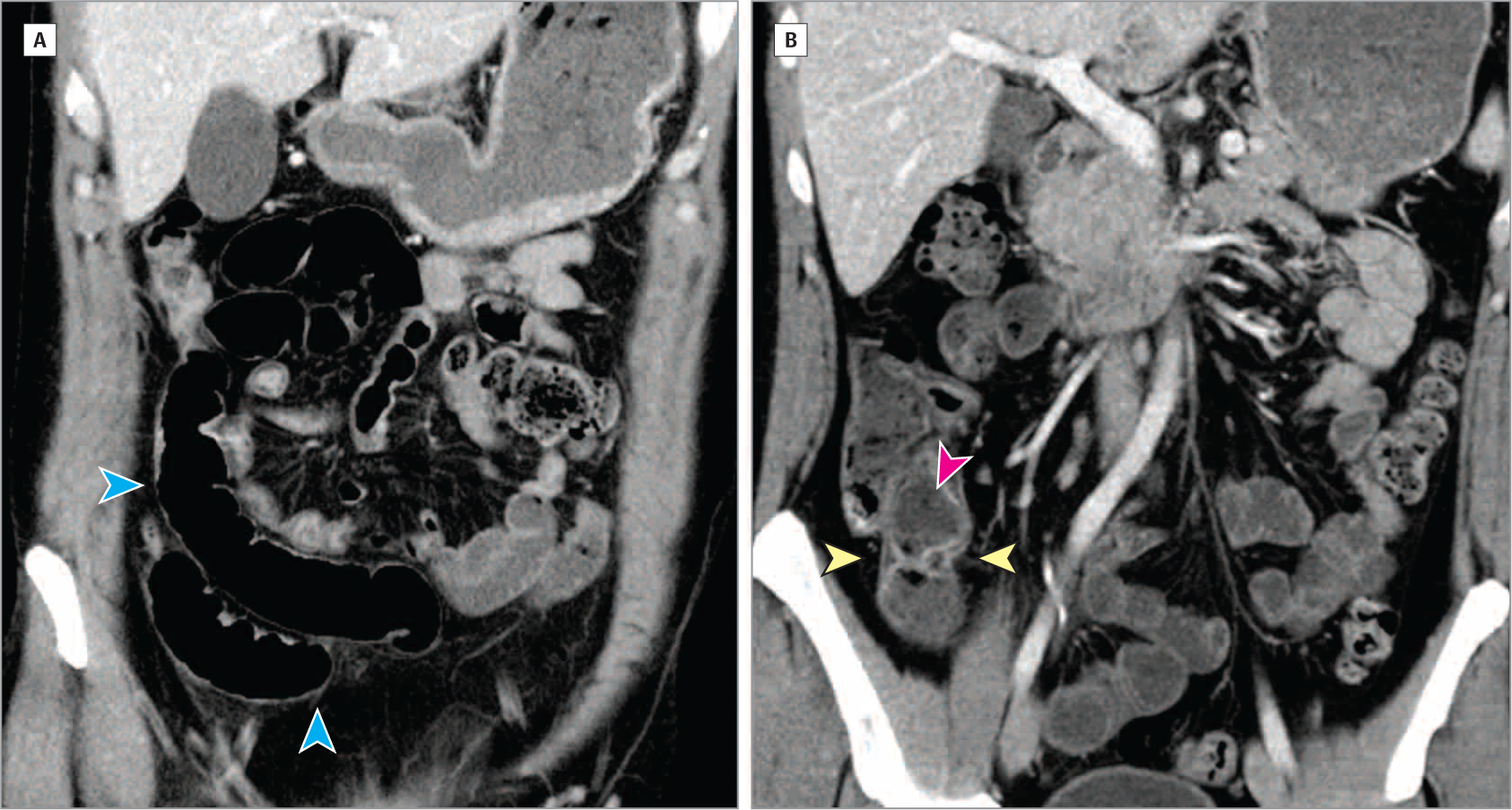
Figure 2. Helical Computed Tomography Imaging of Abdomen and Pelvis.

Coronal computed tomography views show slightly increased enhancement in the mucosa of the terminal ileum near the ileocecal valve with areas of slight wall thickening and mural stratification with predominantly intramural fat (yellow arrowheads), as well as feculent luminal contents (pink arrowhead). More proximal loops of ileum are slightly dilated (blue arrowheads). From Cheifetz AS. Management of acute Crohn disease. JAMA. 2013;309(20):2150–2158. doi:10.1001/jama.2013.4466.
