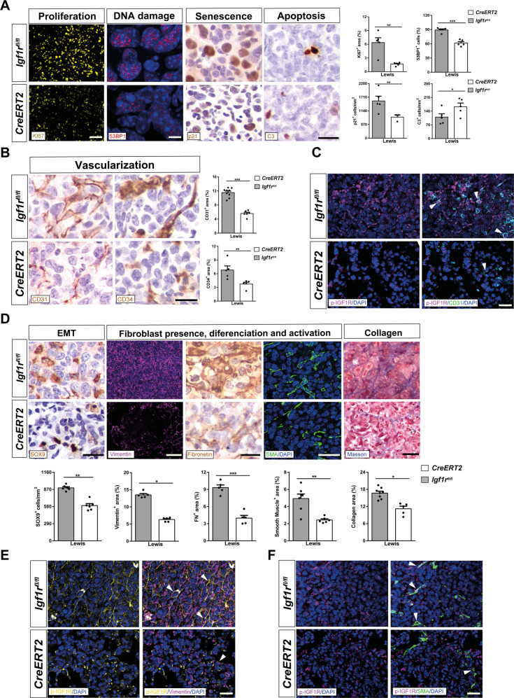
Fig. 5. IGF1R deficiency decreases proliferation, DNA damage, senescence and vascularization, attenuates tumor invasion by reduced EMT and fibrosis, and induces apoptosis upon pulmonary metastasis.
A Representative immunostains and quantifications of Ki67+ (proliferation) (yellow), 53BP1+ (DNA damage) (red) area (%), p21+ (senescence) (brown) and C3+ (apoptosis) (brown) cells per unit area (mm2), as well as B CD31+ (vascularization) and CD34+ (differentiated vascularization) (brown) areas (%) in the lung TME of LLC-challenged CreERT2 vs. Igf1rfl/fl mice (n = 5-7 mice per group; Scale bars: 30 µm in ki67, 8 µm in 53BP1, and 15 µm rest of immunostains). C Representative double immunostains for p-IGF1R and CD31 (magenta and green, respectively; white arrowheads indicate colocalization) in the lung TME of LLC-challenged CreERT2 vs. Igf1rfl/fl mice (n = 3–5 mice per group; Scale bars: 20 µm). D Representative immunostains for SOX9 (EMT) (brown), Vimentin (fibroblast presence) (magenta), Fibronectin (fibroblast differentiation) (brown), and SMA (fibroblast activation) (green), and stains for Masson (collagen content), as well as number of SOX9+ cells per unit area (mm2) and Vimentin+, Fibronectin+, SMA+ and Masson+ areas (%) in the lung TME of LLC-challenged CreERT2 vs. Igf1rfl/f mice (n = 5–6 mice per group; Scale bars: 15, 30, 15, 20 and 30 µm, respectively). E, F Representative double immunostains for p-IGF1R and Vimentin (yellow and magenta; white arrowheads indicate colocalization) and for p-IGF1R and SMA (magenta and green; white arrowheads indicate colocalization) in the lung TME of LLC-challenged CreERT2 vs. Igf1rfl/fl mice (n = 3–5 mice per group; Scale bars: 20 µm). Quantifications were performed randomly in five different fields. Data are expressed as mean ± SEM. *p < 0.05; **p < 0.01; ***p < 0.001 (Mann–Whitney U test or Student´s t-test for comparing two groups).

