Abstract
Background and study aims Data are lacking on the natural history of gastrointestinal tract schwannomas. We aimed to study the natural history of all gastrointestinal schwannomas including location, diagnosis, management, and long-term outcomes.
Patients and methods Patients with a pathological diagnosis of gastrointestinal schwannoma between January 2000 and March 2020 were identified. Data on baseline demographics, presentations, associated malignancies, malignant transformation, treatment, and recurrence were collected.
Results Our cohort consisted of 44 patients with a mean age of 58.6 years, with 63.6 % women and 84.1 % White. The stomach (38.6 %) was the most common location followed by the colorectum (31.8 %). Only 22.7 % of patients were symptomatic and 22.0 % had a personal history of other malignancies. Tissue diagnosis was obtained via endoscopy in 47.7 % and from surgical pathology in 52.3 %. On histology, 65.9 % of the tumors were solid, 11.4 % had mixed features, and 2.3 % had necrosis. SP100 was tested in all but one patient and was positive in all. Mean Ki-67 in 12 patients with tumors measuring ≥ 2 cm was 3.0 % indicating a low proliferation rate. Of the patients, 77.3 % had surgery and 18.2 % underwent endoscopic resection. At a mean follow-up of 5.0 ± 4.31 years, there was no malignant transformation, recurrence or mortality associated with gastrointestinal schwannomas.
Conclusions Gastrointestinal schwannomas are diagnosed in the fifth to sixth decade with predominance in women and Whites. They are benign, mostly asymptomatic, and diagnosed incidentally. Asymptomatic gastrointestinal schwannomas including lesions ≥ 2 cm in size do not appear to need further monitoring or intervention. Patients with them should be counseled to remain up to date with routine screening guidelines pertaining to the colon, breast, and lung cancer due to the high incidence of concomitant malignancy.
Introduction
Schwannomas are slow-growing, mostly benign tumors originating from the Schwann cells that create nerve sheath. Their typical location includes peripheral nerves, the spinal cord, and the central nervous system 1 . They also have been reported to occur at other locations of the body, including the head, neck, mediastinum, retroperitoneum, and pelvis 2 . Gastrointestinal tract schwannomas are rare and represent about 2.0 % to 6.0 % of all mesenchymal tumors 3 . The most common site of gastrointestinal schwannomas is the stomach (60.0 %–70.0%), followed by the colon and rectum (3.0 %) 4 5 6 7 . Small intestinal, esophageal, and pancreatic schwannomas are very sporadic 8 . Although rare, gastrointestinal schwannomas should be considered in the differential diagnosis of gastrointestinal submucosal/mesenchymal lesions such as gastrointestinal stromal tumors (GISTs), leiomyoma, leiomyosarcoma or colorectal cancer, and melanoma 9 10 . It is important to accurately diagnose these lesions as some of these (e. g. GISTs) carry a malignant potential and can recur, while others (schwannomas or leiomyoma) are relatively benign in nature 11 . Preoperative differentiation is difficult as endoscopic and radiological findings are nonspecific, but histological and immunological features are of paramount importance to diagnose and distinguish benign and malignant schwannomas, and other stromal tumors of malignant nature 8 .
Schwannomas in the gastrointestinal tract occur more frequently in the fifth to sixth decade of life and tend to have female predominance 7 8 . Most patients with gastrointestinal schwannomas are asymptomatic and diagnosis often is incidental 8 . With the widespread use of endoscopic and imaging modalities, the incidence of gastrointestinal schwannomas is on the rise but exact data on their prevalence are still lacking. When symptomatic, depending on the location they can present as abdominal pain, bleeding, dyspepsia, obstruction, weight loss or nausea and vomiting 6 7 8 10 12 13 . Gastrointestinal schwannomas are mostly benign with very few cases of malignant transformation 13 14 15 16 . Historically, surgical resection has been the mainstay of therapy 8 15 . But recently, endoscopic resection for small tumors has been shown to be safe and effective with a favorable long-term outcomes 17 . Due to the benign nature of gastrointestinal schwannomas, endoscopic or imaging surveillance has been proposed for small (< 2 cm) schwannomas but there is no clear consensus on these recommendations 17 18 . With the rising use of endoscopic and imaging modalities and morbidity and cost associated with them, there will be an increase in the diagnosis of gastrointestinal schwannomas and we need to have a better understanding of these stromal tumors to outline optimal diagnostic, treatment, and surveillance strategies to avoid any related complications. In this study, we aimed to assess the natural history of all gastrointestinal tract schwannomas including location, presenting symptoms, diagnostic modalities, treatment, recurrence, and long-term outcomes.
Patients and methods
After obtaining approval from the Institutional Review Board, all patients with the histological diagnosis of gastrointestinal schwannoma (esophagus, gastric, pancreas, small bowel and colorectal) at our center were identified from January 2000 to March 2020. Baseline demographics including age, gender, race, smoking status, body mass index (BMI), and family history of any malignancy were collected. Data on concurrent comorbidities including alcohol use, chronic kidney disease (CKD), end-stage renal disease, cirrhosis, chronic obstructive pulmonary disease (COPD), congestive heart failure (CHF), celiac disease, inflammatory bowel disease, and personal history of any kind of malignancy were also collected. Use of certain medications like proton pump inhibitors (PPIs), steroids or any immunosuppressants (immunomodulators and biological therapies) at the time of schwannoma diagnosis was noted. Laboratory data including hemoglobin (Hgb), hematocrit (HCT), platelets (PLT), international normalized ratio, albumin, alpha fetoprotein (AFP), carcinoembryonic antigen (CEA), and cancer antigen 19–9 (CA-19–9); and data on the modalities that aided in the diagnosis including colonoscopy, esophagogastroduodenoscopy (EGD), endoscopic ultrasound (EUS), computed tomography (CT) scans, magnetic resonance imaging (MRI) or magnetic resonance cholangiopancreatography (MRCP) were collected from electronic medical records (EMRs). Locations of the gastrointestinal schwannomas were identified based on imaging, endoscopy or histology samples. Associated symptoms including abdominal pain, dyspepsia, bleeding, pancreatitis, and bowel obstruction were identified from EMRs.
An expert gastrointestinal pathologist reviewed all histological samples to confirm the diagnosis of schwannoma. Presence of cellular atypia, necrosis, cystic, solid, mixed character, and mitotic index on histology samples was also noted. In addition, data on immunological straining for receptor tyrosine kinase (C-Kit), S-100, glial fibrillary acidic protein (GFAP), smooth muscle actin (SMA), Ki-67 proliferation index and CD34 were collected. Information about treatment modalities including surgical interventions or endoscopic therapies was identified. Patients were followed until last follow-up date in the EMR to assess for recurrence and mortality.
Results
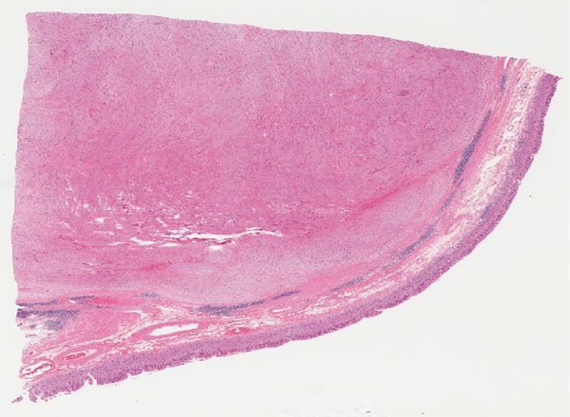
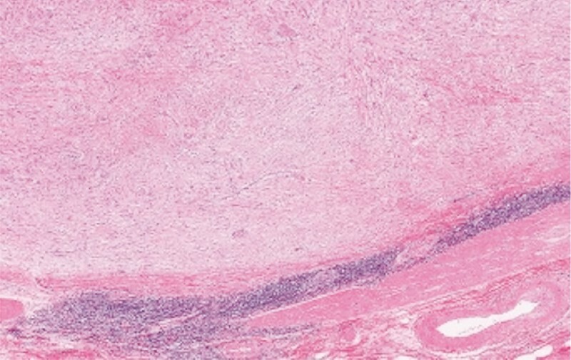
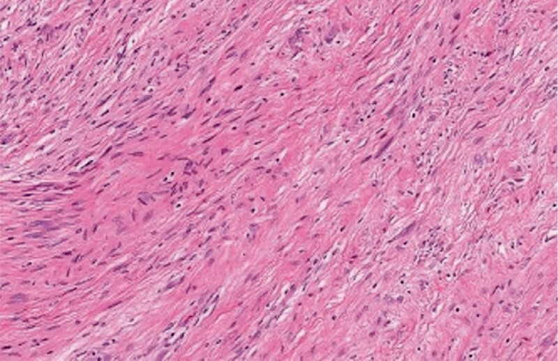
After searching for gastrointestinal schwannomas, a total of 51 patients were identified from the histology database. Of these, two were not schwannomas, one was a duplicate entry, and four had missing data. After exclusions, our final sample consisted of 44 patients with histologically confirmed cases of gastrointestinal schwannoma. Table 1 shows their baseline demographics and comorbidities. The mean age of our cohort was 58.6 years at the time of diagnosis, with 47.7 % in the 51-to-65-year age group and 31.8 % older than 65 years. Of the study population, 63.6 % were women and White (84.1 %) was the most common race, followed by others including Hispanic (9.1%) and Black (6.8 %). Mean BMI in our study population was 29.5 kg/m 2 . Fifty-seven percent were current or former smokers with a mean 12.6 pack years of smoking. Alcohol use was present in 25.0 %, CKD and COPD in 11.4 %, and 2.2 % had CHF. A total of 22.2 % had a personal history of neoplasm including breast (2.2 %), pancreatic (2.2 %), colon (2.2 %), gastric (2.2 %), colonic GIST (2.2 %), lung (2.2 %), non-Hodgkin lymphoma (2.2 %), and renal cancer (4.5 %). Half of them were diagnosed prior to the diagnosis of gastrointestinal schwannoma, four patients had concomitant cancers (pancreas, colon and gastric x 2), and one patient was diagnosed with renal cancer after schwannoma diagnosis. PPIs were used in 47.7 % of patients (52.4 % were on them for ≤ 3 months and 47.6 % were on them for ≥ 3 months) and 9.1 % were on steroids and immunosuppressive medications at the time of diagnosis and resection. Only 22.7 % were symptomatic and the majority were diagnosed incidentally. Abdominal pain and dyspepsia were the most common symptoms (15.9 %), followed by bleeding (9.1 %) and dysphagia (6.8 %). CT scan was the most common initial diagnostic modality (31.8 %) followed by colonoscopy (22.7 %), EUS (18.2 %), upper endoscopy (9.1 %), and MRI (6.8 %). Most of the lesions on imaging were classified as “mass lesion” without any suspicion of schwannoma by the radiologist. There was no statistical difference among various diagnostic modalities in terms of identifying the location of gastrointestinal schwannomas ( P > 0.05). The stomach (38.6 %) was the most common location followed by the colorectum (31.8 %), small bowel (13.6 %), esophagus (13.6 %), and pancreas (2.3 %). There was no statistically significant difference among various age ranges, gender, or races in terms of locations of schwannoma ( P > 0.05). In terms of biopsy confirmation, 52.3 % had surgical specimens and 47.7 % had endoscopic biopsies (22.7 % had EUS-guided biopsy, 15.9 % had EGD biopsy samples, and 9.1 % had biopsies via colonoscopy). Eleven patients were diagnosed with schwannomas before endoscopic or surgical resection. Four had an endoscopic diagnosis and resection at the same time. Gastric schwannomas were the largest with a mean diameter of 37.7 mm, followed by colorectal (24.1 mm), esophagus (22.8 mm), small bowel (21.0 mm), and pancreas (4.0 mm). On histology, 65.9 % of them were solid, 11.4 % had mixed features, and 2.3 % had necrosis but none was cystic ( Fig. 1 , Fig. 2 , Fig. 3 ). On immunology, SP100 was tested in all but one and was positive in all of them. CD34, GFAP, and SMA were positive in only 2.3 % of patients. Of 18 patients with lesions > 2 cm, only 12 had pathological samples to review. The mean Ki-67 in 12 patients with lesions > 2 cm was 3.0 %, indicating a low proliferation rate ( Fig. 4 ). None of the patients were tested for C-Kit ( Table 2 ). Mean Hgb at the time of diagnosis was 12.8 g, mean PLT 251 k/uL, mean BUN 15.5 mg/dL, mean creatinine 0.88 mg/dL, and mean albumin was 4.1 g/dL. None of the patients had elevated AFP or CEA levels. Ca-19–9 was elevated in one patient, but he had concomitant pancreatic cancer.
Table 1. Baseline demographics of patients with gastrointestinal schwannomas.
| N | % | ||
| Age | Years | Mean (SD) = 59.2 (12.6) | |
| Age group | 18–35 | 1 | 2.3 |
| 36–50 | 8 | 18.2 | |
| 51–65 | 21 | 47.7 | |
| > 65 | 14 | 31.8 | |
| Race | White | 37 | 84.1 |
| Black | 3 | 6.8 | |
| Other | 4 | 9.1 | |
| Gender | Female | 28 | 63.6 |
| Male | 16 | 36.4 | |
| BMI | 29.49 | 7.60 | |
| Smoking | Current | 14 | 31.8 |
| Former | 11 | 25.0 | |
| Never | 19 | 43.2 | |
| Pack yeasr of smoking | 12.65 | 20.29 | |
| Comorbidities | Alcohol | 11 | 25.0 |
| CKD | 5 | 11.4 | |
| ESRD | 0 | 0 | |
| Cirrhosis | 0 | 0 | |
| COPD | 5 | 11.4 | |
| CHF | 1 | 2.3 | |
| Cancer | 10 | 22.7 | |
| Type of cancer | Breast | 1 | |
| Colon | 1 | ||
| Colon, GIST | 1 | ||
| Gastric cancer | 1 | ||
| GIST | 1 | ||
| Lung | 1 | ||
| NHL | 1 | ||
| Pancreatic cancer | 1 | ||
| Renal | 2 | ||
| Family history of cancer | 4 | 9.1 % | |
| Colon (2) | |||
| Pancreas (1) | |||
| Prostate (1) | |||
| IBD | Crohnʼs ds | 4 | 9.1 |
| Medications | PPIs | 21 | 47.7 |
| Steroids | 4 | 9.1 | |
| Immune suppression | 4 | 9.1 |
SD, standard deviation; BMI, body mass index; CKD, chronic kidney disease; ESRD, end-stage renal disease; COPD, chronic obstructive pulmonary disease; CHF, chronic heart failure; GIST, gastrointestinal stromal tumor; NHL, non-Hodgkin lymphoma; IBD, inflammatory bowel disease.
Fig. 1.

At low magnification, a well-circumscribed mass lesion is visible underlying the gastric oxyntic mucosa. This lesion appears to be situated within the muscularis propria of the stomach.
Fig. 2.

40 ×: A well-defined lymphoid cuff is present, typical of most schwannomas arising in the gastrointestinal tract. The lesion itself is a spindle cell proliferation with a vaguely fascicular pattern.
Fig. 3.

200 ×: Cytologically, the spindle cells are predominantly bland, although scattered large cells or areas with degenerative type-changes may be seen.
Fig. 4.

Representative area from a Ki-67-stained section from this gastric schwannoma, which showed an overall proliferation index of 5 % (S18–-2347).
Table 2. Clinical and immunohistological characteristics of patients with gastrointestinal schwannomas.
| Schwannoma location | Esophagus | 6 | 13.6 |
| Stomach | 17 | 38.6 | |
| Pancreas | 1 | 2.3 | |
| Small bowel | 6 | 13.6 | |
| Colorectal | 14 | 31.8 | |
| Symptoms | Abdominal pain | 7 | 15.9 |
| Bleeding | 4 | 9.1 | |
| Dyspepsia | 6 | 13.7 | |
| Dysphagia | 3 | 6.8 | |
| Initial identification | CT | 14 | 31.8 |
| Colonoscopy | 11 | 25 | |
| EUS | 8 | 18.2 | |
| EGD | 4 | 9.1 | |
| MRI | 4 | 9.09 | |
| No imaging or endoscopy | 3 | 6.8 | |
| Biopsy sample | Surgical Specimen | 23 | 52.3 |
| EUS | 10 | 22.7 | |
| EGD | 7 | 15.9 | |
| Colonoscopy | 4 | 9.1 | |
| Labs at diagnosis | Lab | Mean (SD) | Median |
| Hgb | 12.8 (1.9) | 13.0 | |
| HCT | 38.8 (5.1) | 39.4 | |
| Platelet | 251 (81) | 254 | |
| BUN | 15.7 (8.5) | 14.0 | |
| Creatinine | 0.88 (0.40) | 0.80 | |
| Albumin | 4.09 (0.60) | 4.3 | |
| Pathology | |||
| Size (mm) | Esophagus | 22.8 (18.3) | 20.0 |
| Stomach | 37.7 (23.6) | 33.5 | |
| Pancreas | 4 (NA) | 4 | |
| Small Bowel | 21 (17.9) | 15.5 | |
| Colorectal | 24.1 (18.7) | 17 | |
| Histology | N (44) | % | |
| Cystic | 0 | 0 | |
| Solid | 29 | 82.9 | |
| Mixed | 5 | 14.3 | |
| Necrosis | 1 | 2.8 | |
| Immunological markers | N (44) | Percentage | |
| S100 | 28 | 63.6 | |
| S100 + Ki-67 | 12 | 27.2 | |
| S100 + GFAP | 1 | 2.3 | |
| S100 + SMA | 1 | 2.3 | |
| S100 + CD34 | 1 | 2.3 | |
| Not available | 1 | 2.3 | |
CT, computed tomography; EUS, endoscopic ultrasound; EGD, esophagogastroduodenoscopy; MRI, magnetic resonance imaging; SD, standard deviation; HgB, hemoglobin; HCT, hematocrit; BUN, blood urea nitrogen.
Surgical interventions were performed in 77.3 % of patients (70.6 % upper gastrointestinal including small bowel and 29.4 % in the colorectum) and 18.2 % (37.5 % upper gastrointestinal and 62.5 % colonic) underwent endoscopic resection. The patient with pancreatic schwannoma underwent a Whipple’s procedure, which confirmed the diagnosis. Surgical pathology showed both schwannoma and pancreatic intraepithelial neoplasia grade 1. All patients with surgical or endoscopic interventions had R0 resection except one with R1 resection in the endoscopy group. The patient with R1 resection had a repeat colonoscopy after 6 months and there was no recurrence or new lesions. One patient is scheduled for surgery and another one is awaiting further discussions with his physician. Only four patients had a lag of more than 30 days after the date of biopsy diagnosis (mean 115.0 days). Over a mean follow-up period of 5.0 ± 4.31 years, there was no malignant transformation, recurrence or mortality associated with gastrointestinal schwannoma. One patient died due to metastatic pancreatic cancer and another one died due to a postsurgical complication of gastric resection for adenocarcinoma ( Table 3 ).
Table 3. Clinical course of gastrointestinal schwannomas.
| N | % | |
| Monitoring | 2 | 4.5 |
| Malignant transformation | 0 | 0 |
| Endoscopic resection | 8 | 18.2 |
| Surgery | 34 | 77.3 |
| Schwannoma-related mortality | 0 | – |
Discussion
Our study suggests that gastrointestinal schwannomas are diagnosed in the fifth to sixth decade with predominance in women and Whites. They are benign, mostly asymptomatic, and diagnosed incidentally. During a mean follow-up period of 5.0 ± 4.31 years, there was no malignant transformation, recurrence or mortality associated with gastrointestinal schwannoma. Asymptomatic gastrointestinal schwannomas including lesions > 2 cm do not appear to need further monitoring or intervention. Patients with them should be counseled to remain up to date with routine screening guidelines pertaining to the colon, breast, and lung cancer due to the high incidence of concomitant malignancy.
Daimaru and colleagues first described schwannomas of the gastrointestinal tract in 1988 as benign tumors with S-100 positivity 19 . Since then, multiple studies have been conducted suggesting no to minimal malignant potential of these mesenchymal tumors of the gastrointestinal tract 4 8 11 13 . In a recent review including 171 patients with gastric schwannomas, four patients with an initial diagnosis of malignant gastric schwannomas died of disease recurrence and three of them had liver metastases. The follow-up among these patients after surgery to recurrence ranged from 12 to 58 months 16 . Our study has a mean follow-up of 5.0 ± 4.31 years and we did not notice any recurrence or mortality in our cohort. Interestingly, Shu et al in his recent study including nine patients with intestinal schwannomas pointed out that most of the malignant schwannomas in the gastrointestinal tract were mainly reported before 2000 when there was limited understanding of gastrointestinal schwannomas. Currently, there are no clear indicators suggesting malignant potential of gastrointestinal schwannomas. Some authors have suggested Ki-67 proliferative index (MIB-1) (> 10 %), nuclear atypia, mitotic activity rate (> 5 HPF), and tumor size (> 5 cm) as high-risk features for malignant potential and recurrence 7 20 21 . Based on these features, there is a concern for malignant gastrointestinal schwannomas. In our cohort, 23 patients had schwannoma ≥ 2 cm and 12 patients with tumors > 2 cm were tested for Ki-67 and had a mean index of 3.0 %. No results were provided for mitotic activity, but none of the patients had any cellular atypia, which suggests the benign nature of gastrointestinal schwannomas. There was no recurrence in our study group, suggesting these parameters might be useful in predicting the malignant potential and should be part of the histological report but the criteria of larger size as alluded by some authors was not applicable in our study, which is the largest to date with long-term follow-up. Future studies with larger cohorts are required to confirm these findings and if there is a concern for malignant tumor, complete resection with an attempt to remove all tumor tissue with negative margins should be performed. In addition, as shown in our study with S-100 positivity in all who were tested, all patients with suspected schwannomas should undergo immunohistochemical testing, as the results can differentiate gastrointestinal schwannoma (strongly positive for vimentin, GFAP and S-100) from other gastrointestinal tract mesenchymal tumors 22 . A preoperative increase in neuron specific enolase (NSE) might contribute to a diagnosis of gastrointestinal schwannomas but data are equivocal on this and need further studies 7 23 24 .
As in prior studies, the majority of our patients with gastrointestinal schwannomas were in their fifth to sixth decade of life with a female predominance 7 8 17 . White (84.0 %) was the most prevalent race in our cohort. Interestingly, there are limited data on race from prior studies. The higher prevalence of Whites in our study could be from the predominantly White patient population seen at our center. Fifty-seven percent of our patients were current or former smokers and we noticed relatively higher rates of smoking in patients with esophageal and gastric schwannomas, but this association was not statistically significant ( P > 0.05). Interestingly, 22.0 % of our cohort had a personal history of various cancers. Whether to screen for other cancers in patients with gastrointestinal schwannomas is a matter of debate and further data are needed to support this proposition. Seventy-seven percent of gastrointestinal schwannomas in our cohort were asymptomatic and were diagnosed incidentally. In a case series by Mekras et al, six of seven gastrointestinal schwannomas were diagnosed incidentally 8 . In another series of 16 patients by Zhai and colleagues, 62.5 % were asymptomatic 17 . With the increasing use of imaging and endoscopic, the prevalence of incidentally diagnosed gastrointestinal schwannomas likely will increase in the near future. In terms of location, the stomach (38.6 %) was the most common location in our cohort followed by the colorectum (31.8 %) and small intestine and esophagus (13.6 %). This is different from prior reports in which gastric schwannomas comprised 57.0 % to 83.0 % of all gastrointestinal schwannomas 7 8 . Data on gastrointestinal schwannoma distribution is not consistent and the difference in our cohort could be from differences in race and environmental factors. Interestingly, 48.0 % of our patients were diagnosed with presurgical biopsies, suggesting a possible improvement in endoscopic biopsy techniques. In difficult cases, EUS can determine location and boundaries of the tumor, take fine-needle aspiration or fine-needle biopsy samples from the lesion and surrounding lymph nodes, and may aid in differentiation from other stromal tumors based on certain characteristics such as tumor capsule, cystic changes, cavity formation, necrosis, and calcification commonly seen on abdominal CT scan 25 .
Seventy-seven percent of the patients in our study underwent surgical resection and 18.0 % had endoscopic resection. Until recently, surgery has been the mainstay of therapy for gastrointestinal schwannomas 7 13 . But with recent advances in endoscopic techniques, endoscopic resection of gastrointestinal tract schwannomas is emerging as an alternative to surgical interventions 6 26 . In a recent study of 16 patients with gastric schwannomas, Zhai and colleagues performed endoscopic submucosal and full thickness resection 26 . Eighty-seven percent of these patients achieved R0 resection without any adverse events and need for surgical resection. At a mean follow-up period of 21.8 months, there was no residue, recurrence or metastasis. Although endoscopic resections seem promising for gastrointestinal schwannomas, they are limited by widespread availability, safety, feasibility (small intestinal tumor), lack of standardized surveillance strategies, high cost of repeat procedures, and patient stress and compliance. They still could be offered to patients with high surgical risks or those reluctant to undergo surgical resection after a detailed discussion about need for long-term surveillance and possibility of recurrence.
Our study has several strengths. First, this is one the largest single-center studies looking at schwannomas of the entire gastrointestinal tract with long-term follow-up. It adds new information about the racial prevalence of gastrointestinal schwannomas in Whites. It underscores the importance of endoscopic and biopsy skills as 48.0 % of our patients were diagnosed with endoscopic biopsies. It concurs with the previous studies that most gastrointestinal schwannomas are benign with female predominance and it is important to perform immunohistochemical analysis to accurately differentiate gastrointestinal schwannomas from other stromal tumors. Furthermore, our study shows that even larger lesions (≥ 2 cm) had a low Ki-67 proliferative index and no recurrence, which adds to the data supporting the benign nature of the lesion regardless of size. Another important finding from our cohort was the high incidence of concomitant cancers, which raises the awareness that these patients should be counseled to remain up to date with routine cancer screening guidelines pertaining to the colon, breast, and lung. In addition to its retrospective design, our study has certain limitations. Being a single-center study introduces “selection bias.” All biopsy specimens were not assessed for NSE and GDAP positivity, Ki-67 proliferative index, and mitotic index, which could have provided more certainty about the benign nature of these tumors. Even though we did not see any malignant transformation, tumor recurrence or mortality associated with gastrointestinal schwannomas, these finding are limited by a relatively short follow-up period of 1,380 days.
Conclusions
In conclusion, our study suggests that gastrointestinal schwannomas are benign tumors, most commonly seen in the fifth to sixth decade with a predominance of female gender and White race. They usually are diagnosed incidentally, are benign and mostly asymptomatic. All biopsy specimens from suspected gastrointestinal schwannomas should undergo immunohistochemical testing, along with assessment for presence of Ki-67 index, cellular atypia, and mitotic activity. Given the benign nature of gastrointestinal schwannomas, asymptomatic patients do not appear to need further monitoring or intervention. In symptomatic patients, surgical or endoscopic interventions can be offered. These patients should be counseled to remain up to date with routine cancer screening guidelines pertaining to the colon, breast, and lung given to the high incidence of concomitant malignancy. Future prospective studies are required to confirm these findings.
Footnotes
Competing interests The authors declare that they have no conflict of interest.
References
- 1.Gubbay A D, Moschilla G, Gray B N et al. Retroperitoneal schwannoma: a case series and review. Aust N Z J Surg. 1995;65:197–200. doi: 10.1111/j.1445-2197.1995.tb00607.x. [DOI] [PubMed] [Google Scholar]
- 2.Almo K M, Traverso L W. Pancreatic schwannoma: an uncommon but important entity. J Gastrointest Surg. 2001;5:359–363. doi: 10.1016/s1091-255x(01)80062-7. [DOI] [PubMed] [Google Scholar]
- 3.Melvin W S, Wilkinson M G. Gastric schwannoma. Clinical and pathologic considerations. Am Surg. 1993;59:293–296. [PubMed] [Google Scholar]
- 4.Zheng L, Wu X, Kreis M E et al. Clinicopathological and immunohistochemical characterisation of gastric schwannomas in 29 cases. Gastroenterol Res Pract. 2014;2014:202960. doi: 10.1155/2014/202960. [DOI] [PMC free article] [PubMed] [Google Scholar]
- 5.Voltaggio L, Murray R, Lasota J et al. Gastric schwannoma: a clinicopathologic study of 51 cases and critical review of the literature. Hum Pathol. 2012;43:650–659. doi: 10.1016/j.humpath.2011.07.006. [DOI] [PMC free article] [PubMed] [Google Scholar]
- 6.Ramai D, Lai J, Changela K et al. Transverse colon schwannoma treated by endoscopic mucosal resection: A case report. Mol Clin Oncol. 2017;7:830–832. doi: 10.3892/mco.2017.1418. [DOI] [PMC free article] [PubMed] [Google Scholar]
- 7.Shu Z, Li C, Sun M et al. Intestinal Schwannoma: A clinicopathological, immunohistochemical, and prognostic study of 9 cases. Gastroenterol Res Pract. 2019;2019:3.414678E6. doi: 10.1155/2019/3414678. [DOI] [PMC free article] [PubMed] [Google Scholar]
- 8.Mekras A, Krenn V, Perrakis A et al. Gastrointestinal schwannomas: a rare but important differential diagnosis of mesenchymal tumors of gastrointestinal tract. BMC Surg. 2018;18:47. doi: 10.1186/s12893-018-0379-2. [DOI] [PMC free article] [PubMed] [Google Scholar]
- 9.Wilde B K, Senger J L, Kanthan R. Gastrointestinal schwannoma: an unusual colonic lesion mimicking adenocarcinoma. Can J Gastroenterol. 2010;24:233–236. doi: 10.1155/2010/943270. [DOI] [PMC free article] [PubMed] [Google Scholar]
- 10.Miettinen M, Shekitka K M, Sobin L H. Schwannomas in the colon and rectum: a clinicopathologic and immunohistochemical study of 20 cases. Am J Surg Pathol. 2001;25:846–855. doi: 10.1097/00000478-200107000-00002. [DOI] [PubMed] [Google Scholar]
- 11.Kwon M S, Lee S S, Ahn G H. Schwannomas of the gastrointestinal tract: clinicopathological features of 12 cases including a case of esophageal tumor compared with those of gastrointestinal stromal tumors and leiomyomas of the gastrointestinal tract. Pathol Res Pract. 2002;198:605–613. doi: 10.1078/0344-0338-00309. [DOI] [PubMed] [Google Scholar]
- 12.Levy A D, Quiles A M, Miettinen M et al. Gastrointestinal schwannomas: CT features with clinicopathologic correlation. AJR Am J Roentgenol. 2005;184:797–802. doi: 10.2214/ajr.184.3.01840797. [DOI] [PubMed] [Google Scholar]
- 13.Morales-Maza J, Pastor-Sifuentes F U, Sanchez-Morales G E et al. Clinical characteristics and surgical treatment of schwannomas of the esophagus and stomach: A case series and systematic review. World J Gastrointest Oncol. 2019;11:750–760. doi: 10.4251/wjgo.v11.i9.750. [DOI] [PMC free article] [PubMed] [Google Scholar]
- 14.Catania G, Puleo C, Cardi F et al. Malignant schwannoma of the rectum: a clinical and pathological contribution. Chir Ital. 2001;53:873–877. [PubMed] [Google Scholar]
- 15.Hu B G, Wu F J, Zhu J et al. Gastric schwannoma: a tumor must be included in differential diagnoses of gastric submucosal tumors. Case Rep Gastrointest Med. 2017;2017:9.615359E6. doi: 10.1155/2017/9615359. [DOI] [PMC free article] [PubMed] [Google Scholar]
- 16.Lauricella S, Valeri S, Masciana G et al. What About Gastric Schwannoma? A Review Article. J Gastrointest Cancer. 2021;52:57–67. doi: 10.1007/s12029-020-00456-2. [DOI] [PubMed] [Google Scholar]
- 17.Zhai Y Q, Chai N L, Li H K et al. Endoscopic submucosal excavation and endoscopic full-thickness resection for gastric schwannoma: five-year experience from a large tertiary center in China. Surg Endosc. 2020;34:4942–4949. doi: 10.1007/s00464-019-07285-w. [DOI] [PubMed] [Google Scholar]
- 18.Tan Y, Tan L, Lu J et al. Endoscopic resection of gastric gastrointestinal stromal tumors. Transl Gastroenterol Hepatol. 2017;2:115. doi: 10.21037/tgh.2017.12.03. [DOI] [PMC free article] [PubMed] [Google Scholar]
- 19.Daimaru Y, Kido H, Hashimoto H et al. Benign schwannoma of the gastrointestinal tract: a clinicopathologic and immunohistochemical study. Hum Pathol. 1988;19:257–264. doi: 10.1016/s0046-8177(88)80518-5. [DOI] [PubMed] [Google Scholar]
- 20.Hornick J L, Bundock E A, Fletcher C D. Hybrid schwannoma/perineurioma: clinicopathologic analysis of 42 distinctive benign nerve sheath tumors. Am J Surg Pathol. 2009;33:1554–1561. doi: 10.1097/PAS.0b013e3181accc6c. [DOI] [PubMed] [Google Scholar]
- 21.Nonose R, Lahan A Y, Santos Valenciano J et al. Schwannoma of the Colon. Case Rep Gastroenterol. 2009;3:293–299. doi: 10.1159/000237736. [DOI] [PMC free article] [PubMed] [Google Scholar]
- 22.Hou Y Y, Tan Y S, Xu J F et al. Schwannoma of the gastrointestinal tract: a clinicopathological, immunohistochemical and ultrastructural study of 33 cases. Histopathology. 2006;48:536–545. doi: 10.1111/j.1365-2559.2006.02370.x. [DOI] [PubMed] [Google Scholar]
- 23.Seno K, Itoh M, Endoh K et al. Schwannoma of the duodenum causing melena. Intern Med. 1994;33:621–623. doi: 10.2169/internalmedicine.33.621. [DOI] [PubMed] [Google Scholar]
- 24.Prevot S, Bienvenu L, Vaillant J C et al. Benign schwannoma of the digestive tract: a clinicopathologic and immunohistochemical study of five cases, including a case of esophageal tumor. Am J Surg Pathol. 1999;23:431–436. doi: 10.1097/00000478-199904000-00007. [DOI] [PubMed] [Google Scholar]
- 25.He M, Zhang R, Zhai F et al. Comparative study on computed tomography features of gastrointestinal schwannomas and gastrointestinal stromal tumors. Zhonghua Wei Chang Wai Ke Za Zhi. 2015;18:1020–1025. [PubMed] [Google Scholar]
- 26.Zhou Y, Zheng S, Ullah S et al. Endoscopic resection for gastric schwannoma: our clinical experience of 28 cases. J Gastrointest Surg. 2020;24:2135–2136. doi: 10.1007/s11605-020-04679-3. [DOI] [PubMed] [Google Scholar]


