Fig. 2.
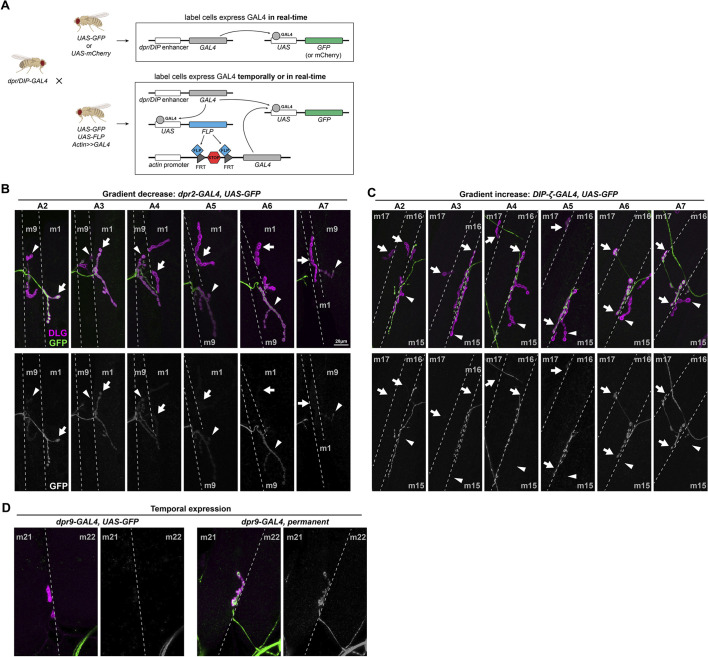
Dpr and DIP genes are expressed in various patterns in MNs. (A) Schematic showing the experimental procedure. Each dpr/DIP-GAL4 line was crossed to a real-time reporter (UAS-GFP or UAS-mCherry) and a permanent reporter [UAS-GFP, UAS-FLP, actin-(FRT.STOP)-GAL4] to reveal the dynamic expression of Dpr and DIP genes. (B) Example of a decrease in expression of dpr2-GAL4 in MN1-Ib (arrows) from anterior hemisegment A2 to posterior hemisegment A7. Note that the expression in nearby MN9-Ib (arrowheads) is also not robust as it was not expressed in A2 and A3 but was expressed in A4 to A7. (C) Example of an increase in expression of DIP-ζ-GAL4 in MN16/17-Ib (arrows) from anterior hemisegment A2 to posterior hemisegment A7. Note that the expression in nearby MN15/16-Ib (arrowheads) was always absent. (D) Example of temporal expression of dpr9-GAL4 in MN21-Ib. MN21-Ib was not labeled by dpr9-GAL4>GFP animals, but 50% of MN21-Ib were labeled in the cross to the permanent reporter. Dashed lines indicate muscle boundaries.

