Abstract
A set of PCR primers targeting 16S rRNA gene sequences was designed, and PCR parameters were optimized to develop a robust and reliable protocol for selective amplification of Escherichia coli 16S rRNA genes. The method was capable of discriminating E. coli from other enteric bacteria, including its closest relative, Shigella. Selective amplification of E. coli occurred only when the annealing temperature in the PCR was elevated to 72°C, which is 10°C higher than the optimum for the primers. Sensitivity was retained by modifying the length of steps in the PCR, by increasing the number of cycles, and most importantly by optimizing the MgCl2 concentration. The PCR protocol developed can be completed in less then 2 h and, by using Southern hybridization, has a detection limit of ca. 10 genomic equivalents per reaction. The method was demonstrated to be effective for detecting E. coli DNA in heterogeneous DNA samples, such as those extracted from soil.
Bacteria of the Enterobacteriaceae are important pathogens causing intestinal and systemic illness of humans and other animals. Recent outbreaks of gastrointestinal diseases focused public attention on one of the more widely known members, Escherichia coli, and the potential problems with strains of this organism as food-borne pathogens. Consumption of water polluted with fecal material is an important exposure pathway, and while monitoring of total coliforms is the standard technique, some research suggests that analysis for E. coli specifically may be a better indicator (3).
Traditional approaches for analysis of E. coli have relied on cultural techniques, and many selective-differential media have been developed. Generally, lactose fermentation is used for differentiation, sodium lauryl sulfate or bile salts are used as a selective agent, and a fluorogenic reaction is used for confirmation. β-Glucuronidase is the target enzyme for confirmation of E. coli, which mediates hydrolysis of 4-methylumbelliferyl-β-d-glucuronide (MUG) to a fluorescent product. However, other Enterobacteriaceae produce β-glucuronidase (e.g., Shigella, Salmonella, and Yersinia), not all strains of E. coli express the uiaA gene that encodes β-glucuronidase, and some Staphylococcus spp. hydrolyze MUG (6, 25). Biochemical analysis for an enzyme associated with a particular pathogenic trait and immunodiagnostic assays for O antigens associated with pathogenic strains have also been developed (11, 16). Again, cross-reactivity limits the utility of these techniques for identification of E. coli.
The emergence of DNA technology has opened new possibilities for development of methods with improved selectivity for E. coli. Relevant techniques include use of PCR and/or hybridization probes to detect E. coli-specific genes encoding invasion proteins (12, 13), toxins (17, 23, 24), catabolic enzymes (4, 8), or structural (lipo)proteins (10, 12). A drawback to these approaches has been that when cross-reactivity tests were done, target sites were often detected not only in E. coli but also in closely related organisms (14, 17, 18). Cross-reaction is particularly problematic with Shigella. This is not surprising insofar as 16S rRNA and other genome-level similarities suggest that Escherichia and Shigella are sufficiently similar for placement in a single genus (5, 19). Nevertheless, for clinical, epidemiological, and historical reasons they are regarded as different genera. Thus, for development of molecular methods, the challenge is to identify sequences conserved within E. coli that may be targeted to minimize false negatives yet can be distinguished from similar sequences likely to be present in Shigella.
The goal of this study was to design a selective and sensitive PCR method for amplification of a 16S rRNA gene region from E. coli. The key performance criterion for the method was to reliably amplify the targeted region from template levels equivalent to 100 or fewer E. coli cells without cross-reaction with similar sequences present in Shigella. An additional consideration was that the method be sufficiently robust for analysis of genomic DNA extracted from water or soil samples as well as that prepared from clinical isolates.
Cultures and DNA template preparation.
Cultures used in this study are listed in Table 1. Genomic DNA for PCR experiments was extracted from the liquid cultures as described by Ausubel et al. (2). DNA concentration and quality were determined by UV light absorbance at 260 and 280 nm and by band intensity densitometry using NIH Image version 1.55 (National Institutes of Health, Bethesda, Md.).
TABLE 1.
Cultures used in these testsa
| Category | Isolatea |
|---|---|
| Enteric bacteria | |
| Escherichia coli | UW8002 (CDC4102-72) |
| UW8009 | |
| UW8101 | |
| UW8204 (WSLH 10555) | |
| UW8410 | |
| UW8P39 | |
| UW8001D (ATCC 12814) | |
| Shigella spp. | S. dysenteriae, serotype 4, UW8P01 |
| S. dysenteriae, serotype 1 (WSLH isolate) | |
| S. flexnerii UW8P02 | |
| S. sonnei UW8P15 | |
| Other enteric bacteria | Citrobacter freundii UW8606 |
| Enterobacter aerogenes UW8411 (ATCC 13048) | |
| Enterobacter nimipressuralis UW8103 (ATCC 09912) | |
| Enterobacter agglomerans UW8710 (ATCC 55046) | |
| Klebsiella pneumoniae UW8215 (WSLH 25400) | |
| Proteus vulgaris UW8068 (WSLH 58224) | |
| Salmonella serovar Typhimurium UW8P14 | |
| Salmonella serovar Typhimurium UW8P40 | |
| Nonenteric gamma Proteobacteria | Pseudomonas aeruginosa UW9020 (ATCC 10145) |
All cultures were obtained from either the University of Wisconsin Department of Bacteriology (UW) or the Wisconsin State Laboratory of Hygiene (WSLH). Some cultures obtained from the UW collection are also on deposit at the American Type Culture Collection (ATCC) or the Centers for Disease Control and Prevention (CDC); the culture identifiers for the latter collections are given in parentheses for cross-reference.
Primers and probes.
Primers targeting hypervariable regions of the E. coli 16S rRNA gene were developed by using PrimerSelect (DNAStar, Madison, Wis.). Three sets of primer pairs were designed and tested: ECP79F (forward, targeting bases 79 to 96; 5′-GAAGCTTGCTTCTTTGCT-3′)-ECR620R (reverse, targeting bases 602 to 620; 5′-GAGCCCGGGGATTTCACAT-3′); ECB75F (forward, targeting bases 75 to 97; 5′-GGAAGAAGCTTGCTTCTTTGCTG-3′-ECR620R (reverse, described above); and ECA75F (forward, targeting bases 75 to 99; 5′-GGAAGAAGCTTGCTTCTTTGCTGAC-3′)-ECR619R (reverse, targeting bases 594 to 619; 5′-AGCCCGGGGATTTCACATCTGACTTA-3′). The optimal melting temperature and expected PCR product sizes for the primer pairs were as follows: ECP79F-ECR620R, 55°C and 541 bp; ECB75F-ECR620R, 59°C and 545 bp; and ECA75F-ECR619R, 60°C and 544 bp. The probe used in Southern hybridization experiments was S-D-Bact-0338-a-A-1 (previously referred to as EUB338 [1]). This probe targeted a 16S rRNA gene sequence conserved in the domain Bacteria and occurring near the center of the PCR products generated by all primer pairs. The oligonucleotide was 5′ labeled with digoxygenin by the supplier (Sigma-Genosys, The Woodlands, Tex.).
PCR and hybridization protocols.
PCR protocols were developed empirically for each primer set to obtain maximum selectivity (E. coli versus Shigella and other enteric bacteria) while retaining sensitivity (desired detection level of 10 fg to 1 pg, ca. 1 to 100 cell equivalents). Optimization focused on levels of primers, DNA polymerase, and MgCl2 in the reaction mixture as well as thermal cycling programs. For ECP79F-ECR620R, the reaction mixture (50 μl, total volume) contained 1:10 dilution of 10× PCR buffer (500 mM KCl, 100 mM Tris-HCl [pH 8.3], 15 mM MgCl2, 0.01% [wt/vol] gelatin), 200 μM each deoxynucleoside triphosphate, 0.6 μM primers, and an appropriate amount of template. The thermal cycling program consisted of a hot start (5 min, 94°C) before 1.25 U of AmpliTaq DNA polymerase (PE Biosystems, Foster City, Calif.) was added per reaction. The reaction was run for 40 cycles with 45-s denaturation (94°C), 45-s annealing (50°C), 1.5-min extension (72°C), and a final extension (5 min, 72°C). For ECA75F-ECR619R and ECB75F-ECR620R, reaction mixtures (50 μl, total volume) contained 1:10 dilution of 10× PCR buffer (100 mM Tris-HCl [pH 9.0], 500 mM KCl, 0.1% Triton X-100), 200 μM each deoxynucleoside triphosphate, 2 mM MgCl2, 0.4 μM primers, bovine serum albumin (40 μg/reaction), and an appropriate amount of template. The thermal cycling program consisted of a hot start (30 s, 94°C) followed by addition of 1.5 U of native Taq DNA polymerase (Promega, Madison, Wis.) per reaction. The thermal cycling program was run for 40 cycles of denaturation (45 s, 94°C) and annealing-extension (45 s, 72°C) and then a final extension (10 min, 72°C).
All of the reactions were done in thin-walled 0.5-ml Eppendorf tubes (USA/Scientific Plastics, Ocala, Fla.) and were assembled in a CloneZone unit (USA/Scientific Plastics) to prevent airborne contamination or template carryover from previous experiments. Thermal cycling was done with a DeltaCycler I unit (Ericomp, San Diego, Calif.). After amplification, 10 μl of each PCR mixture was analyzed by electrophoresis (8 mV/cm, 1 h) in ethidium bromide-stained agarose (2%, wt/vol) gels. A 100-bp ladder (Promega) was included for molecular weight estimation. Gels were placed on an UV transillumination unit (Fotodyne, Hartland, Wis.) for band visualization and photography. Southern hybridization to DNA immobilized on Hybond N+ charged nylon membranes (Amersham, Arlington Heights, Il.) was done according to the manufacturer's directions. Bound probe was detected by chemiluminescence using the Genius system and CPD-Star substrate solution (Boehringer Mannheim, Indianapolis, Ind.). The membrane was then exposed to X-ray film (Eastman Kodak, Rochester, N.Y.), and the film was developed according to the manufacturer's instructions.
Detecting E. coli in soil by PCR.
Soil was sampled from experimental plots located at the Arlington Research Station (Arlington, Wis.) and at the Lakeland Agricultural Complex (Lakeland, Wis.). The Arlington soil was a Plano silt loam (typic argiudoll; pH 6.9, 48 g of organic matter kg−1), while that from Lakeland was a Griswold silt loam (aquic argiudoll; pH 7.0, 42 g of organic matter kg−1). At both sites, the plots were established as continuous corn and had not been amended with animal manure for at least 12 years. The samples collected were kept on ice for transport to the laboratory and then stored frozen at −20°C. Prior to use, the soils were sieved through 2-mm mesh screen and their moisture contents were determined. DNA was extracted by a freeze-thaw method essentially as described by Tsai and Olson (22) except that CaCl2 (final concentration of Ca2+, 90 mM) was substituted for NaCl in the lysis solution. The extracts were further purified by a Wizard kit (Promega). Use of Ca2+ in the lysis solution precipitates humic materials and makes cleanup by the Wizard kit more effective. Concentrations of Ca2+ in extracts were 690 μM or less as determined by atomic absorption spectroscopy and shown not to interfere with the PCR (21). DNA purity and concentrations were determined by UV absorbance at 260 and 280 nm and by gel image analysis with NIH Image version 1.55 software (National Institutes of Health).
Two groups of experiments were done to examine the efficiency of the PCR method for amplification of E. coli 16S rRNA gene sequences from soil. In the first, DNA was extracted from the Arlington and Lakeland soils, and an aliquot of each purified extract (containing approximately 100 ng of DNA) was then spiked with either 1 ng or 1 pg of E. coli genomic DNA and used for PCR. In the second experiment, Lakeland soil (4 g, wet weight) was inoculated with E. coli at densities ranging from 7 × 10⁴ to 7 × 10⁷ cells g−1 (each inoculum level established in triplicate) and then incubated for 1 h. The incubation time in soil was kept relatively short to allow interaction between cells and soil particles while minimizing the extent to which inoculum densities were altered by population growth or death. Plate counts were made on Levine eosin-methylene blue (Difco Laboratories, Detroit, Mich.) agar and Shigella-Salmonella agar (Difco). Plate counts on the two media were not significantly different and confirmed that numbers of cells inoculated into and recovered from soil after 1 h incubation were similar. DNA was extracted from the soils as described above. The amount of DNA recovered from the noninoculated soil was approximately 7 μg g−1, while that extracted from the inoculated soils ranged from ca. 7 to 10 μg g−1. A total of 10 ng of DNA from each extract was used in the PCR tests.
Primer design and PCR development.
The primary criterion for selection of PCR primers was the identification of 16S rRNA gene sequences that could be used to distinguish E. coli from its closest relative, Shigella. The 16S rRNA gene of E. coli differs from that of Shigella flexneri, S. sonnei, S. boydii, and S. dysenteriae by 4, 5, 8, and 17 bases, respectively (7, 26). When the E. coli and Shigella 16S rRNA genes are aligned, only two areas (both within hypervariable regions) have more than two mismatches over 20 contiguous nucleotides, the length of a typical PCR primer. One region spans bases 75 to 100 and was chosen as the target site for the forward primer because of its relatively high number of mismatches with Shigella (Table 2). The other region has substantial disagreement between reported Shigella sequences and was deemed unsuitable. Within the region from bp 75 to 100, a number of primers targeting various stretches of nucleotides were considered, and one (ECP79F) that presented the best balance between the desired selectivity and potential unfavorable secondary structure characteristics was chosen for further study. The reverse primer was ultimately targeted to bases 594 to 620, as mismatches in this region provide selection against other Proteobacteria that may cross-react with the forward primer.
TABLE 2.
Alignment of forward primers with the 16S rRNA gene of E. coli, Shigella, and other enteric bacteria
| Source | Region from bp 75–99 of 16S rRNA gene (5′-3′)a | GenBank Accession no. | ||||||||||||||||||||||||
|---|---|---|---|---|---|---|---|---|---|---|---|---|---|---|---|---|---|---|---|---|---|---|---|---|---|---|
| ECA75F-ECP79Fb | G | G | A | A | G | A | A | G | C | T | T | G | C | T | T | C | T | T | T | G | C | T | G | A | C | |
| E. coli | ||||||||||||||||||||||||||
| rrnA operon | G | G | A | A | G | A | A | G | C | T | T | G | C | T | T | C | T | T | T | G | C | T | G | A | C | CR01508 |
| rrnB operon | G | G | A | A | G | A | A | G | C | T | T | G | C | T | T | C | T | T | T | G | C | T | G | A | C | CR04840 |
| rrnC operon | G | G | A | A | A | C | A | G | C | T | T | G | C | T | C | T | T | T | C | G | C | T | G | A | C | X80723 |
| rrnD operon | G | G | A | A | A | C | A | G | C | T | T | G | C | T | G | T | T | T | C | G | C | T | G | A | C | CR05849 |
| rrnE operon | G | G | A | A | G | A | A | G | C | T | T | G | C | T | T | C | T | T | T | G | C | T | G | A | C | CR01512 |
| rrnG operon | G | G | A | A | G | C | A | G | C | T | T | G | C | T | C | T | T | C | G | C | T | G | A | C | C | R01932 |
| rrnH operon | G | G | A | A | G | A | A | G | C | T | T | G | C | T | T | C | T | T | T | G | C | T | G | A | C | CR01815 |
| Shigella boydii | G | G | A | A | G | C | A | G | C | T | T | G | C | T | G | T | T | T | C | G | C | T | G | A | C | X96965 |
| Shigella dysenteriae | G | G | A | A | G | C | A | G | C | T | T | G | C | T | G | C | T | T | T | G | C | T | G | A | C | X80680 |
| G | A | A | A | G | C | A | G | C | T | T | G | C | T | G | T | T | T | G | C | T | G | A | C | G | X96966 | |
| G | A | A | A | G | C | A | G | C | T | T | G | C | T | G | C | T | T | T | G | C | T | G | A | C | AF207826 | |
| Shigella flexnerii | G | G | A | A | G | C | A | G | C | T | T | G | C | T | G | T | T | T | C | G | C | T | G | A | C | X80679 |
| Shigella sonnei | G | G | A | A | A | C | A | G | C | T | T | G | C | T | G | T | T | T | C | G | C | T | G | A | C | X80726 |
| Salmonella LT2 | G | G | A | A | G | C | A | G | C | T | T | G | C | T | G | C | T | T | T | G | C | T | G | A | C | Z49264 |
| Citrobacter freundii | C | A | G | A | G | G | A | G | C | T | T | G | C | T | C | C | T | T | G | G | G | T | G | A | C | M59291 |
| Edwardsiella tarda | G | G | A | G | A | A | A | G | C | T | T | G | C | T | T | T | C | T | C | C | G | C | T | G | A | AF053975 |
| Enterobacter aerogenes | A | C | A | G | A | G | A | G | C | T | T | G | C | T | C | T | C | G | G | G | T | G | A | C | G | AJ001237 |
| Enterobacter nimipressuralis | A | C | A | G | A | G | A | G | C | T | T | G | C | T | T | C | T | T | C | A | G | G | G | T | G | Z96077 |
| Klebsiella pneumoniae | A | C | A | G | A | G | A | G | C | T | T | G | C | T | C | T | C | G | G | G | T | G | A | C | G | Y17656 |
| Proteus vulgaris | G | G | A | G | A | A | A | G | C | T | T | G | C | T | T | T | C | T | T | G | C | T | G | A | C | J01874 |
| Pseudomonas aeruginosa | G | A | A | G | G | G | A | G | C | T | T | G | C | T | C | C | T | G | G | A | T | C | A | G | C | AJ249451 |
Boldface bases are mismatches.
Primers ECA75F and ECP79F cover bases 75 to 99 and 79 to 96, respectively.
The first set of tests used the primer pair ECP79F-ECR620R and the 40-cycle protocol. Amplification was very effective, and the amount of product accumulated from as little as 50 fg of E. coli genomic DNA (five genomic equivalents) was sufficient to allow easy visualization in ethidium bromide-stained agarose gels. However, in these tests a product of the expected size was also amplified from the water blanks. We subsequently determined that the latter originated from E. coli DNA present in the recombinant polymerase AmpliTaq and found that this problem could be eliminated by treatment of AmpliTaq (DNase digestion, UV light irradiation), use of purified recombinant polymerase (AmpliTaq LD), or using Taq DNA polymerase isolated from Thermus aquaticus. The latter was the most cost- and time-efficient and was used as standard practice.
Use of ECP79F-ECR609 combined with native Taq polymerase in the extended PCR program achieved the desired sensitivity. However, subsequent experiments demonstrated that the PCR protocol was not adequately selective for E. coli. In these specificity tests, there was amplification of the expected 541-bp product from other Enterobacteriaceae (Citrobacter freundii, Proteus vulgaris, Enterobacter aerogenes, and Enterobacter nimipressuralis) and even from Pseudomonas aeruginosa. Amplification could have been driven by the reverse primer alone, but this would result in linear amplification and probably not yield the amount of product observed. The annealing conditions used were optimal for the primers, so it was unlikely that amplification resulted from nonspecific annealing to and extension of regions outside the 16S rRNA gene. Selectivity was not improved by reducing template concentrations from 100 to 1 ng, although at lower levels the amount of PCR product amplified from E. coli template was greater than that from nontarget organisms. A variety of modifications to the PCR program's annealing temperature and the reaction mixture composition (e.g., inclusion of dimethyl sulfoxide or modulation of MgCl2 level) were also tested, but none eliminated the cross-reactivations.
The main constraint for improving PCR selectivity was restriction to a single region within the 16S rRNA gene for which E. coli-specific primers could be targeted. The strategy adopted was to increase stringency by increasing the annealing temperature and by designing longer forward primers (ECA75F and ECB75F) expected to have greater stability at elevated temperatures. Initial PCR experiments with ECA75F-ECR619R and ECB75F-ECR620R were run using the optimal annealing temperatures for each primer pair. Both sets showed good sensitivity, giving detectable amplification from E. coli DNA template levels of at least 500 fg, but cross-reaction with Shigella and other enteric bacteria persisted. Selective amplification of E. coli was attained when the annealing temperature was increased to 72°C, but only with ECA75F-ECR619R. However, while the increase in annealing temperature gave the desired selectivity, it had a serious negative impact on the sensitivity as E. coli template levels needed to be in the nanogram range for detection.
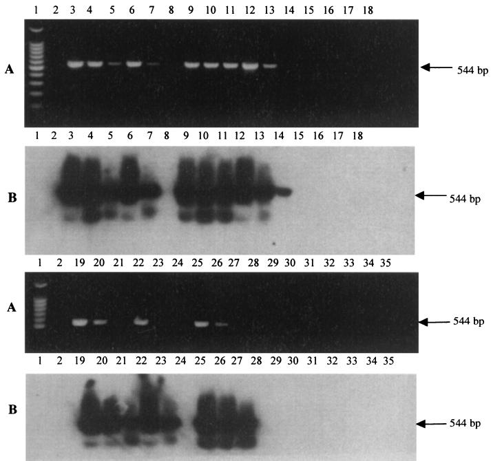
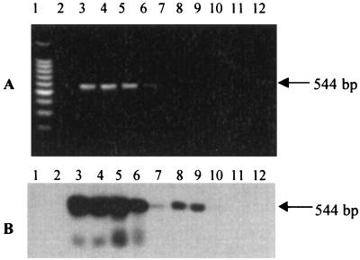
Subsequent tests focused on increasing sensitivity of the PCR by altering amounts of reaction mixture components. First, the MgCl2 concentration was reexamined for its effects on the efficiency of amplification from low template levels (all reactions done in a total volume of 50 μl). Amplification from high amounts of template (100 ng, ca. 108 copies) was efficient with MgCl2 levels ranging from 1.0 to 3.0 mM. However, with low amounts of template (500 fg), amplification was efficient only between 2.0 and 2.5 mM MgCl2. Next, the combination of Taq DNA polymerase and MgCl2 levels that gave the best balance in sensitivity and selectivity (E. coli versus S. sonnei) at low template levels (500 fg, ca. 500 copies) was examined and identified as 1.5 U of Taq and 2.0 mM MgCl2. The selectivity of the optimized PCR protocol was then verified with a battery of different E. coli, Shigella, and Salmonella isolates (Fig. 1). The detection limit of the optimized PCR was established by Southern hybridization as 100 fg of template, which is equivalent to ca. 10 cells of E. coli (Fig. 2).
FIG. 1.
Specificity of primer pair ECA75F-ECR619R in the optimized PCR for amplification of E. coli 16S rRNA. (A) Agarose gel separation of PCR mixtures; (B) Southern hybridization of the gel in panel A to EUB338. Lanes: 1, 100-bp DNA ladder; 2, no template; 3 to 5, 1 ng, 100 pg, and 10 pg of E. coli UW8101 template; 6 to 8, 1 ng, 100 pg, and 10 pg of E. coli UW8002 template; 9 to 11, 1 ng, 100 pg, and 10 pg of E. coli UW8204 template; 12 to 14, 1 ng, 100 pg, and 10 pg of E. coli UW8009 template; 15 and 16, 1 ng and 100 pg of Salmonella serovar Typhimurium UW8P14 template; 17 and 18, 1 ng and 100 pg of Salmonella serovar Typhimurium UW8P40 template; 19 to 21, 1 ng, 100 pg, and 10 pg of E. coli UW8410 template; 22 to 24, 1 ng, 100 pg, and 10 pg of E. coli UW8P39 template; 25 to 27, 1 ng, 100 pg, and 10 pg of E. coli UW8001D template; 28 and 29, 1 ng and 100 pg of Shigella dysenteriae UW8P01 template; 30 and 31, 1 ng and 100 pg of Shigella dysenteriae WSLH template; 32 and 33, 1 ng and 100 pg of Shigella flexnerii UW8P02 template; 34 and 35, 1 ng and 100 pg of Shigella sonnei UW8P15 template. Strain designations refer to those given in Table 1.
FIG. 2.
Sensitivity of primer pair ECA75F-ECR619R in the optimized PCR for amplification of E. coli 16S rRNA DNA template. (A) Agarose gel separation of PCR mixtures; (B) Southern hybridization of the gel in panel A to EUB338. Lanes: 1, 100-bp DNA ladder; 2, no template; 3 to 12, 100 pg, 50 pg, 10 pg, 5 pg, 1 pg, 500 fg, 100 fg, 50 fg, 10 fg, and 5 fg of E. coli genomic DNA.
Specificity was also examined by using BLAST to search GenBank for sequences similar to ECA75F and ECR619R. Vibrio gazogenes and Enterobacter gergoviae have single mismatches to the 5′ end of ECA75F but several mismatches to ECR619R. There were 15 exact matches to ECA75F, 13 of these were bacteria of the Pasteurellaceae family (various species or subspecies of Haemophilus, Mannheimia, and Pasteurella), and two were unknown γ-Proteobacteria. All of these organisms, however, had a number of mismatches to ECR619R. The greatest similarity to our primers was with Erwinia psidii, Pseudomonas flectens, Salmonella enterica serovar Waycross, and S. enterica serovar Chingola, all of which had either a single mismatch or single point deletion with ECA75F and were exact matches to ECR619R. This last group would theoretically pose the greatest potential for cross-reaction. However, these organisms are probably not typical water or soil inhabitants (E. psidii and P. flectens are described as originating from Brazilian guava fruit and an Australian bean, respectively), nor are they common residents of the human digestive tract. Thus, based on both empirical and theoretical testing, the method is expected to be reliable for selective detection of E. coli in soil or water contaminated by human wastes.
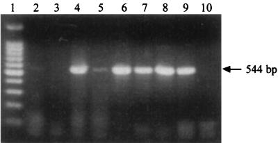
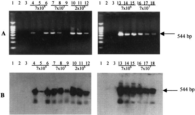
We evaluated the optimized PCR protocol for detection of E. coli in environmental samples. Soil was selected as it is an important reservoir for E. coli, and its chemical complexity provides a good test of the method's robustness. Initial tests to examine matrix effects on the PCR method were done with DNA extracted from soil and spiked with E. coli genomic DNA. These experiments showed that method performance with a heterogeneous mixture was similar to that obtained with pure E. coli genomic DNA: as little as 1 pg of E. coli template added to the PCR mixture gave sufficient product to detect by gel electrophoresis (Fig. 3). The sensitivity was comparable to that obtained using pure E. coli genomic DNA (Fig. 2). In tests to evaluate detection of E. coli DNA extracted from soil inoculated with various densities of E. coli cells, amplification was positive from all DNA extracts, and the expected product was detectable using agarose gels or Southern hybridization (Fig. 4). Furthermore, the lack of amplification from noninoculated soil, which likely had an indigenous population of enteric bacteria (e.g., Klebsiella), suggests that the method provided the necessary selectivity. The main difference between treatments was that with extracts prepared from the two lowest-inoculum densities, PCR products were sometimes detected only by hybridization. This probably reflects variability in E. coli DNA recoveries. Because many variables affect DNA extraction from bacterial cells in soil (e.g., soil type, residence time of cells in the soil, and soil biomass level), the detection limits achievable by this method will vary on a case-by-case basis.
FIG. 3.
Amplification of E. coli 16S rRNA from DNA extracted from Arlington (A) and Lakeland (L) soil and spiked with either 100 ng or 1 pg of E. coli genomic DNA. Lanes: 1, 100-bp DNA ladder; 2, A, nonspiked; 3, L, nonspiked; 4, A, 1-ng spike; 5, A, 1-pg spike; 6, L, 1-ng spike; 7, L, 1-pg spike; 8, 1 ng of E. coli genomic DNA; 9, 1 pg of E. coli genomic DNA; 10, no template.
FIG. 4.
Amplification of E. coli 16S rRNA from nonsterile soil inoculated with various densities of E. coli. (A) Ethidium bromide-stained agarose gel; (B) Southern hybridization of gel from panel A to EUB338. Lanes: 1, 100-bp DNA ladder; 2, no template; 3, 10 ng of DNA extract template from nonsterile, nonseeded soil sample; 4 to 18, 10 ng of DNA extract template from nonsterile soil samples seeded with stationary-phase E. coli at 7 × 10⁴, 7 × 10⁵, 2 × 10⁶, 7 × 10⁶, and 7 × 10⁷ CFU g−1, respectively.
In summary, a sensitive and robust PCR protocol was developed that discriminated E. coli from Shigella and other enteric bacteria based on selective amplification of 16S rRNA gene sequences. To the best of our knowledge, this is the first report of a PCR protocol based on amplification of a 16S rRNA that effectively distinguishes E. coli from these closely related bacteria. For environmental analysis, the extent to which the method's sensitivity of ca. 10 genomic equivalents can be exploited is controlled primarily by the efficiency with which DNA is extracted from the target organism, concentrated, and purified for the samples used in the PCR. For high-volume sample analysis, the protocol could be coupled with rapid methods for PCR product detection such as fluorogen labeling or enzyme-linked immunosorbent assay.
Acknowledgments
This work was supported by funding from the U.S. Department of the Interior, U.S. Geological Survey, Regional Competitive Grants Program (award 1434-HQ-GR-02707) and from the Wisconsin Department of Commerce (award DDG70000228).
REFERENCES
- 1.Amann R I, Krumholz L, Stahl D A. Fluorescent oligonucleotide probing of whole cells for determinative, phylogenetic and environmental studies in microbiology. J Bacteriol. 1990;172:762–770. doi: 10.1128/jb.172.2.762-770.1990. [DOI] [PMC free article] [PubMed] [Google Scholar]
- 2.Ausubel F, Brent R, Kingston R E, Moore D D, Seidman J G, Smith J A, Struhl K, editors. Short protocols in molecular biology. 3rd ed. New York, N.Y: John Wiley & Sons Inc.; 1997. p. 2-11-2-44. [Google Scholar]
- 3.Baudisova D. Evaluation of Escherichia coli as the main indicator of faecal pollution. Water Sci Technol. 1997;35:333–336. [Google Scholar]
- 4.Bouvet O M M, Lenormand P, Guibert V, Grimont P A D. Differentiation of Shigella species from Escherichia coli by glycerol dehydrogenase activity. Res Microbiol. 1995;146:787–790. doi: 10.1016/0923-2508(96)81074-6. [DOI] [PubMed] [Google Scholar]
- 5.Brenner D J, Fanning G R, Miklos G V, Steigerwalt A G. Polynucleotide relatedness among Shigella species. Int J Syst Bacteriol. 1973;23:1–7. [Google Scholar]
- 6.Brenner K P, Rankin C C, Roybal Y R, Stelma G N J R, Scarpino P V, Dufour A P. New medium for the simultaneous detection of total coliforms and Escherichia coli in water. Appl Environ Microbiol. 1993;59:3534–3544. doi: 10.1128/aem.59.11.3534-3544.1993. [DOI] [PMC free article] [PubMed] [Google Scholar]
- 7.Cilia V, LaFay B, Christen R. Sequence heterogeneities among 16S ribosomal RNA genes, and their effect on phylogenetic analysis on the species level. Mol Biol Evol. 1996;13:451–461. doi: 10.1093/oxfordjournals.molbev.a025606. [DOI] [PubMed] [Google Scholar]
- 8.Cleuziat P, Robert-Baudouy J. Specific detection of Escherichia coli and Shigella species using fragments of genes coding for β-glucuronidase. FEMS Microbiol Lett. 1990;72:315–322. doi: 10.1016/0378-1097(90)90324-j. [DOI] [PubMed] [Google Scholar]
- 9.Cone R W, Fairfax M R. Protocol for ultraviolet irradiation of surfaces to reduce PCR contamination. PCR Methods Appl. 1993;3:S15–S17. doi: 10.1101/gr.3.3.s15. [DOI] [PubMed] [Google Scholar]
- 10.Desmarchelier P M, Bilge A P, Fegan N, Mills L, Vary J C, Jr, Tarr P I. A PCR specific for Escherichia coli O157 based on rfb locus encoding O157 lipopolysaccharide. J Clin Microbiol. 1998;36:1801–1804. doi: 10.1128/jcm.36.6.1801-1804.1998. [DOI] [PMC free article] [PubMed] [Google Scholar]
- 11.Fiksdal L, Pommepuy M, Caprais M-P, Midttun I. Monitoring of fecal pollution in coastal waters by use of rapid enzymatic techniques. Appl Environ Microbiol. 1994;60:1581–1584. doi: 10.1128/aem.60.5.1581-1584.1994. [DOI] [PMC free article] [PubMed] [Google Scholar]
- 12.Gannon V P J, D'Souza S, Graham T, King R K, Rahn K, Read S. Use of the flagellar H7 gene as a target in multiplex PCR assays and improved specificity in identification of enterohemorrhagic Escherichia coli strains. J Clin Microbiol. 1997;35:656–662. doi: 10.1128/jcm.35.3.656-662.1997. [DOI] [PMC free article] [PubMed] [Google Scholar]
- 13.Karkkainen U-M, Kauppinen J, Ikaheimo R, Katila M-L, Siitonen A. Rapid and specific detection of three different G adhesion classes of P-fimbriae in uropathogenic Escherichia coli by polymerase chain reaction. J Microbiol Methods. 1998;34:23–29. [Google Scholar]
- 14.Meng J, Zhao S, Doyle M P, Mitchell S E, Kresovich S. A multiplex PCR for identifying shiga-like toxin-producing Escherichia coli O157:H7. Lett Appl Microbiol. 1997;24:172–178. doi: 10.1046/j.1472-765x.1997.00375.x. [DOI] [PubMed] [Google Scholar]
- 15.Newton C R, Graham A, editors. PCR. 2nd ed. Oxford, England: Bios Scientific Publishers; 1997. p. 39. [Google Scholar]
- 16.Orskov F, Orskov I. Escherichia coli serotyping and disease in man and animals. Can J Microbiol. 1992;38:699–704. [PubMed] [Google Scholar]
- 17.Paton A W, Paton J C. Detection and characterization of Shiga toxigenic Escherichia coli by using multiplex PCR assays for stx1, stx2, eaeA, enterohemorrhagic E. coli hlyA, rfb0111, and rfb0157. J Clin Microbiol. 1998;36:598–602. doi: 10.1128/jcm.36.2.598-602.1998. [DOI] [PMC free article] [PubMed] [Google Scholar]
- 18.Pollard D R, Johnson W M, Lior H, Tyler S D, Rozee K R. Rapid and specific detection of verotoxin genes in Escherichia coli by the polymerase chain reaction. J Clin Microbiol. 1990;28:540–545. doi: 10.1128/jcm.28.3.540-545.1990. [DOI] [PMC free article] [PubMed] [Google Scholar]
- 19.Pupo G M, Karaolis D K R, Lan R T, Reeves P R. Evolutionary relationships among pathogenic and nonpathogenic Escherichia coli strains inferred from multilocus enzyme electrophoresis and mdh sequence studies. Infect Immun. 1997;65:2685–2692. doi: 10.1128/iai.65.7.2685-2692.1997. [DOI] [PMC free article] [PubMed] [Google Scholar]
- 20.Sarkar G, Sommer S T. Shedding light on PCR contamination. Nature. 1990;343:27. doi: 10.1038/343027a0. [DOI] [PubMed] [Google Scholar]
- 21.Selbach P A. Ph.D. dissertation. University of Wisconsin, Madison; 1998. [Google Scholar]
- 22.Tsai Y-L, Olson B H. Rapid method for direct extraction of DNA from soil and sediments. Appl Environ Microbiol. 1991;57:1070–1074. doi: 10.1128/aem.57.4.1070-1074.1991. [DOI] [PMC free article] [PubMed] [Google Scholar]
- 23.Tsen H-Y, Jian L-Z. Development and use of a multiplex PCR system for the rapid screening of heat-labile toxin I, heat-stable toxin II and shiga-like toxin I and II genes of Escherichia coli in water. J Appl Microbiol. 1998;84:585–592. doi: 10.1046/j.1365-2672.1998.00385.x. [DOI] [PubMed] [Google Scholar]
- 24.Tsen H-Y, Jian L-Z, Chi W-R. Use of multiplex PCR system for the simultaneous detection of heat labile toxin I and heat stable toxin II genes of enterotoxigenic Escherichia coli in skim milk and porcine stool. J Food Prot. 1998;61:141–145. doi: 10.4315/0362-028x-61.2.141. [DOI] [PubMed] [Google Scholar]
- 25.Venkateswaran K, Murakoshi A, Stake M. Comparison of commercially available kits with standard methods for the detection of coliforms and Escherichia coli in foods. Appl Environ Microbiol. 1996;62:2236–2243. doi: 10.1128/aem.62.7.2236-2243.1996. [DOI] [PMC free article] [PubMed] [Google Scholar]
- 26.Wang R-F, Cao W-W, Cerniglia C E. Phylogenetic analysis and identification of Shigella spp. by molecular probes. Mol Cell Probes. 1997;11:427–432. doi: 10.1006/mcpr.1997.0136. [DOI] [PubMed] [Google Scholar]


