

Abstract
Congenital cleft ear lobe is a rare anomaly, even though the deformities of the ear as a whole are encountered commonly. The deformity results from the failure of fusion of hillocks 1 and 6 during embryonic life. The defective ear lobe carries immense aesthetic importance, especially in females. Several techniques have been used for the reconstruction of the ear lobule with this defect. But utilizing Z-plasty, one of the basic principles of reconstructive surgery, can result in an aesthetically pleasing lobule. The author presents here two such cases in which he could achieve satisfactory results using simple Z-plasty for the reconstruction of ear lobes. Congenital cleft ear lobe is a rare anomaly, even though the deformities of ear as a whole are encountered commonly. The deformity results from the failure of fusion of hillocks 1 and 6 during embryonic life. The defective ear lobe carries immense aesthetic importance, especially in females. Several techniques have been used for reconstruction of ear lobule with this defect. But utilizing Z-plasty, one of the basic principles of reconstructive surgery can result in an aesthetically pleasing lobule. The author presents here two such cases in which he could achieve satisfactory results using simple Z-plasty for reconstruction of ear lobes.
Keywords: Cleft ear lobe, Deformity, Z-plasty
Introduction
Congenital cleft ear lobe is a very rare anomaly we come across. It often occurs due to failure of fusion between hillocks 1 and 6. The pinna of human ear is essentially a vestigial organ having no functional importance, unlike lower animals in whom it helps in directing the sound wave towards the middle ear helping in hearing. But this organ is present in the most visible part of human body (face) and putting different kinds of ornaments adds to the aesthetic value of women in particular. Hence, the reconstruction of this defective organ is of utmost importance with a goal of achieving a near-normal contour and with the minimum visible scar. Several operative techniques have been described in published literature by different authors and some of the principles of cleft lip repair have also been followed. We came across two girls with congenital cleft ear lobule, one 6 years and the other 21 years old, presenting the defect on one side. We used simple Z-plasty with minimal situational variations and obtained appreciable outcomes. The cases are reported after obtaining approval from the institute review board.
Case reports
Case 1
A 6-year-old girl presented to us with a wide notch in the lower margin of the lobule of left ear present since birth. None of the family members had such a type of congenital deformity. She did not have any other congenital anomaly of the face or any other organ. Hence, she was diagnosed as a case of cleft ear lobe affecting the left side. The lobule was reconstructed using Z-plasty on both the lateral and medial surface, the skin flaps being closed with monofilament polyamide(6–0) suture. The sutures were removed on the fifth day and the suture lines were secured further with micropore tape for additional 5 days. The parents were advised to care for prevention of any trauma to the operated part for at least a month. The reconstructed ear lobe was aesthetically acceptable (Fig. 1) and she was advised to avoid ear piercing on that side for at least 3 months, that too not on the suture line.
Fig. 1.
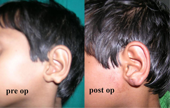
Pre-op & post-op pictures of cleft ear lobe in a 6-year-old girl
Case 2
This was an adult female of 21 years, presented with a similar defect over her left ear lobe present since birth. Her family history was negative, and she also did not have any other congenital anomaly. She was also diagnosed with a case of the cleft ear lobe and treated in a similar manner using Z-plasty on both lateral and medial surfaces. During the follow-up after 3 months of surgery, the contour of the ear lobe was found to be pleasing with the minimal visible scar (Fig. 2).
Fig. 2.

Pre-op & post-op pictures of cleft ear lobe in a 21–year-old girl
Discussion
Congenital anomalies of the lower third of the auricle are seen in 1 every 15,000 live births with cleft ear lobule being the commonest [1, 2]. The auricle develops from six hillocks or mesenchymal proliferations originating from the first and second pharyngeal arches. Hillocks 1,2, 3 and hillocks 4, 5, 6 develop into the parts of the pinna on either side of the external auditory meatus. Failure in fusion of hillocks 1 and 6 result in the cleft ear lobe [3]. In almost all parts of the world, the females used to wear ear rings or some other floral ornaments in the ear lobes to enhance their facial aesthetics. Additionally, any congenital deformity in females carries social stigma and poses a problem to get married. These may be the reasons for the females, in particular, to seek treatment mostly for this deformity, anticipating a near-normal ear lobe. These deformities may be associated with other anomalies in various syndromes like Goldenhar syndrome and JS-X syndrome [4, 5]. Hence, the cases with ear deformities must be investigated to look for other anomalies manifesting a syndrome. Several techniques have been proposed for the surgical correction of these defects [6–10]. Principles used for repair of cleft lip also can be utilized for this purpose. But, following the fundamental principles of reconstructive surgery, a properly planned Z-plasty can result in an aesthetically pleasing ear lobule. The final suture line of this technique also allows the ear piercing at a point away from the suture line of the repair. Though special techniques may be useful for specific types of defects, in our cases Z-plasty was found to be the appropriate procedure in order to achieve a satisfying outcome.
Funding
The author did not receive any funding for this work from any source.
Declarations
Conflict of interest
The authors have no conflicts of interest to declare that arerelevant to the content of this article.
Ethical approval
Ethical approval was obtained for the publication of this case report.
Consent for publication
Consent was obtained from the patients/patients relatives for the publication of their details as a case report.
Footnotes
Publisher's Note
Springer Nature remains neutral with regard to jurisdictional claims in published maps and institutional affiliations.
References
- 1.Conrad K, Reifen E. Congenital cleft ear lobe deformity: a staged reconstruction. J Otolaryngol. 1994;23:19–22. [PubMed] [Google Scholar]
- 2.Park C. Lower auricular malformations: their representation, correction, and embryologic correlation. Plast Reconstr Surg. 1999;104:29–40. doi: 10.1097/00006534-199907000-00005. [DOI] [PubMed] [Google Scholar]
- 3.Fuchs JC, Tucker AS. Development and integration of the ear. Curr Top Dev Biol. 2015;115:213–32. doi: 10.1016/bs.ctdb.2015.07.007. [DOI] [PubMed] [Google Scholar]
- 4.Goswami M, Bhushan U, Jangra B. Goldenhar syndrome: a case report with review. Int J Clin Pediatr Dent. 2016;9(3):278–280. doi: 10.5005/jp-journals-10005-1377. [DOI] [PMC free article] [PubMed] [Google Scholar]
- 5.Hoeve HL, Brooks AS, Smit LS. JS-X syndrome: a multiple congenital malformation with vocal cord paralysis, ear deformity, hearing loss, shoulder musculature underdevelopment, and X-linked recessive inheritance. Int J Pediatr Otorhinolaryngol. 2015;79(7):1164–1170. doi: 10.1016/j.ijporl.2015.05.001. [DOI] [PubMed] [Google Scholar]
- 6.Matsumoto K. Surgical repair of the congenital ear lobe cleft. Br J Plast Surg. 1981;34(4):410–413. doi: 10.1016/0007-1226(81)90046-1. [DOI] [PubMed] [Google Scholar]
- 7.Stanley SS, Thaller SR. Congenital cleft earlobe: technique for repair of a triple-lobe type defect. J Craniofac Surg. 2018;29(8):2310–2311. doi: 10.1097/SCS.0000000000004788. [DOI] [PubMed] [Google Scholar]
- 8.Sinha M. Techniques for correction of acquired or congenital cleft ear lobe repair. J Plast Reconstr Aesthet Surg. 2006;59(9):1009–1010. doi: 10.1016/j.bjps.2005.12.040. [DOI] [PubMed] [Google Scholar]
- 9.Fujiwara T, Matsuo K, Taki K, Noguchi M, Kiyono M. Triangular flap repair of the congenital earlobe cleft. Annal Plast Surg. 1995;34(4):402–405. doi: 10.1097/00000637-199504000-00011. [DOI] [PubMed] [Google Scholar]
- 10.Borman H, Deniz M, Ertas NM, Seyhan T, Caglar B. 7-plasty technique for the surgical treatment of congenital longitudinal ear lobe cleft. J Craniofac Surg. 2008;19(6):1643–1644. doi: 10.1097/SCS.0b013e31818c0306. [DOI] [PubMed] [Google Scholar]