Key Points
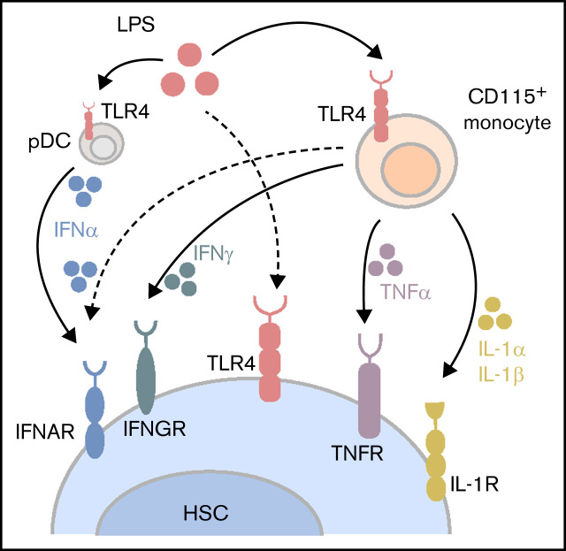
HSCs are transiently activated by acute LPS challenge via direct and indirect mechanisms, including CD115+ monocytic cells in BM.
The combined action of IFNα, IFNγ, TNFα, IL-1α, IL-1β, and other cytokines is required to mediate HSC activation in response to LPS in vivo.
Visual Abstract
Abstract
Infections are a key source of stress to the hematopoietic system. While infections consume short-lived innate immune cells, their recovery depends on quiescent hematopoietic stem cells (HSCs) with long-term self-renewal capacity. Both chronic inflammatory stress and bacterial infections compromise competitive HSC capacity and cause bone marrow (BM) failure. However, our understanding of how HSCs act during acute and contained infections remains incomplete. Here, we used advanced chimeric and genetic mouse models in combination with pharmacological interventions to dissect the complex nature of the acute systemic response of HSCs to lipopolysaccharide (LPS), a well-established model for inducing inflammatory stress. Acute LPS challenge transiently induced proliferation of quiescent HSCs in vivo. This response was not only mediated via direct LPS-TLR4 conjugation on HSCs but also involved indirect TLR4 signaling in CD115+ monocytic cells, inducing a complex proinflammatory cytokine cascade in BM. Downstream of LPS-TLR4 signaling, the combined action of proinflammatory cytokines such as interferon (IFN)α, IFNγ, tumor necrosis factor-α, interleukin (IL)-1α, IL-1β, and many others is required to mediate full HSC activation in vivo. Together, our study reveals detailed mechanistic insights into the interplay of proinflammatory cytokine-induced molecular pathways and cell types that jointly orchestrate the complex process of emergency hematopoiesis and HSC activation upon LPS exposure in vivo.
Introduction
Infection is a major source of stress to the hematopoietic system, leading to exhaustion of blood and immune cells.1,2 Replacement of these cells depends on a small number of multipotent and self-renewing hematopoietic stem cells (HSCs) that predominantly remain in a quiescent state.3,4 In response to infections, quiescent HSCs are efficiently induced to proliferate to counterbalance the loss of blood and immune cells.5-7 The enhanced demand for mature immune cells triggers molecular signals that accelerate hematopoiesis within the bone marrow (BM), so-called emergency hematopoiesis.8 After successful elimination of the infection, homeostasis is reestablished although there is evidence of impaired HSC function after each round of infection.9 Recent studies have shown that HSCs directly respond to infections by detecting infectious particles via Toll-like receptors (TLRs).10-12 Furthermore, we and others could demonstrate that HSCs directly respond to proinflammatory cytokines released during infection, such as interferon (IFN)-α or IFN-γ.5,6
Over the past 2 decades, various groups have investigated the effects of lipopolysaccharide (LPS), a component of the cell membrane of gram-negative bacteria, on the immune system.13 LPS is detected by a receptor complex composed of TLR4, MD-2, and CD14.14 Subsequently, activation of TLR4 mediates the immune response through the production of proinflammatory cytokines and IFNs.15 Functional in vivo studies on the effects of LPS on HSCs have mainly focused on long-term exposure, modeling chronic infections. Sustained LPS exposure impairs the self-renewal and competitive repopulating activity of HSCs,12,16,17 mainly driven by cell-intrinsic LPS-TLR4 signaling in HSCs. However, less is known about the implications of short-term pathogen exposure during acute and limited infectious conditions on HSC function or the interface of HSCs directly sensing infectious particles with a complex network of cytokine-producing immune cells.
Using advanced chimeric and genetic mouse models together with pharmacological interventions, we have dissected the complex nature of the acute systemic response of HSCs to LPS in vivo. LPS challenge transiently activates HSCs via direct LPS-TLR4 conjugation on HSCs and indirect TLR4 signaling in CD115+ monocytic cells, inducing a complex proinflammatory cytokine cascade in BM. In summary, our study provides detailed mechanistic insights into the complex network of proinflammatory cytokines orchestrating the process of HSC activation and emergency hematopoiesis upon LPS exposure in vivo.
Methods
Mice
All mice were housed in individually ventilated cages in the German Cancer Research Center (DKFZ) animal facility. Animal procedures were performed according to protocols approved by the Regierungspräsidium Karlsruhe (no. G266/12, G157/13, G224/14, and G22/17). C57Bl/6 mice are referred to as wild-type (wt; CD45.2+) mice. Ifnar−/−Ifngr−/−,18,19 Sca-1−/−,20 TLR4−/−,21 Tnfrsf1−/−,22 and IL-1R−/−23 mice are crossed on a C57Bl/6 background. CD45.1+ B6.SJL-Ptprca-Pep3b-/BoyJ mice were purchased from Charles River. Myd88−/−24 and Trifmc−/−15 mice were purchased from Jackson Laboratory.
Generation of chimeric mice
BM was isolated and Thy1 depleted. For generation of mixed BM chimeras, BM cells (3 × 10⁶) were diluted in 200 μL of phosphate-buffered saline (PBS) and transplanted IV into lethally irradiated (2 × 500 rad) recipients. Mice were kept on antibiotics containing water (sulfamethoxazol/trimethoprim, Cotrim K ratiopharm, 50 mg/mL, refreshed twice weekly) for 3 weeks. Peripheral blood chimerism was determined after 12 weeks, after which the respective cytokine treatment was administered. Stem cell function and long-term reconstitution ability were tested by injection of 3 × 10⁶ BM cells diluted in 20 μL of PBS into the femur of lethally irradiated mice.
In vivo treatments
All experiments were performed using 8- to 12-week-old mice. Depending on the experiment 0.25 mg/kg of LPS (Escherichia coli 0111:B4; Sigma), 0.75 mg/kg of tumor necrosis factor (TNF)-α (PeproTech), 0.25 mg/kg of interleukin (IL)-1α (PeproTech), or 0.25 mg/kg of IL-1β (PeproTech) dissolved in 200 μL of PBS were injected IP, unless stated differently. Treatment lasted for 18 hours unless indicated differently. Control mice were injected with 200 μL of PBS. TNFα inhibition was induced by intraperitoneal injection of etanercept (EnbrelO, FDA 1998) daily for 10 days (5 mg/kg in 100 μL of PBS on days 1-5; 7.5 mg/kg in 150 μL of PBS on days 6-10). To block IL-1 signaling, 2.5 mg/kg of anakinra (IL-1RA) (PeproTech) was injected IP, dissolved in 200 μL of PBS.
In vivo depletion of myeloid cells
For in vivo depletion of myeloid cells, mice were injected IV with 300 µL of clodronate- (0.25 g/mL) or PBS-loaded liposomes.25 Treatment with LPS was performed 5 days after liposome injection.
Isolation of BM, spleen, and peripheral blood
Cervical dislocation was used for euthanizing mice. Tibiae, femura, coxae, vertebral column, and spleen were withdrawn for experimental analysis. RPMI-1640 medium (Sigma) enriched with 2% v/v fetal calf serum was used for suspension of the cells.
Fluorescence-activated cell sorting
BM cells were stained using CD4, CD8, CD11b, Gr-1, B220, and TER119, cKit, Sca-1, CD150, CD48, and CD34. BM cells were stained for myeloid cells with antibodies against CD11b, Gr-1, and CD115. Antibodies are listed in the supplementary materials. For flow cytometry analysis, the LSRII or LSR Fortessa (BD Biosciences) was used.
Cell cycle analysis
Cell cycle analysis was conducted by icKi67-Hoechst33342 staining. After surface staining, cells were fixed with cytofix-cytoperm buffer (BD Biosciences) and stained with anti-Ki67 for 30 minutes. Prior to fluorescence-activated cell sorting (FACS) analysis, Hoechst 33342 (Molecular Probes, 25 μg/mL) was added for 10 minutes. For the BrdU incorporation assay, BrdU (18 mg/kg, Sigma-Aldrich) was administered IP for 14 hours prior to harvesting the BM. The BD PharmingenTM BrdU Flow Kit protocol was used to stain for BrdU.
Cell sorting
To isolate hematopoietic stem and progenitor cells (HSPCs) (Lin-CD117+CD150+), BM cells were incubated with lineage antibodies (CD4, CD8, CD11b, B220, Gr-1, and TER119), and lineage-positive cells were depleted using Dynabeads Magnetic Beads (Invitrogen). Lineage-negative cells were stained with cKit and CD150. Cell sorting was performed on an FACS Aria I, FACS Aria II, or FACS Aria III (Becton Dickinson) at the DKFZ Flow Cytometry Service Unit.
In vitro cultures
After cell sorting, cells were cultured in StemPro-34 SFM medium (Invitrogen), supplemented with 2 mM of l-glutamine, 100 U/mL of penicillin, 100 mg/mL of streptomycin, IL-11 (20 ng/mL), mFlt3 (30 ng/mL), mTPO (25 ng/mL), and mSCF (50 ng/mL). Cells were incubated for 18 to 20 hours at 37°C, 5% CO2. Cells were cultured in the presence of PBS, LPS (100 ng/mL), or recombinant mIFNα (1000 U/mL).
Colony-forming unit assay
FACS-sorted CD11b+CD115+ monocytic cells were cultured with sorted Lin-c-Kit+CD150+ HSPCs in Stem-Pro 34 SFM medium (Invitrogen) in the presence of PBS or LPS (100 ng/mL). Cells were exposed to LPS either for 18 hours followed by a change of medium or continuously over 7 days. After 7 days, cells were harvested and plated in methylcellulose medium (MethoCult H4434; StemCell Technologies) according to the manufacturer’s instructions. Colonies were evaluated and scored after 8 days.
Enzyme-linked immunosorbent assay
Femura and tibiae were flushed with 400 μL of RPMI. Cytokine levels were measured in these lysates using enzyme-linked immunosorbent assay (ELISA) kits according to the manufacturer’s instruction (TNFα and IFNγ ELISA kits from R&D Systems; IFNα ELISA from Interferon Source [VeriKine]). Absorbance was determined by SpectraMax M5 Microplate Reader (Molecular Devices).
Quantitative real-time PCR
RNA extraction was performed using a PicoPureTM RNA isolation kit (Arcturus) and DNA digestion by RNase-free DNase (Quiagen). cDNA transcription was done by SuperScriptÒVILOTM cDNA Synthesis kit (invitrogen). Quantitative real-time PCR (qRT-PCR) was carried out by Applied Biosystems ViiATM 7 Real-Time PCR System in 384-well plates. Gene expression was normalized to Sdha or Oaz1 levels.
Microarray
Eighteen hours after IFNα (5 × 10⁶U/kg) and LPS (0.25 mg/kg) treatment of wt mice, HSCs were sorted and RNA was isolated. Then 2.5 ng of RNA was used for hybridization onto the Mouse Sentrix-6 Bead-Chip array (Illumina). The Bioconductor package limma was used to perform differential gene expression analysis. Probes with P value <.5 and fold change >=<1.5 were considered top differentially expressed genes. Enrichment analysis was performed using EnrichR, and P values were obtained from the online EnrichR program. Microarray data were deposited in NCBI’s Gene Expression Omnibus under accession number GSE181584.
ProcartaPlex array
One tibia per mouse was crushed in 300 μL of PBS, homogenized, and centrifuged for 5 minutes at 1600 × g. Supernatant was measured on ProcartaPlex Multiplex immunoassay (Mouse monitoring Plex Ref. EPX480-20834-901), conducted according to manufacturer’s instructions (Affymetrix). Results were acquired with Bio-Plex 200 system (Bio-Rad) and analyzed with Bio-Plex Data-pro software.
Statistical analysis
Statistical analyses were performed using GraphPad Prism (GraphPad Software). The error bars shown in the figures represent the standard deviation. In each experiment, the statistical tests used are indicated in the figure legends. Briefly, if only 2 groups were compared, the P value was determined by unpaired t test. If more than 2 groups were compared, P values were determined by analysis of variance (ANOVA) Tukey’s post hoc test.
Results
Acute systemic LPS treatment transiently increases HSC proliferation via TLR4
Counterbalancing the consumption of blood and immune cells when fighting off an infection is an indispensable need for survival. Recent reports have proposed 2 mechanisms of HSC activation to replenish lost immune cells upon inflammatory stress, either by sensing pathogen-associated molecular patterns by the HSCs themselves11,26,27 or indirectly via the production of proinflammatory cytokines in response to invading pathogens.5,6,28 Lipopolysaccharide (LPS) is a well-established model for inducing inflammatory stress in vivo.12,16,17 So far, the LPS-induced response in HSCs has mainly been investigated under conditions of chronic infections, and prolonged exposure to LPS was reported to compromise competitive HSC capacity.12,17
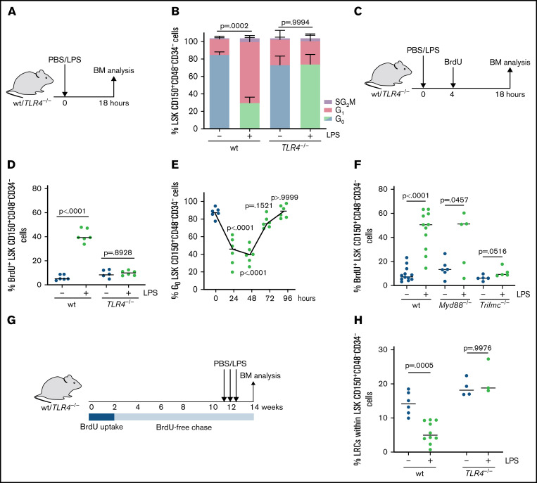
Here, we focus on the acute and immediate HSC response to inflammatory stress. To model the initial infectious phase, we injected wt mice with a single dose of LPS (Figure 1A). Within 18 hours of LPS injection, quiescent HSCs (LSK CD150+CD48-CD34-) entered an active cell cycle (Figure 1B; supplemental Figure 1A-B). In addition, the proliferation was strongly induced upon LPS treatment as assessed by BrdU (5-bromo-2′-deoxyuridine) incorporation assay (Figure 1C-D). In line with recent publications,29 this was accompanied by a decline in blood myeloid cells, whereas lymphoid and erythroid subsets remained unaffected (supplemental Figure 1C). Increased HSC proliferation in response to a single LPS stimulus was transient, with a peak at 48 hours after injection (Figure 1E), suggesting that HSCs return to quiescence after the initial response. Both hematopoietic and nonhematopoietic cells, such as endothelial cells and stroma cells, express TLR4, the pattern recognition receptor for LPS.30,31 In contrast to wt HSCs, LPS did not induce proliferation of HSCs in TLR4-deficient mice, revealing that TLR4 is involved in the HSC early response to inflammatory stress (Figure 1A-D). Of note, TLR4 deficiency did not affect the peripheral immune cell composition in the blood (supplemental Figure 1C). LPS binding to TLR4 is known to activate 2 downstream signaling pathways, MYD88 (myeloid differentiation primary response 88) and TRIF (Toll-like receptor adaptor molecule).32 We found that LPS treatment induced strong cell cycle induction in Myd88−/− mice but not in Trifmc−/− mice, highlighting the indispensable need for TRIF but not MYD88 signaling in mediating the LPS-induced response in HSCs (Figure 1F), which is consistent with previous reports.33 To test whether HSC activation upon acute inflammatory stress was capable of impacting even dormant HSCs, we performed a long-term label-retaining cell (LRC) assay with a 10-day in vivo labeling period with BrdU, followed by a BrdU-free 12-week chase.3,5 After 10 weeks of the BrdU-free chase, mice were injected with LPS 3 times and analyzed at the end of week 12 to determine the BrdU retention upon proliferation (Figure 1G). Although the total number of HSCs in the BM remained unchanged, the number of LRCs in LPS-treated mice was significantly reduced in a TLR4-dependent manner (Figure 1H), indicating that even dormant HSCs respond to LPS.
Figure 1.
Acute systemic LPS treatment transiently increases HSC proliferation via TLR4. (A) Scheme indicating in vivo treatment of wt or TLR4−/− mice with PBS (control) or LPS (0.25 mg/kg) for 18 hours. (B) Cell cycle analysis using intracellular (ic) Ki67-Hoechst 33342 staining (G0 cells icKi67neg Hoechst 33342low; G1 cells icKi67pos Hoechst 33342low; SG2M cells icKi67pos Hoechst 33342hi) of HSCs (LSK CD150+CD48-CD34-) from wt and TLR4−/− mice (n = 3) treated with PBS or LPS (0.25 mg/kg, 18 hours). P values refer to G0 phase and were determined by ANOVA Tukey’s post hoc test. (C) Scheme indicating in vivo BrdU uptake (18 mg/kg, 14 hours) and treatment with PBS (control) or LPS (0.25 mg/kg) of wt or TLR4−/− mice for 18 hours. (D) Fourteen-hour BrdU (18 mg/kg) uptake of HSCs (LSK CD150+CD48-CD34-) from PBS- (control) or LPS- (0.25 mg/kg, 18 hours) treated wt and TLR4−/− mice (n = 6). P values were determined by ANOVA Tukey’s post hoc test. (E) Percentage of HSCs (LSK CD150+CD48-CD34-) in G0 phase (icKi67neg Hoechst 33342low) at indicated time points after treatment of wt mice with PBS (control; time point 0 hours; blue dots) or LPS (0.25 mg/kg; green dots) (n = 6). P values were determined by ANOVA Tukey’s post hoc test. (F) Fourteen-hour BrdU (18 mg/kg) uptake of HSCs from wt (n = 12), Myd88−/− (n = 6), or Trifmc−/− (n = 5) mice treated with PBS (control) or LPS (0.25 mg/kg, 18 hours). P values were determined by unpaired t test. (G) Scheme illustrating the in vivo LRC assay: BrdU (18 mg/kg IP) uptake at day 1, subsequent 10-day BrdU pulse-period (1 mg/mL in drinking water), followed by a BrdU-free 12-week chase. Subsequently, mice were treated with PBS (control) or LPS (0.25 mg/kg) every third day. Analysis was done 2 weeks after last LPS injection. (H) Percentage of BrdU+ LRCs of LSK CD150+CD48-CD34- in wt and TLR4−/− mice. Experimental set-up as indicated in Figure 1G (wt: n = 6 PBS; n = 10 LPS; TLR4−/−: n = 4 PBS; n = 3 LPS). P values were determined by ANOVA Tukey’s post hoc test.
Previous studies described an impaired engraftment capacity of HSCs isolated from mice undergoing chronic LPS exposure.12,16,17 However, transplantation of HSCs from mice treated with a single dose of LPS did not reveal great differences in peripheral blood cell reconstitution or the level of engraftment of HSCs (supplemental Figure 2A-C). Furthermore, a single LPS treatment did not significantly alter the colony-forming unit capacity of FAC-sorted wt HSPCs (Lin-cKit+CD150+), whereas multiple LPS administrations did result in a significant reduction (supplemental Figure 2D-E). Together, these data indicated that short-term LPS exposure did not severely impact the HSPC’s differentiation capacity.
Thus, acute in vivo LPS stimulation induced a transient TLR4-dependent proliferation of HSCs without yet impairing their differentiation ability.
HSC activation is mediated by cell-extrinsic TLR4 signaling
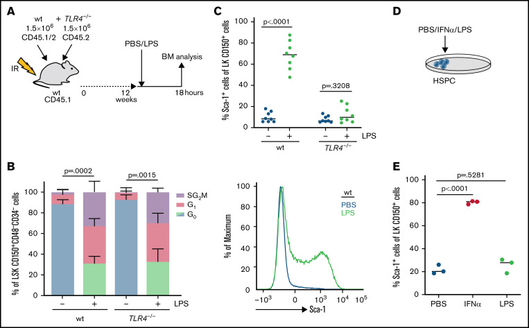
Recent studies suggested that LPS challenges HSCs through direct TLR4 activation.10-12 Because most immune and stroma cells within the BM compartment express TLR4, we wanted to evaluate the contribution of other BM cells to LPS-induced HSC activation. Thus, we generated mixed BM chimeric mice by reconstituting lethally irradiated wt mice (CD45.1) with 50% wt (CD45.1/2) and 50% TLR4−/− (CD45.2) total BM cells to create a mouse in which only half of the hematopoietic cells are able to respond to LPS. In case of indirect LPS-TLR4 signaling on other wt BM cells, HSCs lacking TLR4 should show a response to the LPS treatment. After confirming stable 50% wt–50% TLR4−/− hematopoietic reconstitution 12 weeks after transplantation, chimeric mice were injected with LPS (Figure 2A). As expected, wt HSCs in the chimeric mice responded to LPS similar to the response observed in nonchimeric wt mice (Figure 2B). Unexpectedly, however, in the presence of wt LPS-responsive BM TLR4−/− HSCs also started to proliferate efficiently upon LPS stimulation (Figure 2B), suggesting that the short-term response of HSCs to LPS is, at least partially, mediated via indirect mechanisms involving other LPS-responsive cells in BM.
Figure 2.
HSC activation is mediated by cell-extrinsic TLR4 signaling. (A) Scheme indicating in vivo transplantation: BM from wt (CD45.1/2+) and TLR4−/− mice (CD45.2+) (1.5 × 10⁶ BM cells each) was transplanted into irradiated wt mice (CD45.1+). Mice were treated with PBS (control) or LPS (0.25 mg/kg, 18 hours) 12 weeks after transplantation. (B) Cell cycle analysis (icKi67-Hoechst 33342) of wt and TLR4−/− HSCs (LSK CD150+CD48-CD34-) in mixed BM chimeras after PBS (control) or LPS treatment as indicated in Figure 2A (n = 3). P values refer to G0 phase and were determined by ANOVA Tukey’s post hoc test. (C) Relative Sca-1 expression in LK CD150+ cells (HSPCs) of wt and TLR4−/− mice treated with PBS (control) or LPS (0.25 mg/kg, 18 hours) (n = 8) and representative FACS profile. P values were determined by ANOVA Tukey’s post hoc test. (D) Scheme indicating in vitro treatment or sorted HSPCs (LK CD150+) with PBS (control), IFN-α (1000 U/mL), or LPS (100 ng/mL). (E) Relative Sca-1 expression on sorted HSPCs (LK CD150+) after PBS (control), IFN-α, or LPS in vitro treatment for 18 hours as indicated in Figure 2D (n = 3). P values were determined by ANOVA Tukey’s post hoc test.
To further investigate this, we analyzed the HSC response to LPS in vitro. As shown for other forms of inflammatory stress, LPS did not trigger HSC cell cycle induction in vitro. Therefore, we used Sca-1 expression as a proxy for HSC activation in this setting.34,35 Whereas LPS application triggered efficient, TLR4-dependent Sca-1 induction in HSCs in vivo (Figure 2C), LPS treatment did not affect Sca-1 expression in isolated HSPCs (Lin-CD117+CD150+) in vitro (Figure 2D-E). In contrast, IFNα, a cytokine known to directly act on HSPCs,5 efficiently increased Sca-1 surface expression on HSPCs in vitro (Figure 2D-E), further supporting the model of an indirect effect of LPS on HSPCs.
Systemic LPS challenge induces HSC activation via CD115+ monocytic cells in BM
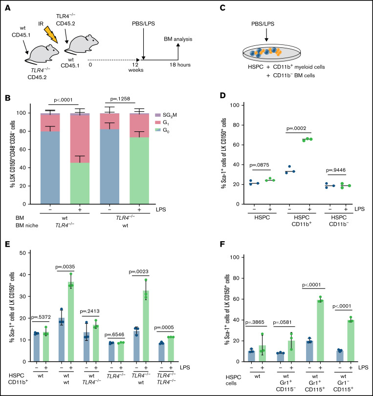
To systematically determine the cell types involved in mediating the indirect LPS effect on HSCs, we created reverse chimeras by transplanting wt BM into TLR4 −/− mice (Figure 3A). LPS treatment of these mice excluded a prominent role of TLR4 signaling in the nonhematopoietic BM niche (Figure 3B) and suggested that BM immune cells are the major mediators responsible for indirect HSC activation upon LPS treatment. To identify the immune cells that mediate the indirect response of HSCs to LPS, we performed in vitro coculture experiments with sorted LK CD150+ HSPCs and different immune cells (Figure 3C-D). Hypothesizing that cytokine secretion by myeloid immune cells upon stress conditions might be the missing link in mediating the HSC’s response to LPS, we cocultured HSPCs with CD11b+ cells (Figure 3C). Indeed, adding CD11b+ myeloid cells to the HSPC culture mimicked the in vivo LPS effect, leading to increased Sca-1 expression on HSPCs (Figure 3D). In contrast, coculturing HSPCs with total BM cells depleted for myeloid cells (CD11b-) failed to achieve this effect (Figure 3D). Moreover, TLR4−/− HSPCs responded to LPS treatment in vitro when cocultured with wt CD11b+ cells, whereas the increase in Sca-1 expression on HSPCs upon LPS application was completely abrogated when wt HSPCs were cocultured with TLR4−/− CD11b+ myeloid cells (Figure 3E). Together, this suggests an important role for CD11b+ myeloid cells in mediating indirect HSC activation upon short-time LPS exposure.
Figure 3.
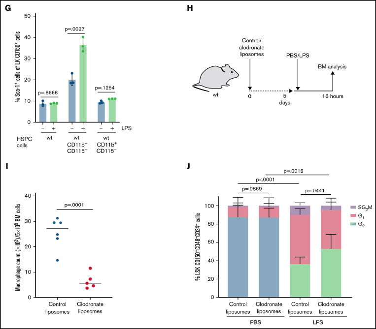
Systemic LPS challenge induces HSC activation via CD115+ monocytic cells in BM. (A) Scheme indicating in vivo transplantation: BM from wt or TLR4−/− mice was transplanted (3 × 10⁶ BM cells) into irradiated TLR4−/− or wt mice. Chimeric mice were treated with PBS (control) or LPS (0.25 mg/kg, 18 hours) 12 weeks after transplantation. (B) Cell cycle analysis (icKi67-Hoechst 33342) of wt and TLR4−/− HSCs (LSK CD150+CD48-CD34-) in forward and reverse chimeric mice after PBS (control) or LPS treatment as indicated in Figure 3A (n = 6). P values refer to G0 phase and were determined by ANOVA Tukey’s post hoc test. (C) Scheme indicating in vitro culturing of sorted HSPCs (LK CD150+) with CD11b+ myeloid cells or CD11b- BM cells in presence of PBS (control) or LPS (100 ng/mL, 18 hours). (D) Relative Sca-1 expression on sorted wt HSPCs (LK CD150+) after in vitro culture for 18 hours in the presence of wt CD11b+ or CD11b- BM cells treated with PBS (control) or LPS (100 ng/mL) as indicated in Figure 3C (n = 3). P values were determined by unpaired t test. (E) Relative Sca-1 expression on sorted wt or TLR4−/− HSPCs (LK CD150+) after in vitro culture for 18 hours in presence of wt or TLR4−/− CD11b+ myeloid BM cells treated with PBS (control) or LPS (100 ng/mL) (n = 3). P values were determined by unpaired t test. (F) Relative Sca-1 expression on sorted wt HSPCs (LK CD150+) after in vitro culture for 18 hours in the presence of wt Gr1+CD115- BM cells, Gr1+CD115+ BM cells, or Gr1-CD115+ BM cells treated with PBS (control) or LPS (100 ng/mL) (n = 3). P values were determined by unpaired t test. (G) Relative Sca-1 expression on sorted wt HSPCs (LK CD150+) after in vitro culture for 18 hours in the presence of wt CD11b+CD115+ BM cells or CD11b+CD115- BM cells treated with PBS (control) or LPS (100 ng/mL) (n = 3). P values were determined by unpaired t test. (H) Scheme indicating in vivo treatment of wt mice with control- or clodronate-loaded liposomes (3.75 g/kg). After 5 days, mice were treated with PBS (control) or LPS (0.25 mg/kg), and subsequent BM analysis was performed 18 hours later. (I) Absolute macrophage count (×103/5×105 BM cells) in BM from mice treated with control- or clodronate-loaded liposomes as indicated in Figure 3H (n = 6). P value was determined by unpaired t test. (J) Cell cycle analysis (icKi67-Hoechst 33342) of HSCs (LSK CD150+CD48-CD34-) from wt mice treated with PBS (control) or LPS (0.25 mg/kg, 18 hours) and pretreated with control- or clodronate-loaded liposomes as indicated in Figure 3H (n = 6). P values refer to G0 phase and were determined by ANOVA Tukey’s post hoc test.
CD11b is a ubiquitous myeloid marker, expressed on granulocytes as well as on cells of the mononuclear phagocyte system, including monocytes, macrophages, and dendritic cells,36 whereas Gr-1 serves as a marker for myeloid differentiation discriminating neutrophils and inflammatory monocytes from resident macrophages. To further characterize the myeloid cell population, we expanded the experimental set-up by discriminating CD115 on Gr-1+, Gr-1-, or CD11b+ cells, exclusively expressed on monocytes and macrophages.37 Remarkably, only CD115+ Gr-1+/− or CD11b+ cells amplified Sca-1 expression levels on HSPCs in response to LPS treatment in vitro (Figure 3F-G). These data suggest that CD115+ monocytic cells are important mediators of the indirect LPS effect on HSCs.
To validate this observation in vivo, wt mice were treated with clodronate-loaded liposomes to deplete monocytic cells in the BM (Figure 3H-I).25,38,39 Clodronate accumulates intracellularly, causing metabolic disorder in phagocytic cells to finally induce pro-apoptotic pathways. Indeed, in vivo depletion of monocytic cells partially rescued the LPS-induced cell cycle increase in HSCs (Figure 3J; supplemental Figure 3A), indicating that monocytic cells are important cellular mediators in the HSC response upon inflammatory stress. Collectively, we show that HSCs both directly and indirectly respond to the acute LPS challenge. The data suggest that this increase in proliferation, at least in part, depends on the presence of CD115+ monocytic cells in BM.
Acute LPS exposure depends on IFN signaling to activate HSCs
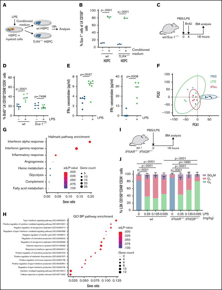
Myeloid and especially monocytic cells function as key players in mediating the immune response to infectious stimuli by accelerating their cytokine secretion.40,41 We hypothesized that cytokine secretion of monocytic cells mediates the indirect response of HSCs to acute LPS exposure. Indeed, both wt and TLR4−/− HSPCs (Lin-CD117+CD150+) cultured in conditioned medium from LPS-treated myeloid-HSPC cocultures showed an increase in Sca-1 expression (Figure 4A-B). The stem cell marker Sca-1 is a well-known IFN-stimulated gene. IFN signaling increases expression of Sca-1 not only on HSCs5,34 but also on common myeloid progenitors and granulocyte-monocyte progenitors that normally do not express Sca-1, shifting these cells into the LSK gate.35 To exclude effects of altered Sca-1 expression levels after LPS challenge on the phenotypic HSC activation (Figure 2C), we omitted Sca-1 in our key analysis. According to our previous data, LK CD150+CD48-CD34- HSCs also transiently entered cell cycle upon LPS treatment (supplemental Figure 3B-D).
Figure 4.
Acute LPS exposure depends on IFN signaling to activate HSCs. (A) Scheme illustrating in vitro culturing of HSPC (LK CD150+) and CD11b+ CD115+ BM cells in presence of LPS (100 ng/mL, 18 hours) and subsequent transfer of conditioned medium to cultured wt or TLR4−/− HSPCs (18 hours). (B) Relative Sca-1 expression in LK CD150+ cells (HSPCs) after in vitro culturing with conditioned medium as indicated in Figure 4A (n = 3). P values were obtained by unpaired t test. (C) Scheme indicating in vivo BrdU uptake (18 mg/kg, 14 hours) and treatment of wt and Sca-1−/− mice with PBS (control) or LPS (0.25 mg/kg) for 18 hours. (D) Fourteen-hour BrdU (18 mg/kg) uptake of HSCs (LK CD150+CD48-CD34-) from wt or Sca-1−/− mice treated with PBS or LPS (0.25 mg/kg) for 18 hours (n = 6). P values were obtained by unpaired t test. (E) IFNα and IFNγ levels determined by ELISA in BM supernatant after in vivo treatment of wt mice with PBS or LPS (0.25 mg/kg, 4 hours) (n = 6). P values were determined by unpaired t test. (F) Principal component analysis and comparison of Illumina Chip array data obtained from HSCs isolated from PBS (control), LPS (0.25 mg/kg), or IFNα (5 × 10⁶U/kg) treated wt mice (18 hours). (G) Hallmark pathway enrichment analysis using EnrichR of 88 differentially expressed genes (DEGs) shared between IFNα– and LPS-treated HSCs. P values were obtained from the online EnrichR program and were not modified. (H) Gene ontology biological processes enrichment analysis using EnrichR of 88 DEGs shared between IFNα– and LPS-treated HSCs. P values were obtained from the online EnrichR program and were not modified. (I) Scheme indicating in vivo treatment of wt and IFNAR−/−IFNGR−/− mice with PBS (control) or LPS (0.25 mg/kg) for 18 hours. (J) Cell cycle analysis (icKi67-Hoechst 33342) of HSCs (LSK CD150+CD48-CD34-) from wt or IFNAR−/−IFNGR−/− mice treated with PBS (control) or LPS (indicated concentrations, 18 hours). Experimental set-up as indicated in Figure 4I (n = 3). P values refer to G0 phase and were determined by ANOVA Tukey’s post hoc test.
Not the increased expression of Sca-1 after IFN signaling as a measure of response of the cells but rather its presence on the cells is essential for the proliferation of HSCs, demonstrated by the HSC’s inability to induce proliferation upon IFN treatment when lacking Sca-15. Interestingly, HSCs lacking Sca-1 also no longer responded to acute LPS treatment in vivo (Figure 4C-D), hinting at a role for IFN signaling in the HSC response to LPS. In line with this observation, BM supernatants from LPS-treated mice contained increased serum levels of IFNα and IFNγ (Figure 4E), both cytokines with an accounted role in HSC activation.5,6 Furthermore, global gene expression profiling of HSCs from mice treated with LPS or IFNα showed a strong overlap in the transcriptional responses (Figure 4F), pointing toward important similarities in the signaling cascades underlying LPS- and IFNα–induced stem cell activation. Strikingly, among the 200 differentially expressed genes, we detected an overlap of 88 genes shared between HSCs of LPS- and IFNα–treated mice, with IFN signaling pathways scoring among the top overlapping biological processes (Figure 4G-H). To determine the role of IFN signaling in the HSC response to LPS, mice lacking receptors for both IFNα and IFNγ (Ifnar−/−Ifngr−/−) were injected with increasing concentrations of LPS (Figure 4I). Upon LPS stimulation, Ifnar−/−Ifngr−/− HSC showed a dose-dependent reduction in proliferation compared with wt HSC, most aberrant at lower concentrations of LPS (Figure 4J), indicating that IFN signaling is part of the LPS-induced response of HSCs in vivo.
LPS-induced cytokine cascade promotes HSC proliferation
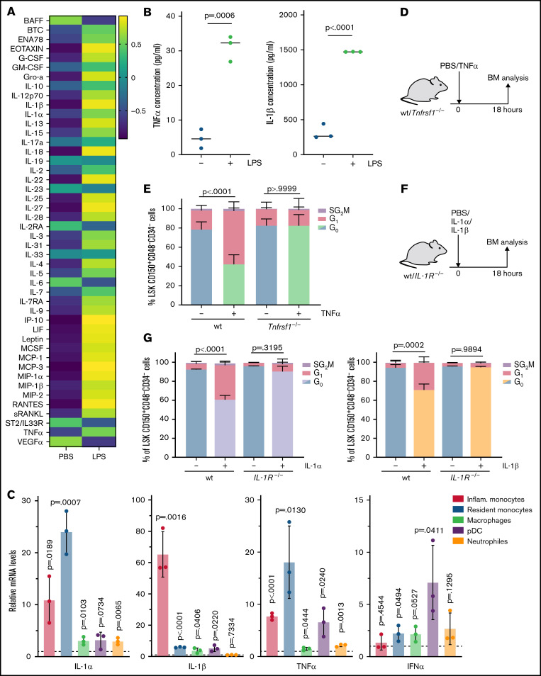
Uncovering that IFN signaling accounts for parts of the HSC response to LPS challenge, other cytokines are most likely involved as well. Thus, to get an overview of the LPS-induced changes in cytokine composition, we performed an unbiased immune response profiling with a ProcartaPlex Array. Remarkably, besides our observed changes in the IFN levels (Figure 4E), LPS induced a strong change in chemokine and cytokine levels in BM (Figure 5A). Among the upregulated cytokines, common key proinflammatory cytokines produced by monocytic cells during inflammation were found, such as TNFα and IL-1, which were confirmed by ELISAs (Figure 5B).
Figure 5.
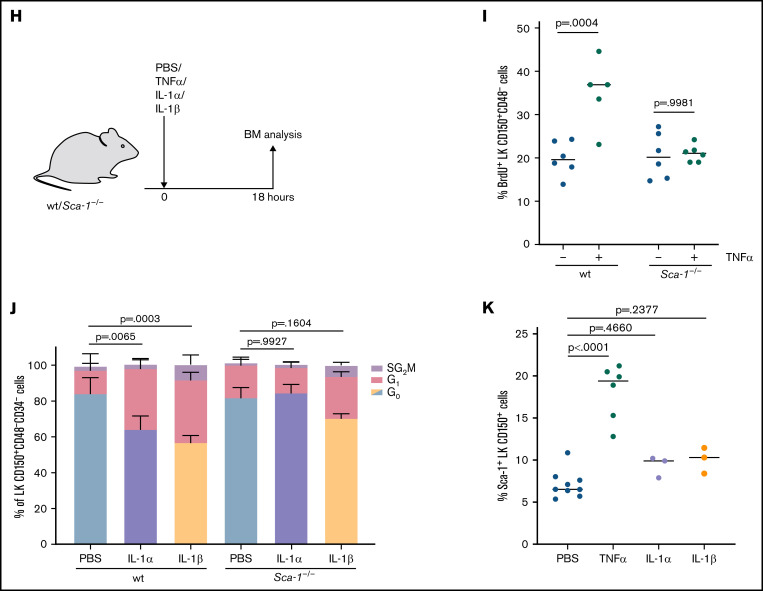
LPS-induced activation of quiescent HSCs depends on Sca-1. (A) Heatmap visualizing the ProcartaPlex Multiplex immunoassay on BM supernatant from wt mice treated with PBS (control) and LPS (0.25 mg/kg, 18 hours) (n = 4). (B) TNFα and IL-1β levels determined by ELISA in BM supernatants after in vivo treatment of wt mice with PBS (control) or LPS (0.25 mg/kg, 4 hours) (n = 3). P values were obtained by unpaired t test. (C) qRT-PCR analyzing cytokine gene expression in indicated cell populations from wt mice (n = 3) treated with PBS (control) or LPS (0.25 mg/kg, 4 hours). Populations are defined as high inflammatory monocytes (CD11b+Cd115+Ly6C+), resident monocytes (CD11b+CD115+Ly6C-), macrophages (Gr1-CD115intF4/80+), and neutrophils (CD11b+Gr1+CD115-). P values were obtained by unpaired t test. (D) Scheme indicating in vivo treatment of wt and Tnfrsf1−/− mice with PBS (control) or TNFα (0.75 mg/kg, 18 hours). (E) Cell cycle analysis (icKi67-Hoechst 33342) of HSCs from wt or Tnfrsf1−/− mice treated with PBS or TNFα (0.75 mg/kg, 18 hours) as indicated in Figure 5D (n = 6). P values refer to G0 phase and were determined by ANOVA Tukey’s post hoc test. (F) Scheme indicating in vivo treatment of wt and IL-1R−/− mice treated with PBS (control) or IL-1α or IL-1β (each 0.25 mg/kg) for 18 hours. (G) Cell cycle analysis (icKi67-Hoechst 33342) of HSCs (LSK CD150+CD48-CD34-) from wt or IL-1R−/− mice treated with PBS (control), IL-1α, or IL-1β (each 0.25 mg/kg, 18 hours) as indicated in Figure 5F (n = 3). P values refer to G0 phase and were determined by ANOVA Tukey’s post hoc test. (H) Scheme indicating treatment of wt and Sca-1−/− mice with PBS (control), TNFα (0.75 mg/kg), IL-1α, or IL-1β (each 0.25 mg/kg) for 18 hours. (I) Fourteen-hour BrdU (18 mg/kg) uptake of HSCs (LK CD150+CD48-CD34-) from wt or Sca-1−/− mice (n = 6) treated with PBS or TNFα (0.75 mg/kg, 18 hours). P values were determined by ANOVA Tukey’s post hoc test. (J) Cell cycle analysis (icKi67-Hoechst 33342) of HSCs (LK CD150+CD48-CD34-) from wt or Sca-1−/− mice treated with PBS, IL-1α, or IL-1β (each 0.25 mg/kg, 18 hours) (n = 3). P values refer to G0 phase and were determined by ANOVA Tukey’s post hoc test. (K) Sca-1 expression on HSPCs (LK CD150+) from wt mice treated with PBS, TNFα (0.75 mg/kg, 18 hours) (n = 6), IL-1α, or IL-1β (each 0.25 mg/kg, 18 hours) (n = 3). P values were determined by ANOVA Tukey’s post hoc test.
To analyze whether monocytes showed increased transcriptional expression of these proinflammatory cytokines in response to LPS treatment, gene expression analysis was performed in inflammatory (CD11b+CD115+Ly6C+) and resident monocytes (CD11b+CD115+Ly6C-) as well as other immune cell populations with a known function in cytokine secretion. Remarkably, IL-1α, IL-1β, and TNFα were highly expressed in both inflammatory and resident monocytes (Figure 5C). In contrast, expression of these cytokines was significantly lower in macrophages, dendritic cells, and neutrophils, which also failed to upregulate Sca-1 expression on HSPCs upon in vitro LPS treatment (supplemental Figure 3E). IFNα was highly expressed in plasmacytoid dendritic cells, known IFNα–producing cells in BM. Thus, these data suggest increased cytokine production by monocytic cells in BM in response to LPS. TNFα, IL-1α, and IL-1β themselves induced a dose-dependent and transient proliferation of HSCs, similar to IFNs (Figure 5D-G). These results not only highlight the complex network of proinflammatory cytokines mediating HSC activation upon LPS challenge but also suggest a role for CD115+ monocytic cells in producing proinflammatory cytokines to mediate the indirect LPS effect on HSCs.
Interestingly, accelerated cell cycle activity upon short-term stimulation with TNFα, IL-1α, and IL-1β was blocked in mice lacking Sca-1 (Figure 5H-J) although IL-1α and IL-1β did not significantly increase Sca-1 expression on HSCs (Figure 5K). These results again suggest that the expression of Sca-1 rather than the upregulation of Sca-1 is indispensable for the proliferation response of HSCs.
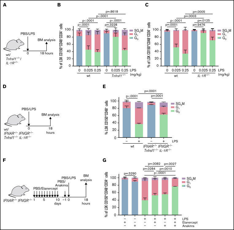
As for LPS and other cytokines, besides HSCs many other BM cells express the receptors for both TNF-α (Tnfrsf1) and IL-1 (IL-1R), opening up the question whether HSCs respond to cytokine exposure via direct signaling downstream of the receptor or via indirect mechanisms. To address this ambiguity, mixed BM chimeras comprising 50% wt and 50% Tnfrsf1−/− or IL-1R−/− BM were injected with the respective cytokine 16 weeks after transplantation (supplemental Figure 3F). Whereas IL-1R−/− HSCs in a wt microenvironment did not show a significant response to IL-1α (supplemental Figure 3H), indicating a direct effect of IL-1α on stem cell proliferation, IL-1R−/− and Tnfrsf1−/− HSCs showed a marginal increase in proliferation upon IL-1β or TNFα exposure in the presence of wt BM, suggesting a combination of direct and indirect effects (supplemental Figure 3G,I). Elucidating their role in the LPS-induced stress response of HSCs, mice lacking either the TNF receptors (Tnfrsfa1−/−) or the IL-1 receptor (IL-1R−/−) were exposed to increasing doses of LPS (Figure 6A). Comparable to the Ifnar−/−Ifngr−/− mice, treatment of Tnfrsf1−/− and IL-1R−/− mice with LPS resulted in a dose-dependent reduction in cell cycle induction in HSCs compared with treatment of wt mice (Figure 6B-C). Together, our data indicate that a proinflammatory signaling cascade including IFNs, TNFα, and IL-1 mediates the indirect HSC response to acute LPS exposure.
Figure 6.
An LPS-induced cytokine cascade promotes HSC proliferation in vivo. (A) Scheme indicating in vivo treatment of wt and Tnfrsf1−/− or IL-1R−/− mice with PBS (control) or increasing concentrations of LPS for 18 hours. (B) Cell cycle analysis (icKi67-Hoechst 33342) of HSCs (LSK CD150+CD48-CD34-) from wt and Tnfrsf1−/− mice treated with PBS (control) or LPS (indicated concentrations, 18 hours) (n = 6). P values refer to G0 phase and were determined by ANOVA Tukey’s post hoc test. (C) Cell cycle analysis (icKi67-Hoechst 33342) of HSCs (LSK CD150+CD48-CD34-) from wt and IL-1R−/− mice treated with PBS or LPS (indicated concentrations, 18 hours) (n = 3). P values refer to G0 phase and were determined by ANOVA Tukey’s post hoc test. (D) Scheme indicating in vivo treatment of IFNAR−/− IFNGR−/− Tnfrsf1−/− IL-1R−/− mice with PBS (control) or LPS (0.25 mg/kg) for 18 hours. (E) Cell cycle analysis (icKi67-Hoechst 33342) of HSCs (LSK CD150+CD48-) from IFNAR−/− IFNGR−/− Tnfrsf1−/− IL-1R−/− mice treated with PBS (control) or LPS (0.25 mg/kg, 18 hours) as indicated in Figure 6D (n = 3). P values refer to G0 phase and were determined by ANOVA Tukey’s post hoc test. (F) Scheme indicating in vivo treatment of IFNAR−/− IFNGR−/− mice with TNFα inhibitor etanercept (5 mg/kg in 100 μL of PBS on days 1-5; 7.5 mg/kg in 150 μL of PBS on days 6-10) and subsequent PBS (control) or LPS (0.25 mg/kg) treatment for 18 hours. IL-1 inhibitor anakinra (2.5 mg/kg) was injected 1 hour before PBS/LPS treatment. (G) Cell cycle analysis (icKi67-Hoechst 33342) of HSCs (LSK CD150+CD48-CD34-) from IFNAR−/− IFNGR−/− mice treated with etanercept/anakinra as indicated in Figure 6F and treated with PBS (control) or LPS (0.25 mg/kg, 18 hours) (n = 3). P values refer to G0 phase and were determined by ANOVA Tukey’s post hoc test.
To validate this hypothesis, we generated quadruple knockout mice lacking the receptors for IFNα, IFNγ, TNFα, and IL-1 (Figure 6D). Compared with wt mice, the LPS-induced activation of HSCs was significantly reduced in these quadruple knockout mice (Figure 6E). To further validate this finding, Ifnar−/−Ifngr−/− mice were treated with the TNFα inhibitor etanercept and the IL-1 antagonist anakinra before exposure to LPS (Figure 6F). As expected, Ifnar−/−Ifngr−/− HSCs showed increased proliferation in response to LPS; however, the effect of LPS on quiescent HSCs was partially rescued upon combined etanercept and anakinra treatment (Figure 6G). Whereas these genetic and inhibitor experiments showed a significant reduction of the LPS response, absence of these 4 major cytokine signaling pathways did not fully rescue the cell cycle induction of HSCs. The cytokine profiling of the BM serum upon LPS already showed that besides IFNα, IFNγ, TNFα, and IL-1, many other cytokines were secreted (Figure 5A). So far, their effects on HSC proliferation mostly remain elusive. Nevertheless, our data highlight the complex nature of the direct and indirect LPS-induced signaling cascade mediating stem cell activation in vivo, in which monocytic BM cells and a complex cytokine signaling network play an important role.
Discussion
In this study, we revealed mechanistic insights into the complex network of proinflammatory cytokines mediating the hematopoietic response to inflammatory stress. Contrary to studies reporting impaired competitive fitness of HSCs during chronic infections,12,16,17 our data suggest that engraftment and differentiation capacity of HSCs are not yet affected during acute and restrained stress conditions. In line with findings by others, part of this response was driven by a direct sensing mechanism, in which HSPCs themselves recognize pathogen-associated molecular patterns via TLR4 expression on their surface.12,28 However, even HSCs lacking TLR4 proliferated upon LPS treatment in presence of wt BM, hinting at indirect signaling through BM immune cells. Indeed, indirect TLR4 signaling in CD115+ monocytic cells resulted in increased cytokine expression in these cells, inducing a complex proinflammatory cytokine cascade in BM mediating HSC activation. Monocytes are part of the first line of defense against invading pathogens,37 and CX3CR1+ monocytes were found to colocalize with HSPCs in close proximity to blood vessels in BM.42 In line with our observations, these monocytes secreted proinflammatory cytokines after sensing bacteria via their TLRs. However, whereas the total HSC number remained unaffected when depleting these monocytes, the number of hematopoietic progenitor cells was diminished, hinting at a role of monocytes in steady-state hematopoiesis. In agreement with these findings, we revealed CD115+ monocytes as key players in HSC activation upon LPS challenge. Therefore, this distinct cell population might orchestrate part of the complex process of emergency hematopoiesis and granulopoiesis in response to inflammatory stimuli.30
Proinflammatory cytokines are well-known mediators of stress hematopoiesis.11,26-28 Immune cytokine profiling of BM from LPS-treated mice uncovered multiple induced proinflammatory cytokines and chemokines potentially involved in the response of HSCs to LPS challenge. By dissecting the proinflammatory cascade downstream of LPS-TLR4 signaling, we uncovered the combined action of not only IFNα and IFNγ but also TNFα, IL-1α, and IL-1β in promoting full HSC activation in response to inflammatory stress. Using an advanced knockout mouse model lacking the receptors for IFNα, IFNγ, TNFα, and IL-1, we not only highlighted the complex signaling pathways resulting in emergency hematopoiesis but also suggested the presence of an even larger proinflammatory cytokine network underlying the response to pathogenic insults.
In contrast to previous reports, this study deepens our understanding of the HSC response during the initial phase of inflammatory stress induced by LPS.12 Pointing out the impact of exposure time to infectious stimuli is of great importance because sustained TLR4 activation is associated with stem cell exhaustion.12,16,17 From a clinical perspective, this is also of high relevance because chronic inflammatory conditions are suggested to promote clonal hematopoiesis of indeterminate potential, a precursor state for hematopoietic malignancies.43,44 Clonal hematopoiesis is closely linked to aging, which by itself displays a chronic inflammatory phenotype building the basis for the emerging field of inflamm-aging.45 In addition, prolonged infectious and inflammatory states have been directly associated with an increased risk of developing hematopoietic malignancies.46,47 Furthermore, BM failure syndromes after allogeneic hematopoietic stem cell transplantation, associated with significant morbidity and mortality, are mediated by chronic stimulation of several inflammatory molecules such as IFNγ and TNFα.48 Thus, these clinical aspects highlight the importance of immediate containment of inflammatory conditions to prevent HSC defects and malignant transformation associated with chronic exposure. In summary, our study revealed detailed mechanistic insights into the interplay of direct and indirect proinflammatory cytokine-induced molecular pathways that jointly orchestrate the complex process of HSC activation in response to acute inflammatory stress.
Supplementary Material
The full text version of the article contains a data supplement.
Acknowledgments
The authors thank the staff at the DKFZ Flow Cytometry Core Facility, the microarray unit of the DKFZ Genomics and Proteomics Core facility, and the DKFZ Animal Laboratory Services for their assistance and expertise.
This work was supported by research funding from the Dietmar Hopp Foundation, Jose Carreras Leukemia foundation, and SFB873 and FOR2033 NicHem funded by the Deutsche Forschungsgemeinschaft (DFG) (M.A.G.E.).
Authorship
Contribution: U.M.D., R.L., S. Sujer, and M.A.G.E. conceived and designed the study; U.M.D., R.L., S. Sujer, Y.D., S. Sood, F.G., A.K., P.W., S.B., H.J.U., S.H., and M.A.G.E. acquired data and analyzed and interpreted data; U.D., R.L., and M.A.G.E. drafted the manuscript; and all authors revised the manuscript for important intellectual content and approved the final version submitted for publication.
Conflict-of-interest disclosure: The authors declare no competing financial interests.
Correspondence: Marieke Alida Gertruda Essers, Im Neuenheimer Feld 280, 69120 Heidelberg, Germany; e-mail: m.essers@dkfz.de.
References
- 1.Mogensen TH. Pathogen recognition and inflammatory signaling in innate immune defenses. Clin Microbiol Rev. 2009;22(2):240-273. [DOI] [PMC free article] [PubMed] [Google Scholar]
- 2.Takeuchi O, Akira S. Pattern recognition receptors and inflammation. Cell. 2010;140(6):805-820. [DOI] [PubMed] [Google Scholar]
- 3.Wilson A, Laurenti E, Oser G, et al. Hematopoietic stem cells reversibly switch from dormancy to self-renewal during homeostasis and repair. Cell. 2008;135(6):1118-1129. [DOI] [PubMed] [Google Scholar]
- 4.Trumpp A, Essers M, Wilson A. Awakening dormant haematopoietic stem cells. Nat Rev Immunol. 2010;10(3):201-209. [DOI] [PubMed] [Google Scholar]
- 5.Essers MA, Offner S, Blanco-Bose WE, et al. IFNalpha activates dormant haematopoietic stem cells in vivo. Nature. 2009;458(7240):904-908. [DOI] [PubMed] [Google Scholar]
- 6.Baldridge MT, King KY, Boles NC, Weksberg DC, Goodell MA. Quiescent haematopoietic stem cells are activated by IFN-gamma in response to chronic infection. Nature. 2010;465(7299):793-797. [DOI] [PMC free article] [PubMed] [Google Scholar]
- 7.Hirche C, Frenz T, Haas SF, et al. Systemic virus infections differentially modulate cell cycle state and functionality of long-term hematopoietic stem cells in vivo. Cell Rep. 2017;19(11):2345-2356. [DOI] [PubMed] [Google Scholar]
- 8.Manz MG, Boettcher S. Emergency granulopoiesis. Nat Rev Immunol. 2014;14(5):302-314. [DOI] [PubMed] [Google Scholar]
- 9.Walter D, Lier A, Geiselhart A, et al. Exit from dormancy provokes DNA-damage-induced attrition in haematopoietic stem cells. Nature. 2015; 520(7548):549-552. [DOI] [PubMed] [Google Scholar]
- 10.Nagai Y, Garrett KP, Ohta S, et al. Toll-like receptors on hematopoietic progenitor cells stimulate innate immune system replenishment. Immunity. 2006;24(6):801-812. [DOI] [PMC free article] [PubMed] [Google Scholar]
- 11.Megías J, Yáñez A, Moriano S, O’Connor JE, Gozalbo D, Gil ML. Direct Toll-like receptor-mediated stimulation of hematopoietic stem and progenitor cells occurs in vivo and promotes differentiation toward macrophages. Stem Cells. 2012;30(7):1486-1495. [DOI] [PubMed] [Google Scholar]
- 12.Takizawa H, Fritsch K, Kovtonyuk LV, et al. Pathogen-induced TLR4-TRIF innate immune signaling in hematopoietic stem cells promotes proliferation but reduces competitive fitness [published correction appears in Cell Stem Cell. 2020;27(1):177]. Cell Stem Cell. 2017;21(2): 225-240.e5. [DOI] [PubMed] [Google Scholar]
- 13.Boettcher S, Manz MG. Sensing and translation of pathogen signals into demand-adapted myelopoiesis. Curr Opin Hematol. 2016;23(1):5-10. [DOI] [PubMed] [Google Scholar]
- 14.Takeda K, Kaisho T, Akira S. Toll-like receptors. Annu Rev Immunol. 2003;21(1):335-376. [DOI] [PubMed] [Google Scholar]
- 15.Yamamoto M, Sato S, Hemmi H, et al. Role of adaptor TRIF in the MyD88-independent Toll-like receptor signaling pathway. Science. 2003; 301(5633):640-643. [DOI] [PubMed] [Google Scholar]
- 16.Chen C, Liu Y, Liu Y, Zheng P. Mammalian target of rapamycin activation underlies HSC defects in autoimmune disease and inflammation in mice. J Clin Invest. 2010;120(11):4091-4101. [DOI] [PMC free article] [PubMed] [Google Scholar]
- 17.Esplin BL, Shimazu T, Welner RS, et al. Chronic exposure to a TLR ligand injures hematopoietic stem cells. J Immunol. 2011;186(9):5367-5375. [DOI] [PMC free article] [PubMed] [Google Scholar]
- 18.Müller U, Steinhoff U, Reis LF, et al. Functional role of type I and type II interferons in antiviral defense. Science. 1994;264(5167):1918-1921. [DOI] [PubMed] [Google Scholar]
- 19.Huang S, Hendriks W, Althage A, et al. Immune response in mice that lack the interferon-gamma receptor. Science. 1993;259(5102):1742-1745. [DOI] [PubMed] [Google Scholar]
- 20.Ito CY, Li CY, Bernstein A, Dick JE, Stanford WL. Hematopoietic stem cell and progenitor defects in Sca-1/Ly-6A-null mice. Blood. 2003;101(2):517-523. [DOI] [PubMed] [Google Scholar]
- 21.Poltorak A, He X, Smirnova I, et al. Defective LPS signaling in C3H/HeJ and C57BL/10ScCr mice: mutations in Tlr4 gene. Science. 1998; 282(5396):2085-2088. [DOI] [PubMed] [Google Scholar]
- 22.Peschon JJ, Torrance DS, Stocking KL, et al. TNF receptor-deficient mice reveal divergent roles for p55 and p75 in several models of inflammation. J Immunol. 1998;160(2):943-952. [PubMed] [Google Scholar]
- 23.Glaccum MB, Stocking KL, Charrier K, et al. Phenotypic and functional characterization of mice that lack the type I receptor for IL-1. J Immunol. 1997;159(7):3364-3371. [PubMed] [Google Scholar]
- 24.Adachi O, Kawai T, Takeda K, et al. Targeted disruption of the MyD88 gene results in loss of IL-1- and IL-18-mediated function. Immunity. 1998; 9(1):143-150. [DOI] [PubMed] [Google Scholar]
- 25.van Rooijen N, Bakker J, Sanders A. Transient suppression of macrophage functions by liposome-encapsulated drugs. Trends Biotechnol. 1997;15(5):178-185. [DOI] [PubMed] [Google Scholar]
- 26.Liu A, Wang Y, Ding Y, Baez I, Payne KJ, Borghesi L. Cutting edge: hematopoietic stem cell expansion and common lymphoid progenitor depletion require hematopoietic-derived, cell-autonomous TLR4 in a model of chronic endotoxin. J Immunol. 2015;195(6):2524-2528. [DOI] [PMC free article] [PubMed] [Google Scholar]
- 27.Massberg S, Schaerli P, Knezevic-Maramica I, et al. Immunosurveillance by hematopoietic progenitor cells trafficking through blood, lymph, and peripheral tissues. Cell. 2007;131(5):994-1008. [DOI] [PMC free article] [PubMed] [Google Scholar]
- 28.Takizawa H, Boettcher S, Manz MG. Demand-adapted regulation of early hematopoiesis in infection and inflammation. Blood. 2012;119(13): 2991-3002. [DOI] [PubMed] [Google Scholar]
- 29.Tak T, van Groenendael R, Pickkers P, Koenderman L. Monocyte subsets are differentially lost from the circulation during acute inflammation induced by human experimental endotoxemia. J Innate Immun. 2017;9(5):464-474. [DOI] [PMC free article] [PubMed] [Google Scholar]
- 30.Boettcher S, Gerosa RC, Radpour R, et al. Endothelial cells translate pathogen signals into G-CSF-driven emergency granulopoiesis. Blood. 2014;124(9):1393-1403. [DOI] [PMC free article] [PubMed] [Google Scholar]
- 31.Shi C, Pamer EG. Monocyte recruitment during infection and inflammation. Nat Rev Immunol. 2011;11(11):762-774. [DOI] [PMC free article] [PubMed] [Google Scholar]
- 32.O’Neill LA, Bowie AG. The family of five: TIR-domain-containing adaptors in Toll-like receptor signalling. Nat Rev Immunol. 2007;7(5):353-364. [DOI] [PubMed] [Google Scholar]
- 33.Kawai T, Takeuchi O, Fujita T, et al. Lipopolysaccharide stimulates the MyD88-independent pathway and results in activation of IFN-regulatory factor 3 and the expression of a subset of lipopolysaccharide-inducible genes. J Immunol. 2001;167(10):5887-5894. [DOI] [PubMed] [Google Scholar]
- 34.Pietras EM, Lakshminarasimhan R, Techner JM, et al. Re-entry into quiescence protects hematopoietic stem cells from the killing effect of chronic exposure to type I interferons. J Exp Med. 2014;211(2):245-262. [DOI] [PMC free article] [PubMed] [Google Scholar]
- 35.Demerdash Y, Kain B, Essers MAG, King KY. Yin and yang: the dual effects of interferons on hematopoiesis. Exp Hematol. 2021;96:1-12. [DOI] [PMC free article] [PubMed] [Google Scholar]
- 36.Sasmono RT, Oceandy D, Pollard JW, et al. A macrophage colony-stimulating factor receptor-green fluorescent protein transgene is expressed throughout the mononuclear phagocyte system of the mouse. Blood. 2003;101(3):1155-1163. [DOI] [PubMed] [Google Scholar]
- 37.Auffray C, Sieweke MH, Geissmann F. Blood monocytes: development, heterogeneity, and relationship with dendritic cells. Annu Rev Immunol. 2009;27(1):669-692. [DOI] [PubMed] [Google Scholar]
- 38.Chow A, Lucas D, Hidalgo A, et al. Bone marrow CD169+ macrophages promote the retention of hematopoietic stem and progenitor cells in the mesenchymal stem cell niche. J Exp Med. 2011;208(2):261-271. [DOI] [PMC free article] [PubMed] [Google Scholar]
- 39.Tyner JW, Uchida O, Kajiwara N, et al. CCL5-CCR5 interaction provides antiapoptotic signals for macrophage survival during viral infection. Nat Med. 2005;11(11):1180-1187. [DOI] [PMC free article] [PubMed] [Google Scholar]
- 40.Fitzgerald-Bocarsly P, Dai J, Singh S. Plasmacytoid dendritic cells and type I IFN: 50 years of convergent history. Cytokine Growth Factor Rev. 2008;19(1):3-19. [DOI] [PMC free article] [PubMed] [Google Scholar]
- 41.Wajant H, Pfizenmaier K, Scheurich P. Tumor necrosis factor signaling. Cell Death Differ. 2003;10(1):45-65. [DOI] [PubMed] [Google Scholar]
- 42.Lee S, Kim H, You G, et al. Bone marrow CX3CR1+ mononuclear cells relay a systemic microbiota signal to control hematopoietic progenitors in mice. Blood. 2019;134(16):1312-1322. [DOI] [PMC free article] [PubMed] [Google Scholar]
- 43.Zhang CRC, Nix D, Gregory M, et al. Inflammatory cytokines promote clonal hematopoiesis with specific mutations in ulcerative colitis patients. Exp Hematol. 2019;80:36-41.e3. [DOI] [PMC free article] [PubMed] [Google Scholar]
- 44.Sezaki M, Hayashi Y, Wang Y, Johansson A, Umemoto T, Takizawa H. Immuno-modulation of hematopoietic stem and progenitor cells in inflammation. Front Immunol. 2020;11:585367. [DOI] [PMC free article] [PubMed] [Google Scholar]
- 45.Kovtonyuk LV, Fritsch K, Feng X, Manz MG, Takizawa H. Inflamm-aging of hematopoiesis, hematopoietic stem cells, and the bone marrow microenvironment. Front Immunol. 2016;7:502. [DOI] [PMC free article] [PubMed] [Google Scholar]
- 46.Kristinsson SY, Landgren O, Samuelsson J, Björkholm M, Goldin LR. Autoimmunity and the risk of myeloproliferative neoplasms. Haematologica. 2010;95(7):1216-1220. [DOI] [PMC free article] [PubMed] [Google Scholar]
- 47.Kristinsson SY, Björkholm M, Hultcrantz M, Derolf AR, Landgren O, Goldin LR. Chronic immune stimulation might act as a trigger for the development of acute myeloid leukemia or myelodysplastic syndromes. J Clin Oncol. 2011;29(21):2897-2903. [DOI] [PMC free article] [PubMed] [Google Scholar]
- 48.Masouridi-Levrat S, Simonetta F, Chalandon Y. Immunological basis of bone marrow failure after allogeneic hematopoietic stem cell transplantation. Front Immunol. 2016;7:362. [DOI] [PMC free article] [PubMed] [Google Scholar]
Associated Data
This section collects any data citations, data availability statements, or supplementary materials included in this article.


