Abstract
Honey bees are important pollinators for the conservation of the ecosystem and agricultural products and provide a variety of products important for human use, such as honey, pollen, and royal jelly. Sacbrood disease (SD) is a devastating viral disease in Apis cerana; an effective preventive measure for SD is urgently needed. In this study, the relationship between the gut microbiome of honey bees and SD was investigated by pyrosequencing. Results revealed that sacbrood virus (SBV)-resistant A. cerana strains harbour a unique acetic acid bacterium, Bombella intestini, and the lactic acid bacteria (LAB) Lactobacillus (unclassified)_uc, Bifidobacterium longum, B. catenulatum, Lactococcus lactis, and Leuconostoc mesenteroides in larvae and Hafnia alvei, B. indicum, and the LAB L. mellifer and Lactobacillus HM215046_s in adult bees. Changes in the gut microbiome due to SBV infection resulted in loss of bacteria that could affect host nutrients and inhibit honey bee pathogens, such as Gilliamella JFON_s, Gilliamella_uc, Pseudomonas putida, and L. kunkeei in A. cerana larvae and Frischella_uc, Pantoea agglomerans, Snodgrassella_uc, and B. asteroides in adult bees. These findings provide important information for the selection of probiotics for A. cerana larvae and adults to prevent pathogenic infections and keep honey bees healthy.
Subject terms: Infectious diseases, Viral infection, Diseases, Microbiology, Microbial communities, Microbiome
Introduction
Honey bees are important insects acting as pollinators as well as producers of various beneficial products, such as honey, propolis, pollen, royal jelly, and bee venom1,2. Sacbrood virus (SBV) causes serious damage to the honey bee industry, especially to Apis cerana, in Southeast Asian countries including Thailand and Vietnam and East Asian countries such as Taiwan, China and Korea3–7. In Korea, SBV was first detected in 20088, and since 2009, sacbrood disease (SD) has resulted in severe losses in Korean apiaries due to colony collapse disorders, similar to that observed in other Southeast Asian countries and China in 1970s9,10; SD continued to be the highest economic loss-causing disease in A. cerana apiaries even in 2021. Therefore, for the development of SBV-resistant honey bee strains, there is a need for research and development of various preventive measures, such as probiotics, to conserve A. cerana and preserve species-diversity among honey bees.
In a previous study, surviving strains of A. cerana from SD-affected apiaries were collected, and breeding techniques were applied to produce two lines of A. cerana with resistance to SBV11, R (with individual immunity) and H (with social immunity). Hybridisation of these two lines was demonstrated to confer high resistance to SBV in South Korea by increasing hygienic behavior and brood survival rate12. Furthermore, SBV is known to exhibit high-affinity binding to the gut of honey bees4,13, and the gut microbiota may influence SBV infection and SD progression14–18. Therefore, in this study, guts from SBV-resistant and -susceptible A. cerana were collected and identification of the microbiome was performed by pyrosequencing. The differences in the microbiota of SBV-resistant and -susceptible strains of A. cerana could provide helpful information on SBV resistance in this species, which could prove helpful in the development of efficient candidates for honey bee probiotics for SD.
Results
Gut microbiome of SBV-susceptible A. cerana
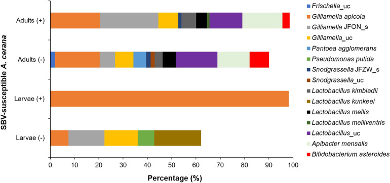
The gut microbiota prevalent in healthy larvae of SBV-susceptible A. cerana included the phyla Proteobacteria and Firmicutes, genera Gilliamella, Pseudomonas, Lachnospiraceae_uc, and Lactobacillus, and species L. kunkeei (19.29%), Gilliamella JFON (uncultured species) (14.81%), Gilliamella_uc (13.71%), G. apicola (7.51%), and Pseudomonas putida (6.81%) (Fig. 1; Table S1). SBV infection resulted in the decline of bacterial diversity in A. cerana larvae (p = 0.0001). The gut microbiome of SD-affected larvae was found to predominantly consist of G. apicola (98.20%) and only a minute percentage of the LAB L. kimbladii (0.02%) (Fig. 1; Tables S1 and S2).
Figure 1.
Gut microbiome of SBV-susceptible Apis cerana. Bacterial species were identified from the gut of sacbrood virus (SBV)-susceptible A. cerana adults and larvae from both healthy (−) and sacbrood disease (SD)-affected (+) honey bees collected from Cheongju province. (“uc” indicates unclassified species; “JFON_s” and “JFZW_s” indicate uncultured species of genera Gilliamella and Snodgrassella, respectively).
The diversity and abundance of the gut microbiome in adult honey bees were higher than those in larvae. The gut microbiota prevalent in A. cerana adults of SBV-susceptible colonies included the phyla Proteobacteria (43.04%), Firmicutes (25.76%), Bacteroidetes (13.24%), and Actinobacteria (8.08%), genera Frischella, Gilliamella, Pantoea, Snodgrassella, Lactobacillus, Apibacter, and Bifidobacterium, and species G. apicola (18.35%), P. agglomerans (5.25%), Frischella_uc (1.97%), Snodgrassella JFZW (uncultured species) (1.84%), A. mensalis (13.24%), and LAB—B. asteroides (8.08%), L. kimbladii (3.27%), and L. mellis (5.35%) (Fig. 1; Table S1). Species composition of the gut microbiota in diseased A. cerana adults was not notably changed in comparison with that in healthy adults (p = 0.3103); the changes were identified by an increase in the abundance of Gilliamella JFON_s, L. kimbladii, and A. mensalis, slight decrease in the abundance of L. mellis, Lactobacillus_uc, and B. asteroides, and the loss of Frischella_uc and P. agglomerans (Fig. 1; Table S1).
Gut microbiome of SBV-resistant A. cerana
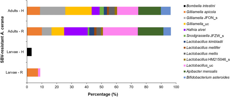
The gut microbiota of SBV-resistant A. cerana larvae of R and H strains belonged to only two phyla, Proteobacteria and Firmicutes. The gut microbiota of SBV-resistant larvae showed a lower diversity than that of SBV-susceptible larvae. The difference of gut microbiota was significant between SBV-resistant H strain and SBV-susceptible strain (p = 0.0001). The major species in the R strain were G. apicola and Lactobacillus_uc, and only Bombella intestini was identified in the H strain (Fig. 2; Table S1).
Figure 2.
Gut microbiome of SBV-resistant A. cerana. The gut microbiota of adults and larvae of two artificially bred A. cerana strains, R (individual immunity) and H (social immunity), was identified and confirmed to be capable to resist SBV (“uc” indicates unclassified species; “JFON_s” and “JFZW_s” indicate uncultured species of genera Gilliamella and Snodgrassella, respectively; HM215046_s indicates the NCBI accession number of Lactobacillus species).
The gut microbiota prevalent in SBV-resistant A. cerana adults in R and H strains comprised the same phyla, namely Proteobacteria, Firmicutes, Bacteroidetes, and Actinobacteria. Further, gut microbiota of SBV-resistant strains was not significantly different from that of susceptible strains, p = 0.6839 and 0.5862 between susceptible strains and resistant R strain and between susceptible strains and resistant H strain, respectively. In comparison with the gut microbiome of SBV-susceptible strains, SBV-resistant strains showed three new species, H. alvei, L. mellifer, and Lactobacillus HM215046_s, and lacked Frischella_uc and P. agglomerans. In addition, Gilliamella JFON_s (17.01%) and Gilliamella_uc (17.66%) were more abundant in the resistant H strain than in the resistant R strain exhibiting Gilliamella JFON_s and Gilliamella_uc at 6.45% and 8.48%, respectively, and those in the susceptible strains were 6.37% and 7.53%, respectively. G. apicola was dominantly detected in the susceptible strain (18.35%). However, it was detected at only 9.98% and 8.71% in R- and H-resistant strain, respectively (Fig. 2; Table S1).
Lactic acid bacteria in A. cerana
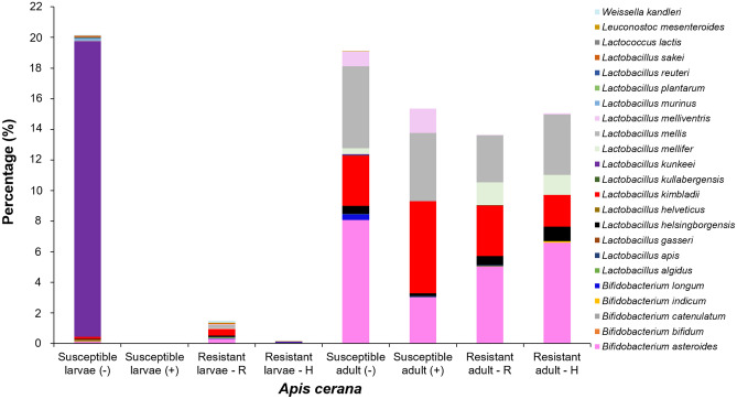
The LAB species L. kunkeei (19.29%) was predominant in the gut of SBV-susceptible A. cerana larvae, while B. asteroides (8.08%), L. mellis (5.35%), and L. kimbladii (3.27%) were identified in the gut of SBV-susceptible adults. There was a major loss of LAB in the gut of SBV infected larvae; only one LAB species, L. kimbladii (0.022%), was detected in the diseased larvae. Similarly, in diseased adults, loss of the LAB species B. longum, L. kullabergensis, L. kunkeei and Leuconostoc mesenteroides and a decrease in the abundance of B. asteroides and L. mellis to 3.02% and 4.44%, respectively, were observed. However, the abundance of L. kimbladii and L. melliventris in diseased adults increased from 3.27% and 0.98% to 6.03% and 1.59%, respectively (Fig. 3; Table S2).
Figure 3.
Lactic acid bacteria (LAB) in the gut of A. cerana. LAB were identified from the gut of adults and larvae of SBV-susceptible and -resistant strains (R and H) of A. cerana. The (−) and (+) symbols indicate healthy and SD-affected honey bees, respectively.
Although the number of LAB in the larvae of SBV-resistant A. cerana was lower than that in susceptible larvae, the difference was not remarkable. B. longum was present in both R and H strains. LAB identified in larvae of only the R strain were L. apis, L. mellifer, L. melliventris, Lactococcus lactis, and Leuconostoc mesenteroides, and LAB in larvae of only the H strain comprised B. catenulatum. There was no notable difference between the SBV-susceptible and -resistant adults in terms of species number and abundance of LAB (Fig. 3; Table S2).
Diversity analysis of gut bacterial communities
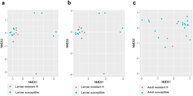
The difference in biodiversity of gut microbiome between SBV-resistant and SBV-susceptible honey bee was shown by alpha diversity with Simpson parameters. The p-value of diversity index between the larvae of SBV-resistant R strain and susceptible strain was 0.0059 (Fig. 4a), and between resistant H strain and susceptible strain was 0.091 (Fig. 4b). Meanwhile, the p-value of comparison between resistant adult and susceptible adult was 0.006 (Fig. 4c). Furthermore, the gut bacterial community in larvae of SBV-resistant strain and susceptible strain (Fig. 5a,b), and in adult bee of the two strains (Fig. 5c) can be distinguished using non-metric multidimensional scaling (NMDS) based on Bray–Curtis indices. The NMDS stress value of resistant larvae strain R and H compare to susceptible strain was 0.0978 and 0.0979, respectively, and the value of adult bee comparison was 0.1059. Alpha diversity and NMDS of other comparisons were shown in Figs. S1, S2, and S3.
Figure 4.
Alpha diversity boxplots designed with Simpson index was used to compare the gut microbiome between SBV-resistant and -susceptible honey bee strain. The comparison of microbiome between larvae of resistant strain R and that of susceptible strain (a), and between resistant strain H and susceptible strain (b). The difference in gut microbiome between adult bee of resistant train H and susceptible strain was also shown (c).
Figure 5.
Gut bacterial communities of larvae and adult bee of SBV-resistant strain compared to SBV-susceptible strain were analysed by the non-metric multidimensional scaling (NMDS). The analysis was done for comparison of microbiome of SBV-resistant larvae strain R (a) and strain H with SBV-susceptible strain (b), and of adult bee of resistant strain H and susceptible strain (c).
Discussion
In this study, we found differences in the microbiota of SBV-susceptible and -resistant A. cerana in Korea for the first time. The microbiota of SBV-resistant A. cerana larvae included Bombella intestini and Lactobacillus_uc, which were absent in SBV-susceptible A. cerana larvae. Members of the core gut community in honey bees have been known to be S. alvi (class: Betaproteobacteria; family: Neisseriales), G. apicola, F. perrara (Gammaproteobacteria; Orbales), Alphaproteobacteria, and Lactobacillae (Firmicutes; Lactobacillaceae)19. Acinetobacter sp., Fructobacillus fructosus, Commensalibacter intestine, and L. kimbladii have been identified in the gut microbiome of A. mellifera in a previous study19–22. Thus, Bombella intestini was a species unique to SBV-resistant A. cerana, when compared with A. mellifera and SBV-susceptible A. cerana. Bombella intestini is an endosymbiotic acetic acid bacterium found in bumble bees (Bombus bimaculatus)23,24, which was also identified in A. cerana for the first time in this study.
SBV has been known as a gut-affinity virus, leading to damage of the gut of larvae and subsequent rotting of the diseased larvae6. One of the preventive measures for honey bee diseases is probiotics, which provide nutrients, protect the attachment of pathogens to cell surfaces, and create an acidic environment that is harsh for the survival of pathogens20,25–27. Therefore, probiotics could have positive effects on the survival of SBV-infected A. cerana. The unique microbiota in SBV-resistant adults in comparison with the SBV-susceptible adults included H. alvei and the LAB L. mellifer, Lactobacillus HM215046_s, and B. indicum. H. alvei was assumed to be an opportunistic pathogen of honey bees28. However, the function of this bacterium in SBV-resistant adult bees remains unclear. Besides, LAB unique to the larvae of SBV-resistant A. cerana compared to those of susceptible strains were B. catenulatum, B. longum, L. apis, L. mellifer, L. melliventris, Lactococcus lactis, and Leuconostoc mesenteroides. LAB produce organic acids, known as anti-microbial metabolites, inhibiting the growth of spoilage and pathogenic microorganisms29. Therefore, microbiota unique to SBV-resistant A. cerana in each developmental stage could be useful for the development of probiotics for disease prevention in honey bee larvae and adults.
LAB play important roles in the production and preservation of honey bee nutrients30. In addition, several studies have showed that LAB was helpful in increasing the size of honey bee colony by increasing the egg-laying capacity of the queen31,32 and resistance to honey bee diseases such as nosemosis31,33,34 and varroosis31. Common LAB in all the larvae and adults of A. cerana in this study were B. asteroides, L. helsingborgensis, L. kimbladii and L. mellis, of which L. helsingborgensis and L. kimbladii were also isolated from A. mellifera and described for the first time in 201422. They showed the ability to produce acid from d-glucose, d-fructose, d-mannose, N-Acetylglucosamine, arbutin, salicin, and d-tagatose22. However, further studies on the usefulness of the LAB species unique to SBV-resistant A. cerana might be important to understand whether they provide practical resistance against SBV infection.
Comparison of the gut microbiota of healthy and SD-affected larvae in SBV-susceptible A. cerana revealed that SD progression resulted in the loss of Gilliamella JFON_s, Gilliamella_uc, Pseudomonas putida, and L. kunkeei. Some of these bacterial species have been identified to have important functions in the gut of honey bees. For instance, Gilliamella spp. are endosymbionts and play a role in degrading polysaccharides that could affect the absorption of host nutrients35, Pseudomonas putida has the ability to degrade neonicotinoid insecticides36, and L. kunkeei is known to inhibit opportunistic pathogens37. Furthermore, SBV infection in adult bees resulted in the loss of Frischella_uc, P. agglomerans, Snodgrassella_uc, B. longum, L. kullabergensis, and L. kunkeei and a decrease in the abundance of B. asteroides and L. mellis in the gut. Frischella spp., such as F. perrara, is known to stimulate the immune system of A. mellifera38, and P. agglomerans acts as a biocontrol agent against fire blight and human facultative pathogens39. G. apicola, L. kimbladii and B. asteroides produce acidic products via fermentation40,41. G. apicola was also demonstrated to have the ability to break down various carbohydrates that are potentially toxic to honey bees42. A. mensalis, Snodgrassella spp., and F. perrara were found to stimulate the immune system of A. mellifera38. However, the mutualistic interaction between the host and the Apibacter spp. remained unclear43,44. Other LAB species are capable of digesting polysaccharides and producing bioactive compounds that possess the potential to act as antimicrobials35,40,45. Therefore, supplements of these bacteria as probiotics could be helpful to maintain a healthy gut environment and provide efficient protection against SD.
There was no common essential microbiota in the larvae. However, in adults, the common microbiota between susceptible and resistant strains were seen; these included G. apicola, Gilliamella JFON_s, Gilliamella_uc, Snodgrassella JFZW_s, L. kimbladii, L. mellis, A. mensalis, and B. asteroids. The common microbiota in adults are vital for survival46. Evidence suggests that differences in the gut microbiota could have originated from differences in the natural environment and the queen lineages of honey bees46–50. The comparison of the gut microbiome from healthy, susceptible adult honey bees collected from two provinces, Jeju and Cheongju, showed that the major bacterial species present in the gut of adults were not notably different (p = 0.493). However, the diversity of the gut microbiome in adults was higher in Cheongju than in Jeju (Fig. S4a). Interestingly, considerable differences were observed in the gut microbiome of larvae collected from the two provinces (p = 0.0436; Fig. S4b). There were also differences observed in the gut microbiome collected in different seasons. The identification of the gut microbiome collected in June and October in Cheongju province was not significantly different, p = 0.5802 and 0.2039 for adult and larvae, respectively. The results revealed that P. agglomerans was predominant in adults in October, while G. apicola was predominant in June (Fig. S5a). In case of the larval gut, L. kunkeei, Gilliamella_uc, and Snodgrassella_uc were found in October and Gilliamella_uc and G. apicola in June (Fig. S5b). Therefore, further studies are required to understand the influence of environmental or natural factors, such as type of pollen and nectar in sampling sites, on the actions of gut bacteria in honey bees and interaction between the host and the bacteria.
In conclusion, the gut microbiota unique to SBV-resistant A. cerana was identified. This study revealed that the SBV infection resulted in the loss of gut microbiota that could affect host nutrients and inhibit certain opportunistic pathogens in A. cerana. The results of this study can provide important information for designing and developing developmental stage- and strain-based probiotics that could be formulated including the essential common bacterial group, specific species in resistant strains, and the important common LAB group. The probiotics could be important to protect A. cerana from pathogenic infections, and for further research on preventing severe SD outbreaks and economic losses to apiaries.
Materials and methods
Selection of apiaries
Three apiaries of SBV-susceptible A. cerana and one of SBV-resistant A. cerana were selected in the regions with 4 clear seasons. Two of these apiaries were free of SD and located in Cheongju and Jeju, at coordinates 36°31′13.1″ N–127°29′29.7″ E, and 33°15′50.3″ N–126°19′53.9″ E, respectively. The third apiary was SD-inflicted and located in Cheongju (35°30′11.7″ N–127°28′16.1″ E) (Table 1), and the fourth one with two SBV-resistant colonies was located in Wanju (35°90′39″ N–127°16′22″ E). Collection of SD-free samples from Cheongju was done in June and October and in March for SD-affected ones. SD-free honey bees from Jeju were collected in June, and SBV-resistant samples from Wanju were collected in May. The two lines of SBV-resistant A. cerana, developed by the National Institute of Agricultural Science, were the R strain with individual immunity against SBV infection and H strain with social immunity against SBV infection. All the colonies of sample collection were confirmed with free of other pathogens using the LiliF™ SBV/KSBV/DWV/BQCV reverse transcription, real-time polymerase chain reaction (RT-qPCR) Kit, LiliF™ ABPV/KBV/IAPV/CBPV RT-qPCR Kit (iNtRON Biotechnology, Inc., Seongnam, Korea), and the POBGEN™ Bee Pathogen Detection Kits (DB-A2 and DB-B2) (POSTBIO Inc., Guri, Korea).
Table 1.
Characteristics and sacbrood disease status of honey bee samples for gut microbiome analysis.
| Characteristics | Region | Sacbrood disease | SBV resistance | Collection time | Hives (n) | Larvae (n) | Adults (n) |
|---|---|---|---|---|---|---|---|
| SBV susceptibility | Cheongju | − | − | June | 6 | 11 | 18 |
| October | 4 | 4 | 8 | ||||
| Jeju | − | − | June | 1 | 1 | 5 | |
| Cheongju | + | − | March | 3 | 5 | 5 | |
| SBV resistance, H strain | Wanju | − | + | May | 1 | 3 | 3 |
| SBV resistance, R strain | 1 | 3 | 3 | ||||
| Total | 16 | 27 | 42 | ||||
SD colonies were identified by clinical signs, which were the irregular capping of combs, the pulled-out larvae out of hives, shrunk or rotten larvae, and larvae inclined to the cell wall. The presence of SBV in larvae and adult bees was confirmed by SBV detection by real-time PCR using specific primers (forward primer: 5′-AGA AGT TTT GGT GTA TAT GCG AGG-3′ and reverse primer: 5′-CTG CGC AGT TTC ATC TTC ATC TTC-3′, and probe 5′-HEX-AAA TAG ACC AAG AAG GGA ATC AGA TAA TCC-BHQ-1-3′)10.
Collection of samples
Larvae and adults of SBV-susceptible and -resistant A. cerana were collected and transported to the laboratory in refrigerated conditions (4 °C). Guts were isolated from both adults and larvae and stored in Eppendorf tubes at − 20 °C before sending them for microbiome analysis by pyrosequencing. Number of larvae and adults collected from each colony varied from one to three depending on the quality of extracted gut and sequencing result. Information of collected samples is shown in Table 1.
Extraction of nucleic acid
The collected gut samples were added to Lysing Matrix E tubes containing ceramic beads (MP Biochemicals GmbH, Eschwege, Germany). After adding PBS (400 μl) to the samples, they were homogenised with a Precellys 24 Tissue Homogenizer (Bertin Instruments, Montigny-le-Bretonneux, France). Nucleic acid was extracted using the FastDNA Spin Kit for Soil (MP Biochemicals GmbH, Eschwege, Germany) following the manufacturer’s instructions.
PCR amplification and illumina sequencing
PCR amplification was performed using primers targeting V3 to V4 regions of the 16S rRNA gene using the extracted DNA. Primers of 341F (5′-TCGTCGGCAGCGTC-AGATGTGTATAAGAGACAG-CCTACGGGNGGCWGCAG-3′) and 805R (5′-GTCTCGTGGGCTCGG-AGATGTGTATAAGAGACAG-GACTACHVGGGTATCTAATCC-3′) were used for amplification of the target gene. Amplification was carried out under the following conditions: initial denaturation at 95 °C for 3 min, followed by 25 cycles of denaturation at 95 °C for 30 s, primer annealing at 55 °C for 30 s, and extension at 72 °C for 30 s, with a final elongation at 72 °C for 5 min. Next, secondary amplification for attaching the Illumina NexTera barcode was performed using the i5 forward primer (5′-AATGATACGGCGACCACCGAGATCTACAC-XXXXXXXX-TCGTCGGCAGCGTC-3′; X indicates the barcode region) and i7 reverse primer (5′-CAAGCAGAAGACGGCATACGAGAT-XXXXXXXX-AGTCTCGTGGGCTCGG-3′). The conditions for secondary amplification were identical to those for the first one, except that the amplification cycle was set to 8.
The PCR product was confirmed by using 2% agarose gel electrophoresis and visualised under a Gel Doc system (BioRad, Hercules, CA, USA). The amplified products were purified with the QIAquick PCR purification kit (Qiagen, Valencia, CA, USA). Equal concentrations of purified products were pooled together and short fragments (non-target products) were removed using the Ampure beads kit (Agencourt Bioscience, Beverly, MA, USA). The quality and product size were assessed using a Bioanalyzer 2100 (Agilent, Palo Alto, CA, USA) using a DNA 7500 chip. Mixed amplicons were pooled and sequencing was carried out at ChunLab, Inc. (Seoul, Korea), using the Illumina MiSeq Sequencing system (Illumina, San Diego, CA, USA) according to the manufacturer’s instruction.
MiSeq pipeline method
Processing of raw reads was conducted via a quality check (QC) and filtering of low quality (< Q 25) reads by Trimmomatic 0.3251. After the QC pass, paired-end sequence data were merged together using PandaSeq52. Primers were then trimmed with ChunLab’s in-house program at a similarity cut off of 0.8. Sequences were denoised using Mothur’s53 pre-clustering program, which merged sequences and extracted unique sequences allowing up to 2 differences between sequences. The EzTaxon database was used for taxonomic assignment using BLAST 2.2.22 and pairwise alignment was used to calculate similarity54,55. Uchime and the non-chimeric 16S rRNA database from EzTaxon were used to detect chimeras on reads that had a best hit similarity rate of less than 97%56. Sequence data were then clustered using CD-Hit and UCLUST, and alpha diversity analysis was carried out57. It is to be noted that in this database, the uncultured phylotype is tentatively given the hierarchal name assigned to the DDBJ/ENA/GenBank accession number with the following suffixes: “_s” (for species), “_g” (genus), “_f” (family), “_p” (phylum)58.
Diversity analysis
Alpha diversity with Simpson index was used to compare gut microbial diversities between group of collected honey bee, and the p value was calculated with Wilcoxon t-test. The difference of gut bacterial community between SBV-resistant and -susceptible honey bee strain was also determined by NMDS distancing by Bray–Curtis index. The analysis was done by using Vegan community ecology package version 2.5-7, and visualized by using ggplot2 package version 3.3.5.
Statistical analysis
Comparison of microbiota in larvae with SD to larvae without SD, in adult with SD to adult without SD, in SBV-resistant larvae to SBV-susceptible larvae, in resistant adult to susceptible adult, and the abundance of different microbiotic strains in different seasons and different regions was done using Mann–Whitney U tests (non-parametric) from program PAST version 4.03. The differences between samples were considered to be significant when p < 0.05.
Supplementary Information
Acknowledgements
We are grateful to Tae Jun Hwang for his invaluable contributions to data acquisition.
Author contributions
Conception and design: Y.S.C.1, D.V.Q., and M.-S.Y., Article writing: Y.S.C.1, B.-R.Y., and A.-T.T., Performed the experiments: B.-R.Y., M.-S.Y., and Y.S.C.1, Data analysis and interpretation: A.-T.T. Y.S.C.2, M.Y.L., B.Y.K., M.S., S.-S.Y., D.V.Q., and Y.S.C.1, and all authors reviewed the manuscript.
Funding
This work was supported by the Animal and Plant Quarantine Agency, the Republic of Korea (Project No. B-1543081-2019-21-03).
Data availability
All data generated or analysed during this study are included in this published article and its Supplementary Information files.
Competing interests
The authors declare no competing interests.
Footnotes
Publisher's note
Springer Nature remains neutral with regard to jurisdictional claims in published maps and institutional affiliations.
These authors contributed equally: Bo-Ram Yun and A-Tai Truong.
Contributor Information
Mi-Sun Yoo, Email: msyoo99@korea.kr.
Dong Van Quyen, Email: dvquen@gmail.com.
Yun Sang Cho, Email: choys@korea.kr.
Supplementary Information
The online version contains supplementary material available at 10.1038/s41598-022-13535-0.
References
- 1.Furst MA, McMahon DP, Osborne JL, Paxton RJ, Brown MJ. Disease associations between honey bees and bumble bees as a threat to wild pollinators. Nature. 2014;506:364–366. doi: 10.1038/nature12977. [DOI] [PMC free article] [PubMed] [Google Scholar]
- 2.Li G, et al. The wisdom of honey bee defenses against environmental stresses. Front. Microbiol. 2018;9:722. doi: 10.3389/fmicb.2018.00722. [DOI] [PMC free article] [PubMed] [Google Scholar]
- 3.Aruna R, Srinivasan MR, Balasubramanian V, Selvarajan R. Complete genome sequence of sacbrood virus isolated from Asiatic honey bee Apis cerana indica in India. VirusDisease. 2018;29:453–460. doi: 10.1007/s13337-018-0490-0. [DOI] [PMC free article] [PubMed] [Google Scholar]
- 4.Hu Y, et al. A comparison of biological characteristics of three strains of Chinese sacbrood virus in Apis cerana. Sci. Rep. 2016;6:37424. doi: 10.1038/srep37424. [DOI] [PMC free article] [PubMed] [Google Scholar]
- 5.Huang WF, et al. Phylogenetic analysis and survey of Apis cerana strain of sacbrood virus (AcSBV) in Taiwan suggests a recent introduction. J. Invertebr. Pathol. 2017;146:36–40. doi: 10.1016/j.jip.2017.04.001. [DOI] [PubMed] [Google Scholar]
- 6.Shan L, et al. Chinese sacbrood virus infection in Asian honey bees (Apis cerana cerana) and host immune responses to the virus infection. J. Invertebr. Pathol. 2017;150:63–69. doi: 10.1016/j.jip.2017.09.006. [DOI] [PubMed] [Google Scholar]
- 7.Nai YS, et al. The seasonal detection of AcSBV (Apis cerana sacbrood virus) prevalence in Taiwan. J. Asia Pac. Entomol. 2018;21:417–422. doi: 10.1016/j.aspen.2018.02.003. [DOI] [Google Scholar]
- 8.Choe SE, et al. Analysis of the complete genome sequence of two Korean sacbrood viruses in the Honey bee, Apis mellifera. Virology. 2012;432:155–161. doi: 10.1016/j.virol.2012.06.008. [DOI] [PubMed] [Google Scholar]
- 9.Choe SE, et al. Prevalence and distribution of six bee viruses in Korean Apis cerana populations. J. Invertebr. Pathol. 2012;109:330–333. doi: 10.1016/j.jip.2012.01.003. [DOI] [PubMed] [Google Scholar]
- 10.Choe SE, et al. Genetic and phylogenetic analysis of South Korean sacbrood virus isolates from infected honey bees (Apis cerana) Vet. Microbiol. 2012;157:32–40. doi: 10.1016/j.vetmic.2011.12.007. [DOI] [PubMed] [Google Scholar]
- 11.Vung NN, et al. Breeding and selection for resistance to sacbrood virus for Apis cerana. Korean J. Apic. 2017;32:345–352. doi: 10.17519/apiculture.2017.11.32.4.345. [DOI] [Google Scholar]
- 12.Vung NN, Choi YS, Kim I. High resistance to sacbrood virus disease in Apis cerana (Hymenoptera: Apidae) colonies selected for superior brood viability and hygienic behavior. Apidologie. 2020;51:61–74. doi: 10.1007/s13592-019-00708-6. [DOI] [Google Scholar]
- 13.Sun L, et al. Preparation and application of egg yolk antibodies against Chinese sacbrood virus infection. Front. Microbiol. 2018;9:1814. doi: 10.3389/fmicb.2018.01814. [DOI] [PMC free article] [PubMed] [Google Scholar]
- 14.Ludvigsen J, Porcellato D, Amdam GV, Rudi K. Addressing the diversity of the honeybee gut symbiont Gilliamella: Description of Gilliamella apis sp. nov., isolated from the gut of honeybees (Apis mellifera) Int. J. Syst. Evol. Microbiol. 2018;68:1762–1770. doi: 10.1099/ijsem.0.002749. [DOI] [PubMed] [Google Scholar]
- 15.Maes PW, Rodrigues PA, Oliver R, Mott BM, Anderson KE. Diet-related gut bacterial dysbiosis correlates with impaired development, increased mortality and Nosema disease in the honeybee (Apis mellifera) Mol. Ecol. 2016;25:5439–5450. doi: 10.1111/mec.13862. [DOI] [PubMed] [Google Scholar]
- 16.Marche MG, Mura ME, Ruiu L. Brevibacillus laterosporus inside the insect body: Beneficial resident or pathogenic outsider? J. Invertebr. Pathol. 2016;137:58–61. doi: 10.1016/j.jip.2016.05.002. [DOI] [PubMed] [Google Scholar]
- 17.Praet J, et al. Large-scale cultivation of the bumblebee gut microbiota reveals an underestimated bacterial species diversity capable of pathogen inhibition. Environ. Microbiol. 2018;20:214–227. doi: 10.1111/1462-2920.13973. [DOI] [PubMed] [Google Scholar]
- 18.Vasquez A, et al. Symbionts as major modulators of insect health: Lactic acid bacteria and honeybees. PLoS ONE. 2012;7:e33188. doi: 10.1371/journal.pone.0033188. [DOI] [PMC free article] [PubMed] [Google Scholar]
- 19.Donkersley P, Rhodes G, Pickup RW, Jones KC, Wilson K. Bacterial communities associated with honeybee food stores are correlated with land use. Ecol. Evol. 2018;8:4743–4756. doi: 10.1002/ece3.3999. [DOI] [PMC free article] [PubMed] [Google Scholar]
- 20.Erban T, et al. Bacterial community associated with worker honeybees (Apis mellifera) affected by European foulbrood. PeerJ. 2017;5:e3816. doi: 10.7717/peerj.3816. [DOI] [PMC free article] [PubMed] [Google Scholar]
- 21.Maddaloni M, Hoffman C, Pascual DW. Paratransgenesis feasibility in the honeybee (Apis mellifera) using Fructobacillus fructosus commensal. J. Appl. Microbiol. 2014;117:1572–1584. doi: 10.1111/jam.12650. [DOI] [PubMed] [Google Scholar]
- 22.Olofsson TC, Alsterfjord M, Nilson B, Butler È, Vásquez A. Lactobacillus apinorum sp. nov., Lactobacillus mellifer sp. nov., Lactobacillus mellis sp. nov., Lactobacillus melliventris sp. nov., Lactobacillus kimbladii sp. nov., Lactobacillus helsingborgensis sp. nov. and Lactobacillus kullabergensis sp. nov., isolated from the honey stomach of the honeybee Apis mellifera. Int. J. Syst. Evol. Microbiol. 2014;64:3109–3119. doi: 10.1099/ijs.0.059600-0. [DOI] [PMC free article] [PubMed] [Google Scholar]
- 23.Li L, et al. Whole-genome sequence analysis of Bombella intestini LMG 28161T, a novel acetic acid bacterium isolated from the crop of a red-tailed bumble bee, Bombus lapidarius. PLoS ONE. 2016;11:e0165611. doi: 10.1371/journal.pone.0165611. [DOI] [PMC free article] [PubMed] [Google Scholar]
- 24.Li L, et al. Bombella intestini gen. nov., sp. nov., an acetic acid bacterium isolated from bumble bee crop. Int. J. Syst. Evol. Microbiol. 2015;65:267–273. doi: 10.1099/ijs.0.068049-0. [DOI] [PubMed] [Google Scholar]
- 25.Patruica S, Mot D. The effect of using prebiotic and probiotic products on intestinal micro-flora of the honeybee (Apis mellifera carpatica) Bull. Entomol. Res. 2012;102:619–623. doi: 10.1017/S0007485312000144. [DOI] [PubMed] [Google Scholar]
- 26.Ptaszynska AA, Borsuk G, Zdybicka-Barabas A, Cytrynska M, Malek W. Are commercial probiotics and prebiotics effective in the treatment and prevention of honey bee nosemosis C? Parasitol. Res. 2016;115:397–406. doi: 10.1007/s00436-015-4761-z. [DOI] [PMC free article] [PubMed] [Google Scholar]
- 27.Sabate DC, Cruz MS, Benitez-Ahrendts MR, Audisio MC. Beneficial effects of Bacillus subtilis subsp. subtilis Mori2, a honey-associated strain, on honeybee colony performance. Probiotics Antimicrob. Proteins. 2012;4:39–46. doi: 10.1007/s12602-011-9089-0. [DOI] [PubMed] [Google Scholar]
- 28.Tian B, Moran NA. Genome sequence of Hafnia alvei bta3_1, a bacterium with antimicrobial properties isolated from honey bee gut. Genome Announc. 2016;4:e00439-00416. doi: 10.1128/genomeA.00439-16. [DOI] [PMC free article] [PubMed] [Google Scholar]
- 29.Rakhmanova A, Khan ZA, Shah K. A mini review fermentation and preservation: Role of lactic acid bacteria. MOJ Food Process. Technol. 2018;6:414–417. doi: 10.15406/mojfpt.2018.06.00197. [DOI] [Google Scholar]
- 30.Janashia I, et al. Beneficial protective role of endogenous lactic acid bacteria against mycotic contamination of honey bee beebread. Probiotics Antimicrob Proteins. 2018;10:638–646. doi: 10.1007/s12602-017-9379-2. [DOI] [PubMed] [Google Scholar]
- 31.Audisio MC. Gram-positive bacteria with probiotic potential for the Apis mellifera L. honey bee: The experience in the northwest of Argentina. Probiotics Antimicrob. Proteins. 2017;9:22–31. doi: 10.1007/s12602-016-9231-0. [DOI] [PubMed] [Google Scholar]
- 32.Audisio MC, Benitez-Ahrendts MR. Lactobacillus johnsonii CRL1647, isolated from Apis mellifera L. bee-gut, exhibited a beneficial effect on honey bee colonies. Benef Microbes. 2011;2:29–34. doi: 10.3920/BM2010.0024. [DOI] [PubMed] [Google Scholar]
- 33.Baffoni L, et al. Effect of dietary supplementation of Bifidobacterium and Lactobacillus strains in Apis mellifera L. against Nosema ceranae. Benef. Microbes. 2016;7:45–51. doi: 10.3920/BM2015.0085. [DOI] [PubMed] [Google Scholar]
- 34.Maes PW, Rodrigues PA, Oliver R, Mott BM, Anderson KE. Diet-related gut bacterial dysbiosis correlates with impaired development, increased mortality and Nosema disease in the honey bee (Apis mellifera) Mol. Ecol. 2016;25:5439–5450. doi: 10.1111/mec.13862. [DOI] [PubMed] [Google Scholar]
- 35.Zheng H, et al. Division of labor in honey bee gut microbiota for plant polysaccharide digestion. Proc. Natl. Acad. Sci. USA. 2019;116:25909–25916. doi: 10.1073/pnas.1916224116. [DOI] [PMC free article] [PubMed] [Google Scholar]
- 36.Zamule SM, et al. Bioremediation potential of select bacterial species for the neonicotinoid insecticides, thiamethoxam and imidacloprid. Ecotoxicol. Environ. Saf. 2021;209:111814. doi: 10.1016/j.ecoenv.2020.111814. [DOI] [PubMed] [Google Scholar]
- 37.Berrios P, et al. Inhibitory effect of biofilm-forming Lactobacillus kunkeei strains against virulent Pseudomonas aeruginosa in vitro and in honeycomb moth (Galleria mellonella) infection model. Benef. Microbes. 2018;9:257–268. doi: 10.3920/BM2017.0048. [DOI] [PubMed] [Google Scholar]
- 38.Emery O, Schmidt K, Engel P. Immune system stimulation by the gut symbiont Frischella perrara in the honey bee (Apis mellifera) Mol. Ecol. 2017;26:2576–2590. doi: 10.1111/mec.14058. [DOI] [PubMed] [Google Scholar]
- 39.Loncaric I, et al. Typing of Pantoea agglomerans isolated from colonies of honey bees (Apis mellifera) and culturability of selected strains from honey. Apidologie. 2009;40:40–54. doi: 10.1051/apido/2008062. [DOI] [Google Scholar]
- 40.Bottacini F, et al. Bifidobacterium asteroides PRL2011 genome analysis reveals clues for colonization of the insect gut. PLoS ONE. 2012;7:e44229. doi: 10.1371/journal.pone.0044229. [DOI] [PMC free article] [PubMed] [Google Scholar]
- 41.Zheng H, Powell JE, Steele MI, Dietrich C, Moran NA. Honeybee gut microbiota promotes host weight gain via bacterial metabolism and hormonal signaling. Proc. Natl. Acad. Sci. USA. 2017;114:4775–4780. doi: 10.1073/pnas.1701819114. [DOI] [PMC free article] [PubMed] [Google Scholar]
- 42.Zheng H, et al. Metabolism of toxic sugars by strains of the bee gut symbiont Gilliamella apicola. MBio. 2016;7:e01326-16. doi: 10.1128/mBio.01326-16. [DOI] [PMC free article] [PubMed] [Google Scholar]
- 43.Kwong WK, Moran NA. Cultivation and characterization of the gut symbionts of honey bees and bumble bees: Description of Snodgrassella alvi gen. nov., sp. nov., a member of the family Neisseriaceae of the Betaproteobacteria, and Gilliamella apicola gen. nov., sp. nov., a member of Orbaceae fam. nov., Orbales ord. nov., a sister taxon to the order 'Enterobacteriales' of the Gammaproteobacteria. Int. J. Syst. Evol. Microbiol. 2013;63:2008–2018. doi: 10.1099/ijs.0.044875-0. [DOI] [PubMed] [Google Scholar]
- 44.Kwong WK, Steele MI, Moran NA. Genome sequences of Apibacter spp., gut symbionts of Asian honey bees. Genome Biol. Evol. 2018;10:1174–1179. doi: 10.1093/gbe/evy076. [DOI] [PMC free article] [PubMed] [Google Scholar]
- 45.Niode NJ, Salaki CL, Rumokoy LJM, Tallei TE. Lactic Acid Bacteria from Honey Bees Digestive Tract and Their Potential as Probiotics. Atlantis Press; 2020. pp. 236–241. [Google Scholar]
- 46.Martinson VG, Moy J, Moran NA. Establishment of characteristic gut bacteria during development of the honeybee worker. Appl. Environ. Microbiol. 2012;78:2830–2840. doi: 10.1128/AEM.07810-11. [DOI] [PMC free article] [PubMed] [Google Scholar]
- 47.Choi SP, et al. Oral administration of Lactobacillus plantarum CJLP133 and CJLP243 alleviates birch pollen-induced allergic rhinitis in mice. J. Appl. Microbiol. 2018;124:821–828. doi: 10.1111/jam.13635. [DOI] [PubMed] [Google Scholar]
- 48.Dai P, et al. The herbicide glyphosate negatively affects midgut bacterial communities and survival of honey bee during larvae reared in vitro. J. Agric. Food. Chem. 2018;66:7786–7793. doi: 10.1021/acs.jafc.8b02212. [DOI] [PubMed] [Google Scholar]
- 49.Seo PJ, et al. Natural variation in floral nectar proteins of two Nicotiana attenuata accessions. BMC Plant Biol. 2013;13:101. doi: 10.1186/1471-2229-13-101. [DOI] [PMC free article] [PubMed] [Google Scholar]
- 50.Shimomura K, et al. Identification of the plant origin of propolis from Jeju Island, Korea, by observation of honeybee behavior and phytochemical analysis. Biosci. Biotechnol. Biochem. 2012;76:2135–2138. doi: 10.1271/bbb.120580. [DOI] [PubMed] [Google Scholar]
- 51.Bolger AM, Lohse M, Usadel B. Trimmomatic: A flexible trimmer for Illumina sequence data. Bioinformatics. 2014;30:2114–2120. doi: 10.1093/bioinformatics/btu170. [DOI] [PMC free article] [PubMed] [Google Scholar]
- 52.Masella AP, Bartram AK, Truszkowski JM, Brown DG, Neufeld JD. PANDAseq: Paired-end assembler for illumina sequences. BMC Bioinformatics. 2012;13:31. doi: 10.1186/1471-2105-13-31. [DOI] [PMC free article] [PubMed] [Google Scholar]
- 53.Schloss PD, et al. Introducing mothur: Open-source, platform-independent, community-supported software for describing and comparing microbial communities. Appl. Environ. Microbiol. 2009;75:7537–7541. doi: 10.1128/AEM.01541-09. [DOI] [PMC free article] [PubMed] [Google Scholar]
- 54.Altschul SF, Gish W, Miller W, Myers EW, Lipman DJ. Basic local alignment search tool. J. Mol. Biol. 1990;215:403–410. doi: 10.1016/S0022-2836(05)80360-2. [DOI] [PubMed] [Google Scholar]
- 55.Myers EW, Miller W. Optimal alignments in linear space. Comput. Appl. Biosci. 1988;4:11–17. doi: 10.1093/bioinformatics/4.1.11. [DOI] [PubMed] [Google Scholar]
- 56.Edgar RC, Haas BJ, Clemente JC, Quince C, Knight R. UCHIME improves sensitivity and speed of chimera detection. Bioinformatics. 2011;27:2194–2200. doi: 10.1093/bioinformatics/btr381. [DOI] [PMC free article] [PubMed] [Google Scholar]
- 57.Edgar RC. Search and clustering orders of magnitude faster than BLAST. Bioinformatics. 2010;26:2460–2461. doi: 10.1093/bioinformatics/btq461. [DOI] [PubMed] [Google Scholar]
- 58.Kim OS, et al. Introducing EzTaxon-e: A prokaryotic 16S rRNA gene sequence database with phylotypes that represent uncultured species. Int. J. Syst. Evol. Microbiol. 2012;62:716–721. doi: 10.1099/ijs.0.038075-0. [DOI] [PubMed] [Google Scholar]
Associated Data
This section collects any data citations, data availability statements, or supplementary materials included in this article.
Supplementary Materials
Data Availability Statement
All data generated or analysed during this study are included in this published article and its Supplementary Information files.


