Summary:
Chylorrhea, a chylous (white viscid) discharge from the external urethral penile opening, is unrelated to urination and is a rare presentation of lymphangioma circumscriptum in the penis that can significantly impair a patient’s quality of life and psychological well-being. Herein, we present a case of recurrent chylorrhea that was detected using diagnostic tools and treated using the deep external pudendal artery perforator flap in the inner thigh, which is extremely rich in lymphatic vessels and lymph nodes.
Chyluria is a primary chylous (white milky viscid) disorder that is observed during urination, whereas chylorrhea refers to continuous chylous discharge from a urethral opening that is unrelated to urination. Chylorrhea is an extremely rare condition, which particularly occurs due to congenital causes, such as lymphangioma circumscriptum (LC). LC is a rare lymphatic channel anomaly that is presented by multiple thin translucent vesicles, which are most commonly noted in the trunk, axilla, thigh, buttocks, and oral cavity. LC rarely occurs on the penis, which has been reported in approximately 30 cases up to 2018. The discharge can impair the patient’s quality of life, and the lymphatic vesicles may rupture and become a site for bacterial infection, thereby causing cellulitis or lymphangitis.1–4
Conservative management, cryotherapy, microlymphatic venous shunt, sclerotherapy, electrodesiccation, electrocoagulation, and laser coagulation are some of the numerous treatment options available for penile LC. Medical care for LC is not yet established; however, surgical excision is the treatment of choice for LC.4,5
CASE REPORT
A 9-year-old boy presented with vesicular lesions on the penile skin with milky discharge from the skin and urethral opening. The vesicles started to appear 1 year previously and did not involve the glans but increased in number and size. A punch biopsy performed by a dermatologist revealed LC. After 2 weeks, the patient started complaining of chylorrhea as well as an increased size and number of vesicles. No other symptoms or difficulty in urination were encountered. He had no history of trauma or surgery on the abdomen, genitalia, or pelvic area; furthermore, he had no family history or neighbors with cases of filariasis, and so on.
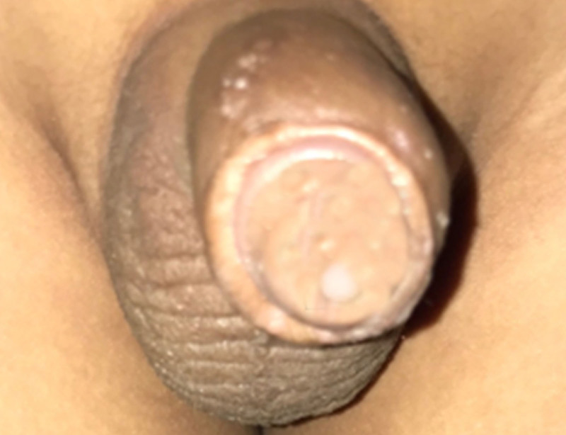
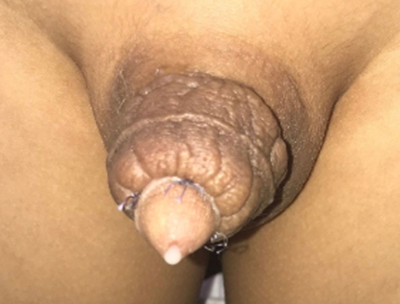
A local examination revealed a vesicular lesion over the penile skin. Gross abnormality was not observed in the scrotum or testis. The white-cloudy copious and continuous chylous discharge was easily visible from the skin vesicles and urethral opening. The patient’s clothes were soaked within half an hour, thereby forcing the patient to wear a diaper, which was changed 2–3 times in 24 hours (Figs. 1, 2).
Fig. 1.
Preoperative view showing the LC lesion on the penile skin with chylus discharge and chylorrhea from urethral opening; the scrotum showed a normal appearance.
Fig. 2.
Postoperative scrotal septal flap showing the presence of recurrent, lymphangioma lesion in transferred scrotal flap and chylorrhea from the urethra.
The laboratory examination and ultrasonography revealed no other abnormalities in the abdomen, pelvis, testis, and scrotum.
Fluid analysis from the urethral and penile skin discharge suggested lymphocytic infiltrate, which was consistent with chylous fluid, and a negative Wuchereria bancrofti antigen.
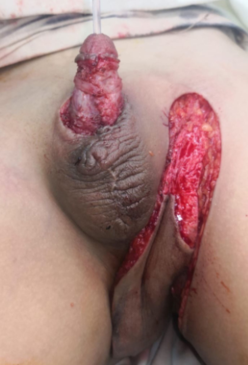
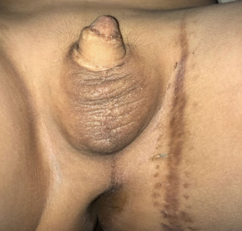
Under general anesthesia, the penile skin was excised to the level of Buck fascia, and reconstruction was performed using a scrotal septal flap to wrap the penis (Fig. 2). The patient had a good early postoperative period, and urethral discharge stopped, with excellent flap survival. Unfortunately, after approximately 5 weeks, new vesicles and chylous discharge started to appear on the scrotal flap and urethral opening. The remaining original scrotal skin was healthy without any clinical changes. Another surgery was performed to remove the lymphoangiomatous scrotal skin. The concept of vascularized lymphatic vessels and lymphatic system flaps from areas rich in lymph nodes and vessels6 was used in this case to treat recurrent penile LC in the upper inner part of the thigh, which is extremely rich in lymphatic vessels that are directly attached to groin lymph nodes. Thus, a deep external pudendal artery perforator flap7 was used to cover the penis. The width of the flap was customized to the penile length when erected, and its length was customized to wrap around the penile girth (Fig. 3). The thin suprafascial flap was elevated, and the proximal part of the flap was de-epithelialized for it to be tunneled to the penile defect; however, it was left as a pedicle flap to maintain its attachment to the original lymphatic system (3). The postoperative follow-up was excellent, where healing was observed in both the donor and recipient sites and chylorrhea was stopped. In the 3-year follow-up, no recurrences were observed and a thin pliable flap was noted (Fig. 4).
Fig. 3.
Deep external pudendal artery perforator flap was elevated and proximal part of the flap was de-epithelialized before tunneling it to the penile defect.
Fig. 4.
Late postoperative results after 3 years showing good healing of the deep external pudendal artery perforator flap with no recurrence; well-healed concealed donor site of deep external pudendal artery perforator flap in the groin crease.
DISCUSSION
Lymphangiomas are hamartomatous lymphatic system malformations that are most commonly found in the head and neck, of which two-thirds are found by 2 years of age. Penile lymphangiomas are underreported because of their hidden site, and they are often unnoticed not only by the physician but also by the patient. Additionally, because of multiple factors, such as misdiagnosis and mistreatment as genital warts, molluscum contagiosum, or gonorrhea, LC has been reported in approximately 30 cases up to 2018.4,8,9
Based on the depth and size of the lymphatic vessel, lymphangiomas are classified into LC (most superficial), cavernous lymphangioma, and cystic hygroma (most deep). Furthermore, based on the causes, they are classified into congenital lymphangiomas, which result from fetal lymph vessels that fail to involute and/or join with the central lymphatic system, and acquired lymphangiomas, which result from trauma, certain infections (cellulitis, neoplastic disease, tuberculosis, and filariasis), radiotherapy, pregnancy, scleroderma, severe phimosis, or sexually transmitted diseases.10
Penile lymphangioma presents with chylous discharge from the urethra, known as chyluria or chylorrhea.10 Chylorrhea leads to psychological problems due to fluid leakage that may affect self-esteem by forcing the patient to wear diapers.
Surgical excision with radical penile skin excision is the treatment of choice.4 However, other treatment modalities, including conservative treatment, laser, radiation, cryotherapy, sclerotherapy, and cautery, have higher recurrence rates, recurrent cellulitis, and lymph fluid leakage.5,10,11 In a review study,4 of 30 patients with LC in penis, 12 were treated via radical excision without recurrence, two were treated via local excision with recurrence (one recurred three times), one was treated using electrocoagulation, four patients were treated using conservative treatment with no new lesion or complication, three patients refused surgery, one patient was treated via four sessions of laser coagulation, one was treated using diathermy coagulation, and six had no history of treatment.
Several reconstructive options are available for penile coverage, including skin graft, scrotal, and local flaps at one or multiple stages.12–14 One of the most important plastic surgery principles in tissue loss is replacing it with a similar tissue, thus preputial skin and local-regional flaps are preferred. The use of skin graft is debatable as the penis should be at full erection when the graft and dressing are applied to prevent graft wrinkling and contracture, penile laxity, which would lead to graft rejection; furthermore, its long-term effect on penile growth is considered. Additionally, the scrotum and local flaps are not always available, particularly with previous surgical procedures. Thus, the donor tissue should most closely replicate the native tissue in function and cosmetic appearance.
In this case, the penile skin was removed, and the coverage was performed using a scrotal flap, and LC recurred on the transferred scrotal flap; thus, a flap coverage with good lymphatic vessels and lymph node for chylous fluid drainage was considered. Therefore, a flap from an area rich in lymphatic vessels and nodes, the groin area and medial aspect of the thigh, was chosen. A deep external pudendal artery perforator flap15 with a pedicle based in the groin was used with excellent function drainage of lymphatic fluid with the normal appearance of the penis. After the long-term 3-year follow-up, no recurrence was observed in the penis or the scrotum.
CONCLUSIONS
Chylorrhea is a rare lymphatic abnormality that may impair the patient’s quality of life. The treatment of choice is radical excision of the affected skin; however, reconstruction using flaps from areas rich in lymphatic vessels and nodes may be an effective solution with good results.
This case report is presented to increase awareness and highlight this extremely rare condition in its presentation, diagnosis, risks, and treatment.
ACKNOWLEDGMENT
We acknowledge all the staff members of Plastic Surgery Department, Faculty of Medicine, Al-Azhar University, Al-Hussain University Hospital, Cairo, Egypt.
ETHICAL APPROVAL STATEMENT
The procedures performed in this study were in accordance with the ethical standards of the institutional research committee (Al-Azhar faculty of medicine ethical committee, Cairo, Egypt) and with the 1964 Helsinki declaration and its later amendments or comparable ethical standards.
PATIENT CONSENT STATEMENT
The patient provided written consent for the use of his image.
Footnotes
Published online 16 June 2022.
Disclosure: The authors have no financial interest to declare in relation to the content of this article.
REFERENCES
- 1.Banadji H, Wahyudi I, Situmorang G, et al. Chylorrhea of external genitalia. J Case Rep. 2017;7:33–36. [Google Scholar]
- 2.Swanson DL. Genital lymphangioma with recurrent cellulitis in men. Int J Dermatol. 2006;45:800–804. [DOI] [PubMed] [Google Scholar]
- 3.Gloviczki P, Noel AA. Lymphatic reconstructions. In: Rutherford RB, ed. Rutherford’s Vascular Surgery. 5th ed. Philadelphia: WB Saunders Company; 2000:2159–2174. [Google Scholar]
- 4.Macki M, Anand SK, Jaratli H, et al. Penile lymphangioma: review of the literature with a case presentation. Basic Clin Androl. 2019;29:1. [DOI] [PMC free article] [PubMed] [Google Scholar]
- 5.Bauer BS, Kernahan DA, Hugo NE. Lymphangioma circumscriptum–a clinicopathological review. Ann Plast Surg. 1981;7:318–326. [DOI] [PubMed] [Google Scholar]
- 6.Chen W, McNurlen M, Ding J, et al. Vascularized lymph vessel transfer for extremity lymphedema—is transfer of lymph node still necessary? Int Microsurg J. 2019;3:1. [Google Scholar]
- 7.Abd Almoktader MA. Deep external pudendal artery perforator (DEPAP) flap with scrotal septal flap phalloplasty: a single-stage sensate phallus. Eur J Plast Surg. 2021;44:345–354. [Google Scholar]
- 8.Ferris DO, Holmes CL. Extensive lymphangioma of scrotum, penis and adjacent areas; report of a case. J Urol. 1947;58:453–457. [DOI] [PubMed] [Google Scholar]
- 9.Ferro F, Spagnoli A, Villa M, et al. A salvage surgical solution for recurrent lymphangioma of the prepuce. Br J Plast Surg. 2005;58:97–99. [DOI] [PubMed] [Google Scholar]
- 10.Adikari S, Philippidou M, Samuel M. A rare case of acquired lymphangioma circumscriptum of the penis. Int J STD AIDS. 2017;28:205–207. [DOI] [PubMed] [Google Scholar]
- 11.Demir Y, Latifoğlu O, Yenidünya S, et al. Extensive lymphatic malformation of penis and scrotum. Urology. 2001;58:106. [DOI] [PubMed] [Google Scholar]
- 12.Mirza B, Ijaz L, Saleem M, et al. Different modalities used to treat concurrent lymphangioma of chest wall and scrotum. J Cutan Aesthet Surg. 2010;3:189–190. [DOI] [PMC free article] [PubMed] [Google Scholar]
- 13.Thakar HJ, Dugi DD, III. Skin grafting of the penis. Urol Clin North Am. 2013;40:439–448. [DOI] [PubMed] [Google Scholar]
- 14.Alwaal A, McAninch JW, Harris CR, et al. Utilities of split-thickness skin grafting for male genital reconstruction. Urology. 2015;86:835–839. [DOI] [PMC free article] [PubMed] [Google Scholar]
- 15.Abd Almoktader MA. Anteriorly based pudendal thigh flap for scrotal reconstruction based on the deep external pudendal artery (DEPA) flap. Eur J Plast Surg. 2016;39:107–112. [Google Scholar]


