Abstract
Portal hypertension is a major complication of cirrhosis characterized by a pathological hepatic venous pressure gradient (HVPG) ≥ 5 mmHg. The structural changes observed in the liver leading to intrahepatic vascular resistance and, consequently, portal hypertension appear in the early stages of cirrhosis. Clinically significant portal hypertension (HVPG ≥ 10 mmHg) is associated with several clinical consequences, such as ascites, hyponatremia, gastroesophageal variceal bleeding, hepatorenal syndrome, cardiopulmonary complications, adrenal insufficiency, and hepatic encephalopathy. The diagnosis and management of these complications depend on their early identification and treatment. Regarding ascites, diuretics are a useful treatment, although plasma sodium levels must be properly controlled to avoid hyponatremia. The management of hypovolemic hyponatremia usually consists in stopping diuretics and the administration of volume. On the contrary, hypervolemic hyponatremia is managed with fluid and sodium restriction. Transjugular intrahepatic portosystemic shunt (TIPS) should be considered in patients with refractory ascites. Primary prophylaxis of variceal bleeding should be based mainly on non-selective beta-blockers. Management of acute gastroesophageal variceal bleeding includes vasoactive drugs and endoscopic band ligation and, in patients at high risk of failure and rebleeding, preemptive use of TIPS. Secondary prophylaxis with a combination of non-selective beta-blockers and endoscopic band ligation is the treatment of choice. This article focuses on the management of ascites, hyponatremia, and gastroesophageal variceal bleeding.
Key Points
| Ascites, hyponatremia, and variceal bleeding are complications associated with advanced chronic liver disease and portal hypertension. |
| Ascites should be managed with dietary sodium intake restriction and diuretics (spironolactone alone or in combination with furosemide). In patients with refractory ascites, transjugular intrahepatic portosystemic shunt (TIPS) should be considered. |
| Hypovolemic hyponatremia is rare and should be managed by stopping diuretics and expanding the plasma volume with isotonic solutions. Hypervolemic hyponatremia should be managed with fluid and dietary sodium restriction. |
| Primary prophylaxis of acute gastroesophageal variceal bleeding should be based on non-selective beta blockers (NSBB). Preemptive use of TIPS in patients with variceal bleeding and high risk of failure and rebleeding should be considered. Secondary prophylaxis should be based on NSBB plus endoscopic band ligation. |
Introduction
Portal hypertension is a major complication of cirrhosis characterized by a pathological increase in the portal pressure gradient. This gradient is defined as the pressure difference between the portal vein (inflow to the liver) and the inferior vena cava (outflow from the liver). A gradient of less than 5 mmHg is considered normal, while ≥ 5 mmHg defines portal hypertension. Clinical complications develop when there is a gradient ≥ 10 mmHg, which defines the so-called clinically significant portal hypertension. Medical management should be aimed at avoiding the progression of liver disease and preventing decompensation [1]. The development of portal hypertension is due in part to the formation of scar tissue that leads to an increase in intrahepatic vascular resistance [1].
Clinically significant portal hypertension is associated with several clinical consequences. Once portal hypertension is established, compensatory mechanisms develop, such as the growth of collateral circulation and an increase in the plasma concentration of vasodilator substances (nitric oxide, prostacyclin, and endocannabinoids) that attempt to reverse the increase in intrahepatic resistance. However, these mechanisms are insufficient and, in fact, produce a decrease in splanchnic and systemic vascular resistance that results in a decrease in the effective arterial blood volume. The kidneys detect this theoretical decrease in volume and activate compensatory mechanisms such as the renin-angiotensin-aldosterone system, the sympathetic system, and antidiuretic hormone. These mechanisms lead to an increase in sodium and water retention that promote the development of ascites [2–4]. The onset of renal sodium retention and ascites is associated with a decrease in the excretion of free water. The retained water dilutes the internal milieu and produces hyponatremia [5, 6].
In addition to ascites and hyponatremia, other complications of portal hypertension include gastroesophageal variceal bleeding, hepatorenal syndrome, cardiopulmonary complications, adrenal insufficiency, or hepatic encephalopathy, among others. In this article, we will focus on the management of ascites, hyponatremia, and gastroesophageal variceal bleeding.
Diagnosis and Management of Ascites
Ascites is due to cirrhosis in approximately 80% of cases [7]. Approximately 20% of patients with cirrhosis have ascites at diagnosis, and 20% of those with ascites die within the first year of diagnosis [8]. To evaluate patients presenting with ascitic decompensation for the first time, it is necessary to perform a complete diagnostic work-up of liver disease and rule out malignancy and other causes of ascites. A diagnostic paracentesis will determine whether the characteristics of the ascitic fluid are compatible with cirrhosis. Typically, in cirrhotic ascites, a low protein concentration (< 15 g/L) and a serum albumin gradient ≥ 11 g/L are detected. In addition, it is also necessary to rule out spontaneous bacterial peritonitis (SBP), which is characterized by a neutrophil count > 250 cells/mm3. It is important to always culture the ascitic fluid to guide the antibiotic treatment of SBP. In other cases, the ascites culture can be positive but the neutrophil count is below < 250 cells/mm3. This scenario is known as bacterioascites, not SBP, and it does not have a clear pathological significance. The need for antibiotic treatment in these cases should be discussed and individualized according to the isolated organism and the clinical condition of the patient [2, 9, 10].
All patients who have recovered from an episode of SBP should receive long-term antibiotic prophylaxis. Importantly, primary SBP prophylaxis should also be initiated in patients with high-risk criteria (low protein ascites < 1.5 g/L and liver failure with bilirubin > 3 mg/dL). A special mention should be made to point out the risk of renal impairment in patients presenting with SBP. In patients with SBP, besides administering antibiotic therapy, it has been demonstrated that the use of intravenous albumin infusion reduces the incidence of renal impairment and in-hospital mortality in comparison with treatment with an antibiotic alone [11].
Focusing on the management of ascites, the first step in its treatment is to establish a low sodium diet. However, it is controversial how strict the salt restriction diet has to be. In very restrictive low sodium diets (< 5 g/day, < 85 mmol sodium/day), a very high rate of adverse events such as hyponatremia, hepatic encephalopathy or hepatorenal syndrome have been reported. Moreover, this diet is difficult to follow and leads to reduced caloric intake. On the other hand, a moderate salt-restricted diet (5–6.5 g/day) could achieve higher adherence and better results, with quicker disappearance of the ascites and, importantly, fewer adverse events [12, 13].
The second step in the management of ascites is the use of diuretics. There are two families of diuretics that have been shown to be highly effective in the management of ascites in patients with cirrhosis: loop diuretics (furosemide) and potassium-sparing diuretics (spironolactone). Furosemide has a more immediate effect and a response rate of 52%. Its maximum recommended dosage is 160 mg/day. Spironolactone has a slower effect (it takes approximately 3–4 days to appreciate its effects), which requires caution when adjusting the dose. It has a response rate of 95%, and the maximum recommended dosage is 400 mg/day [14–16]. It is recommended to start with a combination of spironolactone and furosemide, adjusting their doses according to the response achieved [2]. The goals to be achieved with treatment will vary depending on whether the patient presents with ascites and edema, in which case a reduction of 1 kg of weight per day is intended, or if the patient presents only with isolated ascites, in which case the goal is a reduction of 0.5 kg per day. These goals are set to avoid severe renal vasoconstriction and protect renal function. Spot urine sodium/potassium ratio will allow us to measure the degree of treatment response and whether the patient is correctly following a hyposodic diet. A random spot urine sodium:potassium ratio between 1.8 and 2.5 has a sensitivity of 87.5%, specificity of 56–87.5%, and accuracy of 70–85% in predicting a 24-h urinary sodium excretion of 78 mmol/day (the aim of diuretic therapy is to ensure that urinary sodium excretion exceeds 78 mmol/day) [2].
Despite treatment, approximately 10–20% of patients will develop refractory ascites. Refractory ascites can be diuretic-resistant ascites or diuretic-intractable ascites. Diuretic-resistant ascites is ascites that persists despite a low-sodium diet and a maximum dosage of diuretics (spironolactone 400 mg and furosemide 160 mg per day). Diuretic-intractable ascites appears when the maximum dose of diuretics cannot be reached because the patient develops adverse effects earlier, such as hyponatremia or encephalopathy [2]. In these situations, it is recommended to perform a large volume paracentesis to reduce tension in the abdomen. To minimize the risk of adverse events (injury of the inferior epigastric artery and injury of the liver and spleen), the point of puncture should be at least 8 cm from the middle line and 5 cm above the symphysis in the left lower abdominal quadrant. It is important to be aware that the extraction of more than 5 L of ascites at one time can lead to abrupt hemodynamic changes (circulatory dysfunction), with marked reactivation of the neurohumoural response and increased fluid retention, with consequent renal failure and a risk of hyponatremia [17, 18]. To counteract this, 8 g of albumin must be administered for each liter of ascites removed [19].
Refractory ascites decreases the survival of patients with cirrhosis. The probability of survival is 52% at 1 year and 45% at 2 years [20]. Thus, other therapeutic strategies should be considered, such as liver transplantation. However, this strategy is not suitable for all patients due to long waiting lists and transplant contraindications. In patients with refractory ascites without access to liver transplantation, the placement of transjugular intrahepatic portosystemic shunts (TIPS) is a good alternative. Several studies have shown that TIPS provides better survival and control of ascites than paracentesis in patients with refractory ascites [21, 22]. Importantly, it has been proved that the benefit of TIPS in the setting of ascites is greater in those patients with a lower severity of ascites, not meeting the strict criteria of refractory ascites but of recurrent ascites. Thus, currently TIPS placement is indicated for the management of difficult-to-treat recurrent ascites [23].
Management of Hyponatremia
Hyponatremia is a severe complication of ascites and diuretic treatment. It is defined as a decrease in plasma sodium levels below 135 mmol/L. It can be mild (130–135 mmol/L), moderate (125–129 mmol/L), or severe (< 125 mmol/L). Hyponatremia is a relevant and potentially severe complication that can be associated with hepatorenal syndrome, SBP, refractory ascites, hepatic encephalopathy, and increased mortality [5, 6].
There are three types of hyponatremia: euvolemic, hypovolemic, and hypervolemic hyponatremia. Euvolemic hyponatremia is uncommon in patients with cirrhosis and should be managed based on the specific underlying cause. Hypovolemic hyponatremia, which represents 10% of all hyponatremias in patients with cirrhosis, is caused by an excessive loss of both extracellular volume fluid and sodium, either by excessive diuretic therapy or by conditions leading to dehydration (i.e., diarrhea or vomiting). It is characterized by a low serum sodium concentration and hypovolemia that leads to reduced renal perfusion. The management of hypovolemic hyponatremia consists of stopping diuretics and fluid resuscitation expanding the plasma volume with isotonic solutions [24].
Hypervolemic hyponatremia is much more common in cirrhotic patients. It is caused by hypersecretion of vasopressin and enhanced proximal nephron sodium reabsorption with impaired free-water clearance, leading to dilutional hyponatremia with a high absolute amount of sodium. Its management consists of restricting fluids (1–1.5 L/day) and sodium. Careful replenishment with hypertonic sodium chloride should be reserved only for patients with severe acute symptomatic hyponatremia and always with close monitoring to avoid complications such as central pontine myelinolysis [24, 25].
Management of Gastroesophageal Variceal Bleeding
Variceal bleeding is one of the most severe and feared complications of portal hypertension. Gastroesophageal varices appear in 25–35% of patients with cirrhosis, in 40% of compensated cirrhotic patients, and in 85% of decompensated cirrhotic patients. Variceal bleeding has a high mortality rate. Despite applying the gold-standard therapy, 10–15% of patients with acute variceal bleeding experience treatment failure, 21% rebleed, and 24% die during the first 6 weeks [26].
Portal hypertension is the main driver of varices development and variceal bleeding. Gastroesophageal varices develop when the hepatic venous pressure gradient (HVPG) rises over 10 mmHg. Varices needing treatment are large or have high-risk signs and should be managed with prophylactic treatment to prevent bleeding. When the HVPG is 12 mmHg or higher, the risk of variceal bleeding is greatly increased. During acute variceal bleeding episodes, an HVPG of 20 mmHg and above is associated with a high risk of failure and mortality [27].
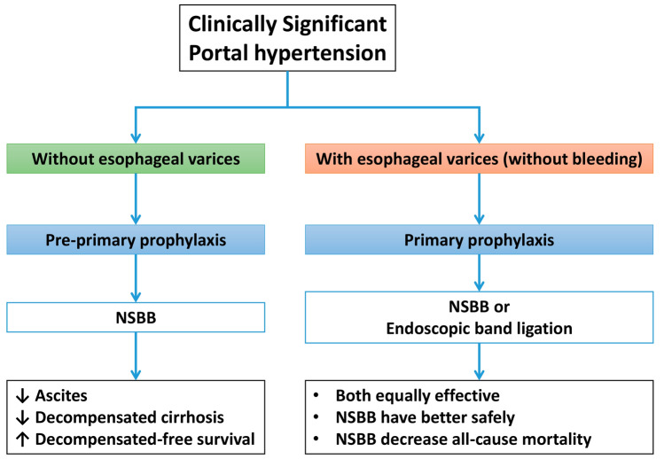
Figure 1 shows an algorithm of prophylaxis for gastroesophageal varices. In compensated patients with clinically significant portal hypertension who have not yet developed varices, long-term prophylactic treatment with noncardioselective beta-blockers (NSBBs) may increase decompensation-free survival, mainly by decreasing the incidence of ascites [28]. In patients who have developed gastroesophageal varices but have not yet presented with bleeding, primary prophylaxis with NSBBs or the use of endoscopic band ligation is recommended. Both have been shown to be equally effective in primary prophylaxis of gastroesophageal variceal bleeding, but only NSBB impacts the natural history of the disease and improves survival with a better safety profile [26, 29]. Table 1 shows a comparison of the dosage and therapeutic goals of beta-blockers and endoscopic band ligation of primary prophylaxis for acute variceal bleeding [27].
Fig. 1.
Prophylaxis of gastroesophageal varices. NSBB noncardioselective beta-blockers
Table 1.
Primary prophylaxis of acute variceal bleeding with beta-blockers
| Therapy | Propranolol | Nadolol | Carvedilol |
|---|---|---|---|
| Recommended dose |
• Begin with 20–40 mg orally twice a day and increase by 20 mg every 2–3 days until reaching the treatment goal. Decrease stepwise if not tolerated • Maximal dosage: 320 mg/day • 160 mg/day in patients with severe ascites |
• Begin with 20–40 mg orally once a day and increase by 20 mg every 2–3 days until reaching the treatment goal. Decrease stepwise if not tolerated • Maximal daily dose: 160 mg/day |
• Start with 6.25 mg once a day • After 3 days, increase to 6.25 mg twice a day • Maximal dose: 12.5 mg/day |
| Therapy goals |
• Resting heart rate 55–60 beats per minute • Maintain systolic blood pressure > 90 mmHg |
• Same as propranolol |
• Systolic blood pressure > 90 mmHg • Heart rate reduction is not used for dose titration |
Regarding the election of NSBB, carvedilol has been demonstrated to have a more potent effect in reducing HVPG and is better tolerated. Also, carvedilol improves outcome and is better at preventing decompensation when compared to the traditional NSBB propranolol and nadolol. In addition, the use of carvedilol has been recently accepted for the prevention of first decompensation in patients with clinically significant portal hypertension, even in the absence of varices [28].
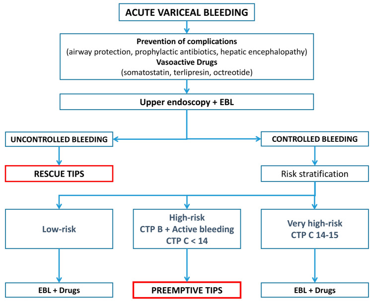
Management of acute variceal bleeding (Fig. 2) includes general management of critically ill patients, careful volume restitution, and antibiotics. Administration of vasoactive drugs such as somatostatin, terlipressin, or octreotide (they have all shown similar efficacy) should be started as soon as possible to decrease the portal pressure. Simultaneously, the prevention of complications such as bronchoaspiration, bacterial infection, hepatic encephalopathy, and acute kidney injury must be ensured. Subsequently, endoscopic evaluation and band ligation should be performed with the aim of controlling the bleeding. If the bleeding cannot be controlled either pharmacologically or endoscopically, the placement of TIPS (rescue TIPS) should be considered [27].
Fig. 2.
Management of acute variceal bleeding. CTP Child–Turcott–Pugh, EBL endoscopic band ligation, TIPS transjugular intrahepatic portosystemic shunt
After controlling the bleeding, the risk of rebleeding must be assessed [27]. Several factors can be used to stratify the risk of rebleeding, but one of the most effective scores to tailor therapy is the Child–Turcott–Pugh (CTP) score and the endoscopic findings. In low-risk patients (CTP A or CTP B without active bleeding at endoscopy), standard secondary prophylaxis with NSBB and endoscopic band ligation is recommended. In high-risk patients (CTP B > 7 + active bleeding or CTP C < 14), preemptive TIPS will be considered as soon as possible with the final aim of preventing failure, rebleeding, and mortality [27, 30, 31]. Finally, in very high-risk patients (CTP C 14–15), the use of preemptive TIPS would be futile if it cannot be followed by liver transplantation, and only secondary prophylaxis with NSBB and band ligation is recommended.
Conclusions
Ascites, hyponatremia, and gastroesophageal variceal bleeding are clinical consequences of portal hypertension. Early identification and the diagnosis of patients at risk is essential to prevent and treat them adequately.
Diagnostic paracentesis is required to determine if ascites is related to portal hypertension rather than cirrhosis, rule out malignancy, and diagnose SBP. Ascites should be managed with dietary sodium intake restriction and diuretics (spironolactone alone or in combination with furosemide). In patients with refractory ascites, TIPS should always be considered.
Accurate assessment of hyponatremia in patients with cirrhosis is always necessary. Hypovolemic hyponatremia should be managed by stopping diuretics and expanding the plasma volume with fluid resuscitation. On the other hand, hypervolemic hyponatremia, the most frequent cause of low sodium in patients with cirrhosis, should be managed with fluid and sodium restriction.
In patients with clinically significant portal hypertension, preprimary prophylaxis with NSBB should be considered, as it may prevent hepatic decompensation. Primary prophylaxis of variceal bleeding with NSBB or band ligation is recommended in patients with high-risk varices. In patients with acute variceal bleeding and a high risk of failure and rebleeding, preemptive TIPS should be considered as early as possible during admission. Secondary prophylaxis for acute variceal bleeding should be based on the combination of NSBB and endoscopic band ligation.
Acknowledgments
The authors would like to thank Fernando Sánchez Barbero PhD on behalf of Springer Healthcare for providing medical writing assistance.
Declarations
Disclosure statement
This article has been published as part of a journal supplement wholly funded by Eisai.
Funding
Medical writing assistance was funded by Eisai Farmacéutica S.A.
Conflict of interest
The authors report no conflict of interest.
Ethics approval
This article is based on previously conducted studies and does not contain any studies with human participants or animals performed by any of the authors.
Consent to participate
Not applicable.
Consent for publication
Not applicable.
Availability of data and material
Not applicable.
Code availability
Not applicable.
Authors’ contributions
All named authors meet the International Committee of Medical Journal Editors (ICMJE) criteria for authorship for this article, take responsibility for the integrity of the work as a whole, and have given their approval for this version to be published.
References
- 1.Simonetto DA, Liu M, Kamath PS. Portal hypertension and related complications: Diagnosis and management. Mayo Clin Proc. 2019;94(4):714–726. doi: 10.1016/j.mayocp.2018.12.020. [DOI] [PubMed] [Google Scholar]
- 2.Aithal GP, Palaniyappan N, China L, Harmala S, Macken L, Ryan JM, et al. Guidelines on the management of ascites in cirrhosis. Gut. 2021;70(1):9–29. doi: 10.1136/gutjnl-2020-321790. [DOI] [PMC free article] [PubMed] [Google Scholar]
- 3.Morali GA, Sniderman KW, Deitel KM, Tobe S, Witt-Sullivan H, Simon M, et al. Is sinusoidal portal hypertension a necessary factor for the development of hepatic ascites? J Hepatol. 1992;16(1–2):249–250. doi: 10.1016/s0168-8278(05)80128-x. [DOI] [PubMed] [Google Scholar]
- 4.Schrier RW, Arroyo V, Bernardi M, Epstein M, Henriksen JH, Rodes J. Peripheral arterial vasodilation hypothesis: a proposal for the initiation of renal sodium and water retention in cirrhosis. Hepatology. 1988;8(5):1151–1157. doi: 10.1002/hep.1840080532. [DOI] [PubMed] [Google Scholar]
- 5.Angeli P, Wong F, Watson H, Gines P, Investigators C. Hyponatremia in cirrhosis: Results of a patient population survey. Hepatology. 2006;44(6):1535–1542. doi: 10.1002/hep.21412. [DOI] [PubMed] [Google Scholar]
- 6.Kim WR, Biggins SW, Kremers WK, Wiesner RH, Kamath PS, Benson JT, et al. Hyponatremia and mortality among patients on the liver-transplant waiting list. N Engl J Med. 2008;359(10):1018–1026. doi: 10.1056/NEJMoa0801209. [DOI] [PMC free article] [PubMed] [Google Scholar]
- 7.Rudler M, Mallet M, Sultanik P, Bouzbib C, Thabut D. Optimal management of ascites. Liver Int. 2020;40(Suppl 1):128–135. doi: 10.1111/liv.14361. [DOI] [PubMed] [Google Scholar]
- 8.Fleming KM, Aithal GP, Card TR, West J. The rate of decompensation and clinical progression of disease in people with cirrhosis: a cohort study. Aliment Pharmacol Ther. 2010;32(11–12):1343–1350. doi: 10.1111/j.1365-2036.2010.04473.x. [DOI] [PubMed] [Google Scholar]
- 9.Gupta R, Misra SP, Dwivedi M, Misra V, Kumar S, Gupta SC. Diagnosing ascites: value of ascitic fluid total protein, albumin, cholesterol, their ratios, serum-ascites albumin and cholesterol gradient. J Gastroenterol Hepatol. 1995;10(3):295–299. doi: 10.1111/j.1440-1746.1995.tb01096.x. [DOI] [PubMed] [Google Scholar]
- 10.Paré P, Talbot J, Hoefs JC. Serum-ascites albumin concentration gradient: a physiologic approach to the differential diagnosis of ascites. Gastroenterology. 1983;85(2):240–244. doi: 10.1016/0016-5085(83)90306-2. [DOI] [PubMed] [Google Scholar]
- 11.Sort P, Navasa M, Arroyo V, Aldeguer X, Planas R, Ruiz-del-Arbol L, et al. Effect of intravenous albumin on renal impairment and mortality in patients with cirrhosis and spontaneous bacterial peritonitis. N Engl J Med. 1999;341(6):403–409. doi: 10.1056/NEJM199908053410603. [DOI] [PubMed] [Google Scholar]
- 12.Gauthier A, Levy VG, Quinton A, Michel H, Rueff B, Descos L, et al. Salt or no salt in the treatment of cirrhotic ascites: a randomised study. Gut. 1986;27(6):705–709. doi: 10.1136/gut.27.6.705. [DOI] [PMC free article] [PubMed] [Google Scholar]
- 13.Bernardi M, Laffi G, Salvagnini M, Azzena G, Bonato S, Marra F, et al. Efficacy and safety of the stepped care medical treatment of ascites in liver cirrhosis: a randomized controlled clinical trial comparing two diets with different sodium content. Liver. 1993;13(3):156–162. doi: 10.1111/j.1600-0676.1993.tb00624.x. [DOI] [PubMed] [Google Scholar]
- 14.Pérez-Ayuso RM, Arroyo V, Planas R, Gaya J, Bory F, Rimola A, et al. Randomized comparative study of efficacy of furosemide versus spironolactone in nonazotemic cirrhosis with ascites. Relationship between the diuretic response and the activity of the renin-aldosterone system. Gastroenterology. 1983;84(5 Pt 1):961–968. doi: 10.1016/0016-5085(83)90198-1. [DOI] [PubMed] [Google Scholar]
- 15.Santos J, Planas R, Pardo A, Durández R, Cabré E, Morillas RM, et al. Spironolactone alone or in combination with furosemide in the treatment of moderate ascites in nonazotemic cirrhosis. A randomized comparative study of efficacy and safety. J Hepatol. 2003;39(2):187–192. doi: 10.1016/s0168-8278(03)00188-0. [DOI] [PubMed] [Google Scholar]
- 16.Angeli P, Fasolato S, Mazza E, Okolicsanyi L, Maresio G, Velo E, et al. Combined versus sequential diuretic treatment of ascites in non-azotaemic patients with cirrhosis: results of an open randomised clinical trial. Gut. 2010;59(1):98–104. doi: 10.1136/gut.2008.176495. [DOI] [PubMed] [Google Scholar]
- 17.Sakai H, Sheer TA, Mendler MH, Runyon BA. Choosing the location for non-image guided abdominal paracentesis. Liver Int. 2005;25(5):984–986. doi: 10.1111/j.1478-3231.2005.01149.x. [DOI] [PubMed] [Google Scholar]
- 18.Joy P, Prithishkumar IJ, Isaac B. Clinical anatomy of the inferior epigastric artery with special relevance to invasive procedures of the anterior abdominal wall. J Minim Access Surg. 2017;13(1):18–21. doi: 10.4103/0972-9941.181331. [DOI] [PMC free article] [PubMed] [Google Scholar]
- 19.Ginès P, Titó L, Arroyo V, Planas R, Panés J, Viver J, et al. Randomized comparative study of therapeutic paracentesis with and without intravenous albumin in cirrhosis. Gastroenterology. 1988;94(6):1493–1502. doi: 10.1016/0016-5085(88)90691-9. [DOI] [PubMed] [Google Scholar]
- 20.Moreau R, Delegue P, Pessione F, Hillaire S, Durand F, Lebrec D, et al. Clinical characteristics and outcome of patients with cirrhosis and refractory ascites. Liver Int. 2004;24(5):457–464. doi: 10.1111/j.1478-3231.2004.0991.x. [DOI] [PubMed] [Google Scholar]
- 21.Narahara Y, Kanazawa H, Fukuda T, Matsushita Y, Harimoto H, Kidokoro H, et al. Transjugular intrahepatic portosystemic shunt versus paracentesis plus albumin in patients with refractory ascites who have good hepatic and renal function: a prospective randomized trial. J Gastroenterol. 2011;46(1):78–85. doi: 10.1007/s00535-010-0282-9. [DOI] [PubMed] [Google Scholar]
- 22.Bureau C, Thabut D, Oberti F, Dharancy S, Carbonell N, Bouvier A, et al. Transjugular intrahepatic portosystemic shunts with covered stents increase transplant-free survival of patients with cirrhosis and recurrent ascites. Gastroenterology. 2017;152(1):157–163. doi: 10.1053/j.gastro.2016.09.016. [DOI] [PubMed] [Google Scholar]
- 23.García-Pagán JC, Saffo S, Mandorfer M, García-Tsao G. Where does TIPS fit in the management of patients with cirrhosis? JHEP Rep. 2020;2(4):100122. doi: 10.1016/j.jhepr.2020.100122. [DOI] [PMC free article] [PubMed] [Google Scholar]
- 24.Bernardi M, Ricci CS, Santi L. Hyponatremia in patients with cirrhosis of the liver. J Clin Med. 2014;4(1):85–101. doi: 10.3390/jcm4010085. [DOI] [PMC free article] [PubMed] [Google Scholar]
- 25.Eisenmenger WJ, Ahrens EH, et al. The effect of rigid sodium restriction in patients with cirrhosis of the liver and ascites. J Lab Clin Med. 1949;34(8):1029–1038. [PubMed] [Google Scholar]
- 26.de Franchis R, Baveno VIF. Expanding consensus in portal hypertension: report of the Baveno VI Consensus Workshop: stratifying risk and individualizing care for portal hypertension. J Hepatol. 2015;63(3):743–752. doi: 10.1016/j.jhep.2015.05.022. [DOI] [PubMed] [Google Scholar]
- 27.Magaz M, Baiges A, Hernández-Gea V. Precision medicine in variceal bleeding: are we there yet? J Hepatol. 2020;72(4):774–784. doi: 10.1016/j.jhep.2020.01.008. [DOI] [PubMed] [Google Scholar]
- 28.Villanueva C, Albillos A, Genescà J, García-Pagan JC, Calleja JL, Aracil C, et al. β-Blockers to prevent decompensation of cirrhosis in patients with clinically significant portal hypertension (PREDESCI): a randomised, double-blind, placebo-controlled, multicentre trial. Lancet. 2019;393(10181):1597–1608. doi: 10.1016/S0140-6736(18)31875-0. [DOI] [PubMed] [Google Scholar]
- 29.Sharma M, Singh S, Desai V, Shah VH, Kamath PS, Murad MH, et al. Comparison of therapies for primary prevention of esophageal variceal bleeding: a systematic review and network meta-analysis. Hepatology. 2019;69(4):1657–1675. doi: 10.1002/hep.30220. [DOI] [PubMed] [Google Scholar]
- 30.García-Pagán JC, Caca K, Bureau C, Laleman W, Appenrodt B, Luca A, et al. Early use of TIPS in patients with cirrhosis and variceal bleeding. N Engl J Med. 2010;362(25):2370–2379. doi: 10.1056/NEJMoa0910102. [DOI] [PubMed] [Google Scholar]
- 31.Hernández-Gea V, Procopet B, Giráldez A, Amitrano L, Villanueva C, Thabut D, et al. Preemptive-TIPS improves outcome in high-risk variceal bleeding: an observational study. Hepatology. 2019;69(1):282–293. doi: 10.1002/hep.30182. [DOI] [PubMed] [Google Scholar]


