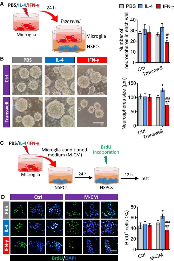
Fig. 5.
Effects of the secretome from IL-4- or IFN-γ-treated microglia on proliferation of NSPCs. A Experimental scheme to monitor microglia-conditioned medium on neurosphere size of adult NSPCs. B Micrographs of adult NSPCs treated with PBS, IL-4 or IFN-γ or co-cultured with PBS-, IFN-γ- or IL-4-treated microglia for 24 h. The histograms represent the number of neurospheres in each well and the neurosphere size. C Experimental scheme to monitor microglia-conditioned medium on proliferation of adult NSPCs. D Fluorescence micrographs of BrdU+ cells from NSPCs exposed to PBS, IL-4 or IFN-γ or microglia-conditioned medium for 24 h. The BrdU+ cells were stained with antibody (green) and nucleus is labeled by DAPI (blue). Scale bar is 10 μm. The histogram represents quantification of percentage of BrdU+ cells from adult NSPCs. Data are showed Mean ± SEM, n = 4–6, *P < 0.05, **P < 0.01 vs PBS group, ##P < 0.01, ###P < 0.001 vs IL-4 group

