

Abstract
Background
As overall cancer survival continues to improve, the incidence of metastatic lesions to the bone continues to increase. The subsequent skeletal related events that can occur with osseous metastasis can be debilitating. Complete and impending pathologic femur fractures are common with patients often requiring operative fixation. However, the efficacy of an intramedullary nail construct, on providing stability, continue to be debated. Therefore, the purpose of this study was to utilize a synthetic femur model to determine 1) how proximal femur defect size and cortical breach impact femur load to failure (strength) and stiffness, and 2) and how the utilization of an IMN, in a prophylactic fashion, subsequently alters the overall strength and stiffness of the proximal femur.
Methods
A total of 21 synthetic femur models were divided into four groups: 1) intact (no defect), 2) 2 cm defect, 3) 2.5 cm defect, and 4) 4 cm defect. An IMN was inserted in half of the femur specimens that had a defect present. This procedure was performed using standard antegrade technique. Specimens were mechanically tested in offset torsion. Force-displacement curves were utilized to determine each constructs load to failure and overall torsional stiffness. The ultimate load to failure and construct stiffness of the synthetic femurs with defects were compared to the intact synthetic femur, while the femurs with the placement of the IMN were directly compared to the synthetic femurs with matching defect size.
Results
The size of the defect invertedly correlated with the load the failure and overall stiffness. There was no difference in load to failure or overall stiffness when comparing intact models with no defect and the 2 cm defect group (p=0.98, p=0.43). The 2.5 cm, and 4.5 cm defect groups demonstrated significant difference in both load to failure and overall stiffness when compared to intact models with results demonstrating 1313 N (95% CI: 874-1752 N; p<0.001) and 104 N/mm (95% CI: 98-110 N/mm; p=0.03) in the 2.5 cm defect models, and 512 N (95% CI: 390-634 N, p<0.001) and 21 N/mm (95% CI: 9-33 N/mm, p<0.001) in the models with a 4 cm defect. Compared to the groups with defects, the placement an IMN increased overall stiffness in the 2.5 cm defect group (125 N/mm; 95% CI:114-136 N/mm; p=0.003), but not load to failure (p=0.91). In the 4 cm defect group, there was a significant increase in load to failure (1067 N; 95% CI: 835-1300 N; p=0.002) and overall stiffness (57 N/mm; 95% CI:46-69 N/mm; p=0.001).
Conclusion
Prophylactic IMN fixation significantly improved failure load and overall stiffness in the group with the largest cortical defects, but still demonstrated a failure loads less than 50% of the intact model. This investigation suggests that a cortical breach causes a loss of strength that is not completely restored by intramedullary fixation.
Level of Evidence: II
Keywords: prophylactic intramedullary nail, pathologic fracture, metastases, musculoskeletal oncology
Introduction
The American Cancer Society estimated that 1,735,350 new cases of cancer would be diagnosed in 2018.1 Approximately, 50% will be one of the various carcinoma subtypes. Carcinoma is the most common cancer to metastasize to bone.2-4 Metastatic disease of bone (MDB) causes skeletal-related events (SRE) including pain, metabolic derangements, and pathologic fractures, requiring extensive and multi-modal healthcare resources.5-8 The most common presenting symptom of MDB is pain and increasing pain can an early indication of impending pathological fractures.7 Diseased bone is weaker than healthy bone and therefore requires less force for a fracture to occur.8
Historically, predictive scoring systems such as the Mirels’ criteria, are most commonly used to assist musculoskeletal oncologists in determining when there is a significant risk of pathologic fracture, and therefore prophylactic internal fixation is warranted.9,10 Newer methods, such as CT-based structural rigidity analysis, have proven to be more accurate in terms of sensitivity, specificity, positive predictive value, and negative predictive value.11 While these measures provide predictably high sensitivity and are helpful to guide surgical decision-making, there continues to be suboptimal specificity that likely results in overtreatment.
The most appropriate fixation method continues to be discussed when addressing both impending and complete pathologic fractures. The choice of reconstruction is ultimately left to the surgeon’s discretion based upon a multitude of clinical factors including histology, location, defect size, bone quality, and estimated overall survival. The femur is the most common location that undergoes MDB that is indicated operative intervention.12-14 The primary methods used to surgically correct or prevent pathologic fractures in the femur are intramedullary nailing (IMN) and endoprosthetic reconstruction (EPR).14 For femoral metastatic fractures, IMN offers many advantages such as reduced invasiveness, sufficient durability, preserved bone stock resulting in additional revision selections, and less cost as compared to EPR.14 However, more extensive bone destruction or more proximal lesion locations in the femur may test the limits of intramedullary stabilization and therefore patient outcome construct durability may be optimized by completely replacing the compromised bone, making EPR justifiable.15,16 However, the size of metastatic lesions that would correlate with risk for fracture as well as the size of the lesion correlated with construct failure is unknown.
The purpose of this study was to determine mechanical data that will guide future studies investigating the role of fracture prediction and fixation choices in MDB. The primary aim was to investigate the effect of the placement of a femoral IMN on failure load and torsional stiffness in femurs with different sized defects. We hypothesized that there would be no difference in decrease in failure load and torsional stiffness with the different sized defects after placement of an IMN.
Methods
A total of 21 4th Generation composite Sawbone femurs, designed to mimic the properties of human bones (Model #3403, Sawbones Inc. Vashon, WA), were used. These models were randomly assigned into four groups: Group 1: no defect (n=3); Group 2: each model consisting of a 2 cm diameter calcar defect with an incomplete cortical breach (n=6); Group 3: each model consisting of a 2.5 cm diameter calcar defect with a complete cortical breach (n=6); Group 4: each model consisting of a 4 cm diameter calcar defect with a complete cortical breach and substantial cancellous involvement (n=6). The medial calcar region was chosen as it is known to be an area more sensitive to changes in strength as compared to other locations in the proximal femur.17 An alignment jig was utilized to position the Sawbones and all defects were centered on the same location consisting of the medial aspect of the calcar: 1 cm anterior to and at the level of the lesser trochanter (Fig. 1). The defects were created using a computer numerical control (CNC) mill (TM-1; HAAS Automation, Inc. Oxnard CA) to trace a circular cutting pattern with the specified diameter. The 2.5 cm diameter defect was selected to be similar in size to the 2 cm defect while including a cortical breach without moving the location of the defect.
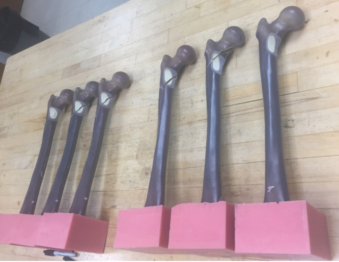
Figure 1.

Origination point of all simulated metastatic lesions investigated in this work. The pink molds held the Sawbones in a reproducible position for machining and the radius of the defect centered at this location was increased to achieve the 2 cm, 2.5 cm and 4 cm lesions tested in this study.
After the establishment of the defects, half of the models in groups 2, 3, and 4 (n=3/group) had an 11.5 x 360 mm, 130-degree cephalomedullary nail with a 100 mm proximal lag screw and single distal locking screw (Smith and Nephew InterTAN, Watford, UK) placed by a board certified, musculoskeletal oncology fellowship trained orthopedic surgeon (BJM). To standardize the nailing technique among specimens, a series of drill guides made from polymethylmethacrylate (PMMA) molds of specific locations on the Sawbones geometry were utilized to ensure consistent locations and trajectories of the trochanteric entry and cephalomedullary lag screw. Separate PMMA molds of the original femoral neck and subtrochanteric geometry were secured around the defect to mechanically stabilize the defect during cephalomedullary screw insertion in order to prevent worsening of the defect prior to biomechanical testing. A total of 3 replicate models for each of the seven groups (intact, 2 cm, 2 cm with IMN, 2.5 cm, 2.5 cm with IMN, 4 cm, 4 cm with IMN) was prepared for mechanical testing.
The femoral models were mechanically tested in offset torsion.18,19 The distal 10 cm of each Sawbone femur was potted in a PMMA block with the axis of the femoral neck in an axial plane parallel to a flat edge of the box. Models were then rigidly clamped with the femoral shaft and femoral neck oriented horizontally, and the femoral head was positioned under the mechanical loading actuator of a servohydraulic mechanical testing machine (MTS Bionix, Eden Prairie, MN). To constrain whole-bone bending during testing while leaving medial-lateral and anterior-posterior translations and all rotations at the proximal end free, a cylindrical horizontal support bar located 11 cm distal to the center of the femoral head was raised until it contacted the bone. A PMMA mold of the Sawbones femoral head geometry was mounted to the mechanical loading actuator on an x-y bearing for application of load to the anterior femoral head (Fig. 2). The loading plate was brought into contact with the femoral head using a compressive force of 50 N. Three preconditioning cycles, consisting of application and removal of 2 mm of femoral head displacement were performed with 30 second rests between cycles. Models were then loaded to failure at a displacement rate of 1 mm/s. Force and displacement data were collected simultaneously at 100 Hz.
Figure 2.

Mechanical testing setup viewed from the side (left) and above (right).
The mode of failure was qualitatively assessed by documenting the location and direction of fracture propagation. Load of failure and overall torsional stiffness were calculated from the measured force-displacement curves. Failure was defined at the instant of maximum force prior Failure was defined as a sudden deviation from the linear load versus the displacement curve. Stiffness was defined as the steepest slope of the linear portion of the force-displacement curve. One-way ANOVA with Tukey’s multiple comparisons were used to evaluate the significance of pairwise differences in failure load and stiffness associated with defect size. Two-way ANOVA with Tukey’s multiple comparisons was used to evaluate differences associated with defect size and the presence/absence of an intramedullary nail. All statistical analysis was performed in GraphPad Prism (v 8.4.3, GraphPad Software LLC, San Diego, CA) with significance defined by multiplicity adjusted p < 0.05.
Results
Intact models (no defect; n=3) failed at an average load of 2701 N (95% CI: 2282-3120 N) with an overall stiffness of 122 N/mm (95% CI: 99-144 N/mm). Models with 2 cm defects, which did not perforate the cortex, demonstrated essentially equivalent loads to failure (2785 N; 95% CI: 2276-3295; p=0.98) and stiffness (112 N/mm; 95% CI: 89-135 N/mm; p=0.43) compared to intact models. The 2.5 cm defect group, which did perforate the cortex, had a load to failure that was 49% of the intact models (1313 N; 95% CI: 874-1752 N; p<0.001). Additionally, the overall stiffness was 85% of the intact models (104 N/mm; 95% CI: 98-110 N/mm; p=0.03). The 4 cm defect group had further reduction in failure load with a load to failure that was 19% of the intact models (512 N; 95% CI: 390-634 N; p<0.001) and an overall stiffness that was 17% of the intact models (21 N/mm; 95% CI: 9-33 N/mm; p<0.001). (Fig. 3)
Figure 3.

Average failure load (top) and torsional stiffness (bottom) values for Sawbones tested to failure in offset torsion. The groups without fixation are shown in solid bars and the groups with IMN fixation are indicated with lighter hashed bars. Error bars indicate 95% confidence intervals, and p values are from Tukey’s multiple comparisons tests.
Placement of an IMN in the 2 cm defect models did not have a significant increase in the overall failure load (3087 N; 95% CI: 2921-3253; p=0.10) or the overall stiffness when compared to previous non-IMN models (125 N/mm; 95% CI: 122-129; p=0.07). IMN fixation of the 2.5 cm defect models had a significant increase in stiffness (125 N/mm; 95% CI:114-136 N/mm) when compared to the non-IMN models (104 N/mm: 95% CI: 98-110 N/mm; p=0.003). However, there was no difference regarding load to failure between these groups (p=0.91). When compared to the non-IMN 4 cm defect models, placement of an IMN in the 4 cm defect models significantly increased both the load to failure (512 N; 95% CI: 390634 N vs. 1067 N; 95% CI: 835-1300 N; p=0.002) and the overall stiffness (21 N/mm; 95% CI: 9-33 N/mm vs 57 N/mm; 95% CI:46-69 N/mm; p=0.001). However, despite these increases relative to the non-IMN models, placement of an IMN in the 4 cm models only achieved 40% of the average failure load and 47% of the average overall stiffness of an intact models. (Fig. 4)
Figure 4.

Aggregate force displacement curves from mechanical testing. There was very consistent mechanical response within each lesion/fixation group allowing for identification of significant differences with a small number of specimens per group.
Discussion
The goal of this work was to investigate the biomechanical relationship between simulated metastatic lesion defects of various sizes, intramedullary fixation, and mechanical failure. Of the three defect sizes tested, only those that perforated the cortex (the 2.5 cm and 4 cm defects) clearly reduced the average failure load and stiffness. IMN fixation increased overall stiffness in the 2.5 cm group, as well as failure load and overall stiffness in the 4 cm defect group. However, failure load in both the IMN stabilized 2.5 cm and 4 cm defect groups remained less than 50% of the intact models. Our data suggest that cortical perforation is a critical event that causes substantial weakening of intact femurs, and fixation with an IMN only partially restores the strength and stiffness.
Lesions located at the calcar of the femur were selected as defects in this location have demonstrated to be more mechanically compromising in comparison to lesions in other locations.17,19-21 Additionally, enlarging lesion size has demonstrated to increase the risk of pathological fracture.22 The results of this biomechanical study demonstrated similar results with decreased load to failure with increasing lesion size located at the calcar of the proximal femur. We found a variable amount of restoration of strength and stiffness after IMN fixation relative to defect size. For the smallest defect, 2 cm (no cortical defect), the changes in load to failure and stiffness were insignificant compared to an intact bone model, both with and without an IMN. This supports the implication that bones lacking a cortical defect retain nearly equivalent mechanical strength of an intact specimen when exposed to this method of mechanical loading. This finding further poses the question whether lesions without cortical deficits need to be prophylactically stabilized.
The 2.5 cm defect models demonstrated an increase in stiffness after stabilization, but not failure load, and failed with a spiral fracture pattern predominantly by torsion with a smaller component of bending (Fig 5). This mechanism of failure would be expected clinically and provides qualitative assurance in our experimental design. In the 4 cm defect model, the IMN significantly improved both strength and stiffness. Prophylactic fixation restored strength and stiffness nearly to the level of the 2.5 cm cortical defect, but not to the same level as the intact model. Overall, these findings suggest that strength and stiffness of simulated bone decrease substantially as lesion size and cortical destruction increase. Intramedullary stabilization can restore strength and failure resistance, but this study demonstrated not more than 50% of an intact model. These results do not allow for generalization into clinical treatment of MDB, however they do suggest that the mechanical properties of femurs with large lesions and cortical deficiencies may not be adequately restored with closed intramedullary nailing.
Figure 5.
Photograph of failure pattern for the 2.5 cm lesions (right) and the 2.5 cm lesions with IMN. The predominant fracture pattern was a spiral through the lesion.
Prior studies have shown strength restoration to a greater extent than ours using simulated metastatic defects with alternate methods of experimental fixation. Kaneko et al. performed a study utilizing human cadavers with a femoral neck inferomedial defect.23 They subsequently filled the defect with bone cement and tested the specimens with single-limb stance-type loading, (rather than offset torsion). They reported at least 85% strength when compared to specimens with no defect. In a study of cadaveric canine femurs with 50% cortical defects, Leggon et al. described the greatest increase in strength came from the utilization of polymethylmethacrylate with a plate and bicortical screws. They demonstrated that this combination retained 56% strength of the intact specimens.24 The 2.5 cm and 4 cm defects in our investigation both demonstrated significant changes after IMN fixation, however the magnitudes differed substantially. The reasons for the limited improvements of strength are unclear, however are likely due to the combination of defect location and mechanical testing. This stresses the IMN construct in the direction of its most substantial deficiency. Specifically, the points of IMN fixation in the bone are limited to screws in the femoral head and distal shaft and does not directly support the defect. Further, our testing in offset torsion exposes the specimens to forces that the IMN is least designed to withstand, as IMN would perform better in axial loading and bending forces. These choices were made specifically to investigate conditions in which IMN fixation may not be mechanically adequate and are at highest risk of failure.
There were several limitations that should be discussed. First, the mechanical tests performed were in a controlled environment established for this work and do not directly translate to clinical practice. Like most biomechanical studies of metastatic lesions, the defects used in this study were created artificially with a regular geometry that by definition cannot replicate the tortuous shapes or encroachment of the lesion into the cortex from the intramedullary space.17,19,20 The Sawbones that we used replicate the mechanical properties of human femurs are designed for use in biomechanical research. This offers an advantage over cadaveric models used in similar experiments in terms of uniformity and reproducibility of the mechanical testing. However, Sawbones lack several potentially important features seen in patients with MDB, namely abnormal bone mineral density secondary to malnutrition or systemic agents and effects of external beam radiation. Another limitation was that defects investigated in this work were created mechanically, which will not fully replicate the often amorphous and asymmetric nature of invasive carcinoma. The offset torsion test was selected for this work to better simulate functional loading of the femur during high demand flexion activity; however, it does not specifically replicate any single physiological activity and does not reflect the only mechanism of fracture. Finally, the number of replicates (n=3) within each experimental group were extremely limited. While this did not facilitate robust statistical analysis, with our standardized procedures for lesion creation and IM nailing, the mechanical response was extremely similar within each group, and the mechanical differences among groups with a cortex perforation were dramatic (Figure 4). Within these limitations, our intent was not to perform an investigation that can directly be applied to clinical practice, but rather to explore the mechanical changes that occur with increasing metastatic lesion size, and the strength that can be recovered under ideal conditions with an intramedullary device.
In conclusion, this study demonstrated that cortical deficiencies, rather than simply lesion size, caused substantial losses in torsional strength. Additionally, IMN fixation did not completely restore the strength and stiffness to that of an intact bone model when loaded in offset torsion. IMN fixation alone does not address cortical breaches that may lead to compromised osseous integrity during activities of daily living that require torsional load of the femur. The clinical significance of incomplete restoration of strength after IMN nailing of proximal femoral defects remains unclear, but it does suggest that additional mechanical investigation to determine a mechanically optimal treatment strategy to definitively manage impending pathologic fractures with large cortical defects in high-stress areas of the proximal femur is warranted. Future investigations should attempt to find a threshold of defect size in which IMN is insufficient to adequately support all activities of daily living for patients with MDB.
References
- 1.Siegel R.L., Miller K.D., Jemal A. Cancer statistics, 2018. CA Cancer J Clin. 2018;68(1):7–30. doi: 10.3322/caac.21442. [DOI] [PubMed] [Google Scholar]
- 2.Virk M.S., Lieberman J.R. Tumor metastasis to bone. Arthritis Res Ther, 2007;9(Supp. 1 Supp. 1):S5. doi: 10.1186/ar2169. [DOI] [PMC free article] [PubMed] [Google Scholar]
- 3.Siegel R.L., Miller K.D., Jemal A. Cancer statistics, 2020. CA Cancer J Clin. 2020;70(1):7–30. doi: 10.3322/caac.21590. [DOI] [PubMed] [Google Scholar]
- 4.Liu D., et al. Prognosis of prostate cancer and bone metastasis pattern of patients: a SEER-based study and a local hospital based study from China. Sci Rep. 2020;10(1):9104. doi: 10.1038/s41598-020-64073-6. [DOI] [PMC free article] [PubMed] [Google Scholar]
- 5.Idota A., et al. Bone Scan Index predicts skeletal-related events in patients with metastatic breast cancer. Springerplus. 2016;5(1):1095. doi: 10.1186/s40064-016-2741-0. [DOI] [PMC free article] [PubMed] [Google Scholar]
- 6.Baek Y.H., et al. Incidence of skeletal-related events in patients with breast or prostate cancer-induced bone metastasis or multiple myeloma: A 12-year longitudinal nationwide healthcare database study. Cancer Epidemiol. 2019. –61.pp. 104–110. [DOI] [PubMed]
- 7.Clain A. Secondary Malignant Disease of Bone. Br J Cancer, 1965;19:15–29. doi: 10.1038/bjc.1965.3. [DOI] [PMC free article] [PubMed] [Google Scholar]
- 8.Coleman R.E. Metastatic bone disease: clinical features, pathophysiology and treatment strategies. Cancer Treat Rev, 2001;27(3):165–76. doi: 10.1053/ctrv.2000.0210. [DOI] [PubMed] [Google Scholar]
- 9.Mirels H. Metastatic disease in long bones: A proposed scoring system for diagnosing impending pathologic fractures. Clin Orthop Relat Res. 1989 2003. pp. S4–13. [DOI] [PubMed]
- 10.Mirels H. Metastatic disease in long bones. A proposed scoring system for diagnosing impending pathologic fractures. Clin Orthop Relat Res, 1989. pp. 256–64. [PubMed]
- 11.Damron T.A., et al. CT-based Structural Rigidity Analysis Is More Accurate Than Mirels Scoring for Fracture Prediction in Metastatic Femoral Lesions. Clin Orthop Relat Res. 2016;474(3):643–51. doi: 10.1007/s11999-015-4453-0. [DOI] [PMC free article] [PubMed] [Google Scholar]
- 12.Bohm P., Huber J. The surgical treatment of bony metastases of the spine and limbs. J Bone Joint Surg Br, 2002;84(4):521–9. doi: 10.1302/0301-620x.84b4.12495. [DOI] [PubMed] [Google Scholar]
- 13.Hage W.D., Aboulafia A.J., Aboulafia D.M. Incidence, location, and diagnostic evaluation of metastatic bone disease. Orthop Clin North Am, 2000;31(4):515–28. doi: 10.1016/s0030-5898(05)70171-1. vii. [DOI] [PubMed] [Google Scholar]
- 14.Tanaka T., et al. Intramedullary nailing has sufficient durability for metastatic femoral fractures. World J Surg Oncol. 2016;14:80. doi: 10.1186/s12957-016-0836-2. [DOI] [PMC free article] [PubMed] [Google Scholar]
- 15.Janssen S.J., et al. Outcome after fixation of metastatic proximal femoral fractures: A systematic review of 40 studies. J Surg Oncol. 2016;114(4):507–19. doi: 10.1002/jso.24345. [DOI] [PubMed] [Google Scholar]
- 16.Steensma M., et al. Endoprosthetic treatment is more durable for pathologic proximal femur fractures. Clin Orthop Relat Res. 2012;470(3):920–6. doi: 10.1007/s11999-011-2047-z. [DOI] [PMC free article] [PubMed] [Google Scholar]
- 17.Kaneko T.S., Skinner H.B., Keyak J.H. Lytic lesions in the femoral neck: Importance of location and evaluation of a novel minimally invasive repair technique. J Orthop Res, 2008;26(8):112732. doi: 10.1002/jor.20555. [DOI] [PubMed] [Google Scholar]
- 18.Johnson J.E., et al. Simulated lesions representative of metastatic disease predict proximal femur failure strength more accurately than idealized lesions. J Biomech. 2020;106:109825. doi: 10.1016/j.jbiomech.2020.109825. [DOI] [PubMed] [Google Scholar]
- 19.Sivasundaram R., et al. The biomechanical effect of proximal tumor defect location on femur pathological fractures. J Orthop Trauma. 2013;27(8):e174–80. doi: 10.1097/BOT.0b013e3182809748. [DOI] [PubMed] [Google Scholar]
- 20.Benca E., et al. Effect of simulated metastatic lesions on the biomechanical behavior of the proximal femur. J Orthop Res. 2017;35(11):2407–2414. doi: 10.1002/jor.23550. [DOI] [PubMed] [Google Scholar]
- 21.Keyak J.H., et al. The effect of simulated metastatic lytic lesions on proximal femoral strength. Clin Orthop Relat Res. 2007;459:139–45. doi: 10.1097/BLO.0b013e3180514caa. [DOI] [PubMed] [Google Scholar]
- 22.Caypinar B., et al. Biomechanical determination of the relationship between femoral neck lesion size and the risk of pathological fracture. Hip Int. 2016;26(2):158–63. doi: 10.5301/hipint.5000309. [DOI] [PubMed] [Google Scholar]
- 23.Kaneko T.S., Skinner H.B., Keyak J.H. Feasibility of a percutaneous technique for repairing proximal femora with simulated metastatic lesions. Med Eng Phys, 2007;29(5):594–601. doi: 10.1016/j.medengphy.2006.06.008. [DOI] [PubMed] [Google Scholar]
- 24.Leggon R.E., Lindsey R.W., Panjabi M.M. Strength reduction and the effects of treatment of long bones with diaphyseal defects involving 50% of the cortex. J Orthop Res, 1988;6(4):540–6. doi: 10.1002/jor.1100060410. [DOI] [PubMed] [Google Scholar]