

Abstract
To seek a new delivery system of vaccine for infectious bronchitis virus (IBV), transgenic potato expressing full-length spike (S) protein of IBV was produced and its immunogenicity in chickens was investigated. One to three copies of S gene of IBV were randomly and stably inserted into potato (Solanum tuberrosum cv. Dongnong 303) genome by Agrobacterium tumefaciens-mediated transformation. Transcription and translation of S gene for IBV were confirmed by Northern blot and Western blot analyses in transgenic plantlets. Chickens immunized orally and intramuscularly with transgenic potato tubers expressing S protein generated the detectable levels of serum neutralizing antibodies and were protected against the challenge with the virulent IBV. In vitro secretion of interleukin 2 and T lymphocyte proliferation of spleen cells from the immunized chickens varied with the dose and the manner of vaccination with S protein derived from transgenic plants. The results indicated that S protein expressed in transgenic plants might be a new source for the production of Coronaviridae IBV vaccine.
Keywords: Infectious bronchitis virus, Spike protein, Transgenic potato, Immunity, Interleukin 2, T lymphocyte
1. Introduction
Infectious bronchitis virus (IBV) is a single-stranded positive sense RNA virus and a prototype of the family Coronaviridae in the new order Nidovirales (Cavanagh, 1997). IBV is the pathogen of chicken infectious bronchitis (IB), which is an acute, highly contagious respiratory disease in young chickens with potential involvement of kidney and reproductive tract (Cavanagh and Naqi, 1997). Typically, the disease has been controlled with the serotype-specific vaccines, however, outbreaks of IB still occur because IBV vaccines offer little or no cross-protection between serologically distinct viruses (Cavanagh, 1997). More than 20 IBV serotypes have been identified worldwide and there are an increasing number of new serotypic variants. Outbreaks of IB are possibly attributed to the variation of IBV and widespread use of live attenuated vaccines (Hofstad, 1981).
IBV genome includes three major structural genes, which encode the spike (S), membrane (M) and nucleocapsid (N) proteins, respectively. The S protein, which forms the peplomers at the surface of the virus, is post-translationally cleaved into the S1 and S2 proteins (Cavanagh et al., 1986). The nucleotide sequences of S and N genes of IBV isolates have been reported previously with the different tissue tropism (Binns et al., 1985, Niesters et al., 1986, Sapats et al., 1996, Zhang et al., 2002, Zhou et al., 2002). These variants are thought to arise from a few amino acid changes in S1 protein and N protein. Recently, new variants were isolated, which differed from all known IBV serotypes (Wang et al., 1994, Williams et al., 1992, Zhou et al., 2003a). Because of the existence of a large number of serotypes, the identification of the regional serotypes plays an important role in determining vaccination regimes. The previous results demonstrated that S protein is responsible for the induction of virus-neutralizing antibodies and protecting the host from the infection of IBV (Cavanagh et al., 1986, Cavanagh, 1997). Chickens immunized with the rS1 protein expressed by baculovirus were able to induce protective immunity (Song et al., 1998). Johnson et al. (2003) constructed a recombinant fowl adenovirus serotype 8 (FAV 8) expressing IBV S1 protein that induced protective immunity against the virulent IBV challenge in commercial broilers. Mice vaccinated with the entire S protein expressed by recombinant vaccinia virus were shown to have virus-neutralizing antibodies to IBV (Tomley et al., 1987). Therefore, S protein of IBV can be used as the desirable immune antigen in the development of the new vaccine.
Recently, transgenic plants have been used as production and delivery systems successfully for pathogen antigens. The plant-expressed antigens generated protective antibodies in immunized animals (Arakawa et al., 1998, Haq et al., 1995, Khandelwal et al., 2003, Marquet-Blouin et al., 2003, Mason et al., 1992, Mason et al., 1996, Streatfield and Howard, 2003, Tuboly et al., 2000, Zhou et al., 2003b). In the present study, we described the generation of the transgenic potato expressing full-length S protein of IBV. Furthermore, the immunogenicity of the potato-derived S protein in chickens was investigated.
2. Materials and methods
2.1. Construction of the plant expression vector
Based on the cDNA sequence of the S gene of IBV strain H52 (Cheng et al., 2002), the open reading frame (ORF) consisting of 3486 nucleotides was obtained by PCR using the following primers, the upstream primer 5′-GCTCTAGAATGTTGGTAACACCTCTT-3′ containing the XbaI site and the downstream primer 5′-CGGGATCCTATTAGGAAGGACGTGGGACT-3′ containing the BamHI site. The PCR was consisted of 35 cycles of denaturation at 94 °C for 45 s, annealing at 56 °C for 90 s and extension at 72 °C for 2 min, finally terminated after extension at 72 °C for 10 min. The amplified cDNA fragment encoding for the full-length S protein was subcloned into the binary vector pBI 121 (Clontech, Franlin Lakes, USA) under the control of cauliflower mosaic virus (CaMV) 35S promoter and sequenced to confirm its identity (Fig. 1 ). The recombinant vector contained the neomycin phosphotransferase gene (nptII), which permitted selection of the transformed plant on medium containing kanamycin. This resultant plant expression vector was then introduced into Agrobacterium tumefaciens strain EHA 105 (Clontech) by triparental mating as described by Ditta et al. (1980).
Fig. 1.

Schematic structure of the recombinant plasmid pBI 121-S used for Agrobacterium-mediated transformation of potato plants. The DNA sequence encoding for S protein of IBV was cloned into the downstream of the CaMV 35S promoter of a binary plasmid pBI 121. This region contains the nptII expression cassette providing kanamycin resistance, the left (LB) and right (RB) borders of transferred DNA for genetic analysis of the transformed plants.
2.2. Production of transgenic potato plants
Sterile potatoes (Solanum tuberosum cv. Dongnong 303) were propagated in vitro by subculturing the top shoots or stem segments including axillary buds in our laboratory. Sterile calli of stem segments excluding axillary bud of potatoes was used for transformation with A. tumefaciens on the method as described elsewhere (Castanon et al., 1999). Transformed plants were regenerated from the calli on Murashige and Skoog (MS) medium containing kanamycin (100 μg ml−1), zeatin (2 mg l−1), 2-naphthaleneacetic acid (20 μg ml−1) and gibberellic acid (20 μg ml−1) at 25 °C under light. Some transgenic plantlets were transferred to the greenhouse under normal light and humidity conditions.
Transgenic plantlets were analyzed for the presence of the foreign cDNA sequence by PCR using the above-mentioned primers. One hundred milligrams fresh plantlets were frozen in liquid nitrogen and grinded to a powder with a mortar and pestle. The powder was suspended in 300 μl extraction buffer containing 50 mM Tris–HCl, 500 mM NaCl, pH 8.0, 50 mM EDTA, 2% SDS (w/v), 1% β-mercaptoethanol (v/v), and 6% polyvinylpyrrolidone (PVP) (v/v), and incubated at 65 °C for 10 min. After a phenol–chloroform–isoamylalcohol (25:24:1) extraction, the supernatant containing DNA was collected and mixed with an equal volume of isopropanol. The pellet was dissolved in TE buffer (10 mM Tris–HCl, 1 mM EDTA, pH 8.0) followed by RNase treatment, and the genomic DNA was stored at −20 °C until being used. Using the genomic DNA from non-transgenic plants as negative control, the determination of the foreign DNA was accomplished by PCR as described above.
Genomic DNA was isolated from both transformed and non-transgenic potato plantlets using CTAB method (Ausubel et al., 1995). Thirty micrograms of the extracted genomic DNA from each sample was digested with BamHI overnight at 37 °C. The digested products were separated by electrophoresis on a 0.8% agarose gel and transferred to a nylon membrane (Amersham-Pharmacia Biotech AB, Uppsala, Sweden). The nylon membrane was hybridized with the 32P-labeled IBV-S1 (part of IBV-S gene) specific cDNA probe as described by Sambrook and Russell (2001).
Total RNA was extracted from fresh potato plantlets with TRIzol regents (GIBCOBRL, Grand island, USA). RNA (20 μg) was separated by electrophoresis on 1.2% formaldehyde/agarose gels, and transferred to a nylon membrane (Amersham-Pharmacia Biotech AB) for hybridization as described by Sambrook and Russell (2001). A 1.7 kb S1 fragment was used as a probe after 32P-labeling with Prime-a-Gene ™DNA Labeling Kit (Promega, USA).
2.3. Detection of S protein in the transgenic potato plant
For the presence of IBV S protein, the extract of the transgenic plants as an antigen in ELISA was determined using IBV specific antibody as described by Zhou et al. (1997). Briefly, 100 mg fresh plantlets from each transgenic and non-transgenic plant were powdered in liquid nitrogen and lysed in 0.05 M bicarbonate buffer (pH 9.6). The 96-well plate (Nunc, Denmark) was coated with the plantlet extracts containing S protein at 4 °C overnight. Purified IBV and the extracts from non-transformed plants were used as positive and negative controls, respectively. The plate was blocked with 5% skimmed milk in PBST (0.01 M PBS containing 0.05% Tween-20, pH 7.4) at 37 °C for 2 h. Then the coated plate was incubated with IBV chicken antiserum (1:500) and alkaline phosphatase-conjugated goat anti-chicken IgG (1:4000, Sigma, MO, USA). Finally, p-nitrophenylphosphate (Sigma) substrate was used for color development, and the optical density (OD) values were measured in ELX800 Automated Microplate Reader (BIO-TEK, Vermont, USA) at 405 nm.
The expression of S protein in the transgenic plants was analyzed by Western blot (Conrad and Fiedler, 1998). Plant extracts were prepared by grinding 100 mg of fresh plantlets in liquid nitrogen. The resulting powder was resuspended in SDS–PAGE sample buffer (100 mM Tris–Cl, 4% SDS, 0.2% bromophenol blue, 200 mM DTT, and 20% glycerol, pH 6.8) and boiled for 10 min. The mixture was centrifuged at 10,000×g for 10 min at 4 °C to remove the insoluble debris. The proteins were separated by 8% SDS–PAGE and blotted to a nitrocellulose membrane (Amersham-Pharmacia Biotech AB). The membrane was blocked with 5% skimmed milk in PBST overnight, and sequentially incubated with the chicken IBV specific antiserum (1:500) for 2 h at 37 °C and goat anti-chicken IgG conjugated alkaline phosphatase (1:4000) for 1 h at 37 °C. The NBT/BCIP (Sigma) were used to develop the color reaction after the membrane was washed five times with PBST.
2.4. Chicken immunization, antibody detection and challenge
SPF fertile eggs of white Leghorn chickens (Beijing Merial Vital Laboratory Animal Technology Co. Ltd, Beijing, China) were hatched at our laboratory, and maintained in strict isolation throughout the study. Fourteen-day-old chicks were divided into seven groups, with six chicks per group. Before oral immunization, chicks were fasted overnight while water was provided ad libitum. Group 1 and 2 chicks were each orally immunized with 2.5 g (6.23 μg of S protein, plant 4#) and 5 g (12.45 μg of S protein, plant 4#) of peeled and sliced transgenic potato tubers, respectively. Chicks in the third group were orally immunized with 5 g of wild potato tubers, Group 4 and 5 chicks were each intramuscularly immunized with 2.5 and 5 g potato tuber extracts, respectively. Chicks in the sixth group was intranasally vaccinated with the commercial modified live IBV vaccine according to the manufacture’s instruction (H52 strain, Hangzhou Jianliang Veterinary Bioproducts Co. Ltd., Hangzhou, China), while the seventh group chicks were each orally immunized with 5 g extraction buffer as control. Under the same feeding condition, all immunizations were administrated at days 0, 7, and 14. Blood samples were collected through wing vein puncture at day 0 and 1 week after each immunization. The VN antibody titer against IBV was measured by the TOC assay with 200 doses of the IBV H52 vaccine strain [103.625 CD50 (ciliostatic dose)] as described by Cook et al. (1976). All chicks were intranasally challenged with 200 μl of the virulent IBV M41 strain (104.89 ELD50/0.1 ml) at the seventh day after third immunization and observed daily for 7 days. The protection was determined by the absence of the characteristic respiratory signs of IB in the challenged chicks at 18–168 h post-inoculated the virulent IBV M41 strain, and the virus was re-isolated from the trachea and feces of the challenge chicks as described previously (Cavanagh and Naqi, 1997).
2.5. In vitro lymphoproliferation
The suspensions of the spleen lymphocytes were prepared as described previously by Zhou et al. (2003c). T lymphocytes proliferation activity in the immunized chickens was screened by MTT test (Hansen et al., 1989). Briefly, spleen lymphocytes from all vaccinated chickens were respectively resuspended at 1×10⁷ cells ml−1 in RPMI 1640 medium (GIBCOBRL) containing 10% fetal calf serum (GIBCOBRL) (v/v), penicillin/streptomycin (200 IU/50 μg ml−1), 12.5 μg ml−1 Concanavalin A (con A, Sigma), and the cells in 96-well plates (Nunc, Denmark) were incubated at 40 °C in 5% CO2 for 48 h. Then, 20 μl MTT (5 mg ml−1) was added and the reaction was stopped 3 h later by adding 100 μl of lysis extraction buffer composed of 10% SDS–0.01 M HCl. The cells continued to be incubated at 40 °C in 5% CO2 for 2 h. The OD values were read in ELX800 Automated Microplate Reader at 490 nm.
2.6. Assay of the immunized chicken IL-2 (chIL-2)
To detect the chIL-2 secreted by lymphocyte from immunized chickens, the suspensions of the spleen lymphocytes were prepared and incubated as the above-mentioned method, and ELISA was accomplished as Miyamoto et al. (2001) described. The 96-well plates were coated with 100 μl of the spleen lymphocyte supernatants (1×10⁷ cells ml−1) in 0.05 M carbonate buffer, pH 9.6 for 18 h at 4 °C. After washed three times with PBST, each well was blocked with 200 μl of PBS containing 5% (w/v) skim milk for 1 h at room temperature. Monoclonal antibody to chIL-2 (kindly provided by Prof. Jimmy Kwang, Temasek Life Sciences laboratory, Singapore University and Nanyang Technological University, Singapore; 100 μl well−1) was added, and the plates were incubated for 2 h at 37 °C. Goat anti-mouse IgG conjugated with horseradish peroxidase (Kirkegaard & Perry Laboratories Inc., USA) diluted in PBS-5% (w/v) skimmed milk (1:5000) was added to each well and the plate was incubated for 1 h at 37 °C. The wells were washed three times, and developed color with 0.04% (w/v) o-phenylenediamine (OPD, AMRESCO, USA) in 0.05 M phosphate–citrate buffer (pH 5.0). Finally, the reaction was stopped with 2 N sulfuric acid. Color changes were monitored using an automated microtiter plate reader at 490 nm absorbance.
3. Results
3.1. Production and genetic analysis of transformed plants
The recombinant plasmid pBI121-S carrying the cDNA coding for the full-length S protein (pBI121-S) was obtained by subcloning the interested gene into a binary plasmid pBI121. The pBI121-S makes the transformants selected on the medium containing kanamycin for the stable integration into the genomic DNA of the potato plants. The transgenic plants resistant to the selective medium showed no morphological changes in comparison with the non-transgenic potato plants in greenhouse. Approximately 94% (48/51) transgenic lines were shown to possessing the IBV S gene when assayed by PCR. Six lines of PCR positive plants were subjected to Southern blot analysis for the copy numbers and the integration pattern of IBV S gene in plants. As shown in Fig. 2 , the hybridized characteristics of the genomic DNA for the transformed potatoes indicated that one to three copies of S gene inserted randomly into the plant genome, while no hybridized signal appeared when the genomic DNA of non-transgenic potato was used as template. Transcription of S gene introduced into the potato plant was monitored by Northern blot analysis using total RNA of five independent PCR-positive transgenic plantlets, four of five lines showed the strong specific bands corresponding to mRNA of S gene and another appeared a weak band, while the control plant did not show any detectable hybridization signal with the specific IBV probe (Fig. 3 ).
Fig. 2.

Genomic DNA from transgenic and non-transgenic potato plants hybridized with a 32P-labeled specific S1 probe. CK+, purified PCR products of IBV S gene (positive control); CK−, genomic DNA from non-transgenic potato was used as a negative control; Lanes 1–6, each lane represents an IBV S gene products expressed in an individual transgenic line.
Fig. 3.

Northern blot analysis of total RNA isolated from the transgenic and non-transgenic plants. RNA was probed with an IBV S1 gene labeled with 32P demonstrating specific mRNAs of IBV S protein. CK−, non-transgenic potato plant was used as a negative control. Each lane represents an individual transgenic plantlet, plant 1# shows a slight hybridization signal.
3.2. Expression of S protein in transgenic potato plants
The S protein polypeptides expressed in the transgenic plants was investigated using ELISA and Western blot. The results indicated that the amount of S protein detected in the individual line was from 2.39 μg g−1 (plant 2#) to 2.53 μg g−1 (plants 1# and 9#) transgenic potato tubers. Nevertheless, no significant differences were appeared in the detected lines. As shown in Fig. 4A , the molecular weight of the plant-synthesized S protein of IBV was found to be approximately 120 kDa. Meanwhile, Data shown in Fig. 4B indicated that the plant-derived S protein retained the reactivity to IBV antisera in Western blot.
Fig. 4.

SDS–PAGE (A) and Western blot (B) analyses of IBV S protein expressed in transgenic plants. (A) Lanes 2–4 indicate the individual transgenic line. Lane M refers to standard molecular marker (14.4–116 kDa). Lane CK−, non-transgenic potato. (B) Lane 4 shows the band (approximately120 kDa) hybridized with IBV antiserum from the transgenic plant no. 4#. No hybridization signal was obtained with IBV antiserum from the transgenic plant no. 3# and non-transgenic plant (lane CK−).
3.3. Induction of chicken’s antibody against IBV and protection
Following the oral or intramuscular inoculations with the transgenic potato tubers, blood samples were collected at specified times, and the serum IBV-neutralizing activity was measured by virus neutralization test in TOC. As shown in Table 1 , the results clearly showed that high levels of neutralizing antibody were present in the sera of chickens immunized with transgenic potato tuber after three immunizations. In the chickens immunized orally with the transgenic potato, the average titers of neutralizing antibodies from third immunization in the TOC reached 1:2455 (group 1) and 1:3447 (group 2), respectively. Titers of groups 4 and 5 were 1:3654 and 1:5754 in intramuscular injection chickens, respectively. As controls, chickens immunized with the live attenuated IBV vaccine had a titer of 1:3108 (group 6). No anti-IBV antibodies were detected in chickens vaccinated with non-transgenic potato (group 3) or buffer (group 7).
Table 1.
Chicken anti-IBV VN antibody response and protection from challenge with virulent IBV
| Group | Immunizationa | Dose (g) | Route | Serum VN titerb |
No. protected/no. challenged | |
|---|---|---|---|---|---|---|
| Second vaccination | Third vaccination | |||||
| 1 | Transgenic potato tubers | 2.5 | Oral | 1:84.4 | 1:2455 | 4/6 |
| 2 | Transgenic potato tubers | 5 | Oral | 1:129.9 | 1:3447 | 4/6 |
| 3 | Wild type tubers | 5 | Oral | 0 | 0 | 0/6 |
| 4 | Transgenic potato tuber extracts | 2.5 | i.m.c | 1:12.6 | 1:3654 | 5/6 |
| 5 | Transgenic potato tuber extracts | 5 | i.m.c | 1:133.4 | 1:5754 | 5/6 |
| 6 | Live attenuated vaccine | One dose | i.n.c | 1:13.0 | 1:3108 | 6/6 |
| 7 | Buffer | 5 | Oral | 0 | 0 | 0/6 |
Each group consisted of six chickens which were immunized three times with transgenic potato tubers.
VN titer was geometry mean titer.
i.m., intramuscular; i.n., intranasal.
Chickens immunized intramuscularly with the extracts of 2.5 or 5 g transgenic potato tubers shared 83.3% (5/6) protection from the challenge of the virulent IBV M41 strain, while those vaccinated with the commercial live IBV vaccine received the full protection (6/6) (Table 1). 66.7% (4/6) of chickens delivered orally by 2.5 or 5 g transgenic potato tubers were also protected against the challenge. No protection was obtained in chickens that received non-transgenic potato or buffer. Furthermore, no virus was isolated in trachea and feces samples collected from these protected chickens at 7 days post-challenge.
3.4. Lymphoproliferative responses and in vitro secretion of chI L-2
At the 14th day post-third immunization, spleen lymphocytes were isolated and used for in vitro proliferation and chIL-2 secretion assays. As shown in Fig. 5, Fig. 6 , the proliferation responses and in vitro chIL-2 secretion of spleen lymphocytes from the vaccinated animals varied with the dose and the manner of vaccination with the recombinant S proteins derived from transgenic plants. Nevertheless, lymphocytes from animals intramuscularly immunized with the extracts of 5 g transgenic potato tubers had the best in vitro proliferation responses and chIL-2 secretions in comparison with that from other animals immunized with transgenic potato tubers or live IBV vaccine. Neither proliferative reactivity nor chIL-2 secretion of lymphocytes was detected from the animals fed with non-transgenic potato tubers.
Fig. 5.
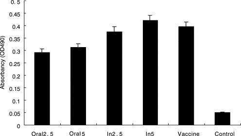
Lymphoproliferation of in vitro culture of spleen cells by MTT. Absorbency (OD490) represents the mean absorbance ± the standard error obtained from three independent assays. Oral2.5 and Oral5 represent chicks immunized orally with 2.5 and 5 g transgenic potato tubers, respectively. In2.5 and In5 indicate chicks vaccinated intramuscularly with the extracts of 2.5 and 5 g transgenic potato tubers, respectively. Vaccine indicates commercial modified live IBV vaccine. Control shows non-transgenic potato plant.
Fig. 6.

Concentration of IL-2 secreted by in vitro culture of spleen cells from the vaccinated chicks at the 14th day after third immunization. Absorbency (OD405) represents the mean absorbance ± the standard error obtained from three independent assays. Oral2.5 and Oral5 represent chicks immunized orally with 2.5 and 5 g transgenic potato tubers, respectively. In2.5 and In5 indicate chicks vaccinated intramuscularly with the extracts of 2.5 and 5 g transgenic potato tubers, respectively. Vaccine, commercial modified live IBV vaccine. Control, non-transgenic potato plant.
4. Discussion
Although other virus polypeptides play a role in protection and recovery from infection in the chickens, the spike protein is an excellent candidate for the development of the novel IBV vaccines. In our present work, the transgenic potato containing the full-length S gene of IBV was obtained using A. tumefaciens-mediated method. Meanwhile, the immunogenicity in chickens was monitored with oral or intramuscular delivery of S protein for IBV expressed in transgenic potato plants. Our results showed that one to three copies of IBV S gene were integrated into potato genome (Fig. 2), and the full-length S gene of IBV was transcribed and expressed in most transgenic potato plants (Fig. 3, Fig. 4). Moreover, we also found the phenomena of transcriptional (plant 1#) and post-transcriptional (plant 3#) gene silencing (TGS and PTGS) in the individual transgenic plant containing S gene of IBV. The previous reports demonstrated that the TGS and PTGS of foreign genes in transgenic plant are related to the directing methylation of homologous nuclear DNA and triggering degradation of homologous RNA in the cytoplasm (Baulcombe, 2000, Matzke et al., 2001). To the best of our knowledge, this is the first report of transgenic plant expressing the full-length S protein of IBV.
By virus neutralization test and re-isolation of IBV from the trachea and feces, we were able to demonstrate that high titer of anti-IBV antibodies was induced in the vaccinated chickens, which can protect the chickens from the infection of the virulent IBV in challenge. The protective effects were influenced by the dose and the manner of vaccination with S proteins derived from transgenic plants (Table 1). Ignjatovic and Galli (1994) reported that four immunizations of chickens with purified S1 protein induced an antibody response that was able to protect at the level of the kidney (80%) and trachea (71%) following challenge with virulent nephropathogenic IBV. Similarly, Song et al. (1998) showed that chickens immunized three times with S1 protein expressed by baculovirus were able to induce protective immunity. Furthermore, the entire S protein of IBV expressed with vaccinia virus was able to induce virus-neutralizing antibodies to IBV in mice (Tomley et al., 1987). Additionally, the recent report also demonstrated that S1 protein of IBV expressed with fowl adenovirus serotype 8 was able to induce protective immunity against virulent challenge in chickens (Johnson et al., 2003). In our present studies, chickens immunized with S protein expressed in transgenic potato plants could be protected well from the virulent IBV challenge, which indicated that transgenic potato expressing S protein of IBV could be the potential of an alternative vaccination strategy against wild-type IBV infection.
Collisson et al. (2000) demonstrated that the induced cell mediated immunity to IBV was believed to be a protective mechanism in IBV infection. In live IBV vaccination, cell mediated immunity is slow to develop and the production of immunity neutralizing IBV infectivity only forms a small proportion of the response (Johnson et al., 2003). In our present work, the remarkable proliferations of T cells and high levels of chIL-2 secretion was detected in immunized chickens vaccinated only thrice with plant-derived antigen at weekly intervals (Fig. 5, Fig. 6), which indicated that the of systemic and cell-mediated immune responses were induced. Therefore, we consider that cell mediated immunity probably plays an important role in protection against the virulent IBV challenge as well as wild type IBV infection.
Transgenic plants for antigen production is a very promising alternative to other methods, though there were some disadvantages needed to overcome. It is the most economic, convenient and safe vaccine production system in the animal industry, which compensates its shortcoming. A reduction or elimination of live IBV vaccines in layers and broilers would be desirable and should reduce the emergence of variants. Furthermore, the combined strategies of induction of a high level of protection afforded by transgenic plant expressing S protein for IBV and good quarantine practices could be a selection of IB disease under control.
Acknowledgements
This work was supported by National Natural Science Foundation of China (Grant No.: 30070570) and Ministry of Education, PR China (Grant No.: 03086).
References
- Arakawa T, Chong D.K, Langridge W.H. Efficacy of a food plant-based oral cholera toxin B subunit vaccine. Nat. Biotechnol. 1998;16:292–297. doi: 10.1038/nbt0398-292. [DOI] [PubMed] [Google Scholar]
- Ausubel, F.M., Brent, R., Kingston, R.E., Moore, D.D., Seidman, J.G., Smith, J.A., Struhl, K., 1995. Short Protocols in Molecular Biology, third ed. John Wiley &Sons Inc., pp. 37–38.
- Baulcombe D.C. Unwinding RNA silencing. Science. 2000;290:1108–1109. doi: 10.1126/science.290.5494.1108. [DOI] [PubMed] [Google Scholar]
- Binns M.M, Boursnell M.E, Cavanagh D, Pappin D.J, Brown T.D. Cloning and sequencing of the gene encoding the spike protein of the coronavirus IBV. J. Gen. Virol. 1985;66:719–726. doi: 10.1099/0022-1317-66-4-719. [DOI] [PubMed] [Google Scholar]
- Castanon S, Marin M.S, Martin-Alonso J.M, Boga J.A, Casais R, Humara J.M, Ordas R.J, Parra F. Immunization with potato plants expressing VP60 protein protects against rabbit hemorrhagic disease virus. J. Virol. 1999;73:4452–4455. doi: 10.1128/jvi.73.5.4452-4455.1999. [DOI] [PMC free article] [PubMed] [Google Scholar]
- Cavanagh D. Nidovirales: a new order comprising Coronaviridae and Arteriviridae. Arch. Virol. 1997;142:629–633. [PubMed] [Google Scholar]
- Cavanagh, D., Naqi, S.A., 1997. Infectious bronchitis. In: Calnek, B.W., Barnes, H.J., Beard, C.W., McDougald, L.R., Saif, Y.M. (Eds.), Diseases of Poultry, 10th ed. Iowa State University Press, Ames, IA, USA, pp. 511–526.
- Cavanagh D, Davis P.J, Darbyshire J.H, Peters R.W. Coronavirus IBV: virus retaining spike glycopolypeptide S2 but not S1 is unable to induce virus-neutralizing or haemagglutination-inhibiting antibody, or induce chicken tracheal protection. J. Gen. Virol. 1986;67:1435–1442. doi: 10.1099/0022-1317-67-7-1435. [DOI] [PubMed] [Google Scholar]
- Cheng L.Q, Zhou J.Y, Shen X.Y, Chen J.G, Chen S.M. Cloning and sequence analysis of H52 isolate of Infectious Bronchitis Virus. J. Zhejiang Univ. (Ser.: Agric. Life Sci.) 2002;28:303–306. [Google Scholar]
- Collisson E.W, Pei J, Dzielawa J, Seo S.H. Cytotoxic T lymphocytes are critical in the control of infectious bronchitis in poultry. Dev. Comp. Immunol. 2000;24:187–200. doi: 10.1016/s0145-305x(99)00072-5. [DOI] [PubMed] [Google Scholar]
- Conrad U, Fiedler U. Compartment-specific accumulation of recombinant immunoglobulins in plant cells: an essential tool for antibody production and immunomodulation of physiological functions and pathogen activity. Plant Mol. Biol. 1998;38:101–109. [PubMed] [Google Scholar]
- Cook J.K, Darbyshire J.H, Peter R.W. The use of chicken trachea organ culture for the isolation and assay of infectious bronchitis virus. Arch. Virol. 1976;50:109–118. doi: 10.1007/BF01318005. [DOI] [PubMed] [Google Scholar]
- Ditta D, Stanfield S, Corbin D, Helinskim D.R. Broad host range DNA cloning system for Gram-negative bacteria: construction of a gene bank of Rhizobium meliloti. Proc. Natl. Acad. Sci. U.S.A. 1980;77:7347–7351. doi: 10.1073/pnas.77.12.7347. [DOI] [PMC free article] [PubMed] [Google Scholar]
- Hansen M.B, Nielsen S.E, Berg K. Re-examination and further development of a precise and rapid dye method for measuring cell growth/cell kill. J. Immunol. Methods. 1989;119:203–210. doi: 10.1016/0022-1759(89)90397-9. [DOI] [PubMed] [Google Scholar]
- Haq T.A, Mason H.S, Clements J.D, Arntzen C.J. Oral immunization with a recombinant bacterial antigen produced in transgenic plants. Science. 1995;268:714–716. doi: 10.1126/science.7732379. [DOI] [PubMed] [Google Scholar]
- Hofstad M.S. Cross-immunity in chickens using seven isolates of avian infectious bronchitis virus. Avian Dis. 1981;25:650–654. [PubMed] [Google Scholar]
- Ignjatovic J, Galli L. The S1 glycoprotein but not the N or M proteins of avian infectious bronchitis virus induces protection in vaccinated chickens. Arch. Virol. 1994;138:117–134. doi: 10.1007/BF01310043. [DOI] [PMC free article] [PubMed] [Google Scholar]
- Johnson M.A, Pooley C, Ignjatovic J, Tyack S.G. A recombinant fowl adenovirus expressing the S1 gene of infectious bronchitis virus protects against challenge with infectious bronchitis virus. Vaccine. 2003;21:2730–2736. doi: 10.1016/s0264-410x(03)00227-5. [DOI] [PubMed] [Google Scholar]
- Khandelwal A, Lakshmi S.G, Shaila M.S. Oral immunization of cattle with hemagglutinin protein of rinderpest virus expressed in transgenic peanut induces specific immune responses. Vaccine. 2003;21:3282–3289. doi: 10.1016/S0264-410X(03)00192-0. [DOI] [PMC free article] [PubMed] [Google Scholar]
- Marquet-Blouin E, Bouche F.B, Steinmetz A, Muller C.P. Neutralizing immunogenicity of transgenic carrot (Daucus carota L.)-derived measles virus hemagglutinin. Plant Mol. Biol. 2003;51:459–469. doi: 10.1023/A:1022354322226. [DOI] [PMC free article] [PubMed] [Google Scholar]
- Mason H.S, Lam D.M, Arntzen C.J. Expression of hepatitis B surface antigen in transgenic plants. Proc. Natl. Acad. Sci. U.S.A. 1992;89:11745–11749. doi: 10.1073/pnas.89.24.11745. [DOI] [PMC free article] [PubMed] [Google Scholar]
- Mason H.S, Ball J.M, Shi J.J, Jiang X, Estes M.K, Arentzen C.J. Expression of Norwalk virus capsid protein in transgenic tobacco and potato and its oral immunogenicity in mice. Proc. Natl. Acad. Sci. U.S.A. 1996;93:5335–5340. doi: 10.1073/pnas.93.11.5335. [DOI] [PMC free article] [PubMed] [Google Scholar]
- Matzke M.A, Matzke A.J, Pruss G.J, Vance V.B. RNA-based silencing strategies in plants. Curr. Opin. Genet. Dev. 2001;11:221–227. doi: 10.1016/s0959-437x(00)00183-0. [DOI] [PubMed] [Google Scholar]
- Miyamoto T, Lillehoj H.S, Sohn E.J, Min W. Production and characterization of monoclonal antibodies detecting chicken interleulkin-2 and the development of an antigen capture enzyme-linked immunosorbent assay. Vet. Immunol. Immunopathol. 2001;80:245–257. doi: 10.1016/s0165-2427(01)00273-2. [DOI] [PubMed] [Google Scholar]
- Niesters H.G, Lenstra J.A, Spaan W.J, Zijderveld A.J, Bleumink-Pluym N.M, Hong F, van Scharrenburg G.J, Horzinek M.C, van der Zeijst B.A. The peplomer protein sequence of the M41 strain of coronavirus IBV and its comparison with Beaudette strains. Virus Res. 1986;5:253–263. doi: 10.1016/0168-1702(86)90022-5. [DOI] [PMC free article] [PubMed] [Google Scholar]
- Sambrook, J., Russell, D.W., 2001. Southern blotting and northern hybridization. In: Molecular Cloning: A Laboratory Manual, third ed. Cold Spring Harbor Laboratory Press, Cold Spring Harbor, New York, pp. 6.39–6.58, 7.21–7.75.
- Sapats S.I, Ashton F, Wright P.J, Ignjatovic J. Novel variation in the N protein of avian infectious bronchitis virus. Virology. 1996;226:412–417. doi: 10.1006/viro.1996.0670. [DOI] [PMC free article] [PubMed] [Google Scholar]
- Song C.S, Lee Y.J, Lee C.W, Sung H.W, Kim J.H, Mo I.P, Izumiya Y, Jang H.K, Mikami T. Induction of protective immunity in chickens vaccinated with infectious bronchitis virus S1 glycoprotein expressed by a recombinant baculovirus. J. Gen. Virol. 1998;79:719–723. doi: 10.1099/0022-1317-79-4-719. [DOI] [PubMed] [Google Scholar]
- Streatfield S.J, Howard J.A. Plant-based vaccines. Int. J. Parasitol. 2003;33:479–493. doi: 10.1016/s0020-7519(03)00052-3. [DOI] [PubMed] [Google Scholar]
- Tomley F.M, Mockett A.P, Boursmell M.E, Binns M.M, Cook J.K, Brown T.D, Smith G.L. Expression of the infectious bronchitis virus spike protein by recombinant vaccinia virus and induction of neutralizing antibodies in vaccinated mice. J. Gen. Virol. 1987;68:2291–2298. doi: 10.1099/0022-1317-68-9-2291. [DOI] [PubMed] [Google Scholar]
- Tuboly T, Yu W, Bailey A, Degrandis S, Du S, Erickson L, Nagy E. Immunogenicity of porcine transmissible gastroenteritis virus spike protein expressed in plants. Vaccine. 2000;18:2023–2028. doi: 10.1016/s0264-410x(99)00525-3. [DOI] [PubMed] [Google Scholar]
- Wang L, Junker D, Hock L, Ebiary E, Collison E.W. Evolutionary implications of genetic variations in the S1 gene of infectious bronchitis virus. Virus Res. 1994;34:327–338. doi: 10.1016/0168-1702(94)90132-5. [DOI] [PMC free article] [PubMed] [Google Scholar]
- Williams A.K, Wang L, Sneed L.W, Collisson E.W. Comparative analyses of the nucleocapsid genes of several strains of infectious bronchitis virus and other coronaviruses. Virus Res. 1992;25:213–222. doi: 10.1016/0168-1702(92)90135-V. [DOI] [PMC free article] [PubMed] [Google Scholar]
- Zhang D.Y, Zhou J.Y, Chen J.G, Ding H.M, Shen X.Y. Cloning and sequence analysis of nucleocapsid gene of X-strain of nephripathogenic Infectious Bronchitis Virus. Chin. J. Vet. Sci. 2002;22:529–532. [Google Scholar]
- Zhou J.Y, Cheng L.Q, Shen X.Y. Cloning and sequencing of S gene of novel variant of infectious bronchitis virus ZJ971 isolate in China. Agric. Sci. Chin. 2002;1:101–107. [Google Scholar]
- Zhou J.Y, Zhang D.Y, Ding H.M, Cheng L.Q, Chen J.G. Genetic variance analysis of nucleocapsid protein gene of infectious bronchitis viruses with different tissue tropism. Chin. J. Virol. 2003;19:59–63. [Google Scholar]
- Zhou J.Y, Wu J.X, Cheng L.Q, Zheng X.J, Gong H, Shang S.B, Zhou E.M. Expression of immunogenic S1 glycoprotein of infectious bronchitis virus in transgenic potatoes. J. Virol. 2003;77:9090–9093. doi: 10.1128/JVI.77.16.9090-9093.2003. [DOI] [PMC free article] [PubMed] [Google Scholar]
- Zhou J.Y, Chen J.G, Wang J.Y, Kwang J. Cloning and genetic evolution analysis of chIL-2 gene of Chinese local breeds. Prog. Biochem. Biophys. 2003;30:384–389. [Google Scholar]
- Zhou X.P, Liu Y.L, Calvert L, Munoz C, Otim-Nape W, Robinson D.J, Harrison B.D. Evidence that DNA-A of a geminivirus associated with severe cassava mosaic disease in Uganda has arisen by interspecific recombination. J. Gen. Virol. 1997;78:2101–2111. doi: 10.1099/0022-1317-78-8-2101. [DOI] [PubMed] [Google Scholar]