Abstract
Many clostridial proteins are poorly produced in Escherichia coli. It has been suggested that this phenomena is due to the fact that several types of codons common in clostridial coding sequences are rarely used in E. coli and the quantities of the corresponding tRNAs in E. coli are not sufficient to ensure efficient translation of the corresponding clostridial sequences. To address this issue, we amplified three E. coli genes, ileX, argU, and leuW, in E. coli; these genes encode tRNAs that are rarely used in E. coli (the tRNAs for the ATA, AGA, and CTA codons, respectively). Our data demonstrate that amplification of ileX dramatically increased the level of production of most of the clostridial proteins tested, while amplification of argU had a moderate effect and amplification of leuW had no effect. Thus, amplification of certain tRNA genes for rare codons in E. coli improves the expression of clostridial genes in E. coli, while amplification of other tRNAs for rare codons might not be needed for improved expression. We also show that amplification of a particular tRNA gene might have different effects on the level of protein production depending on the prevalence and relative positions of the corresponding codons in the coding sequence. Finally, we describe a novel approach for improving expression of recombinant clostridial proteins that are usually expressed at a very low level in E. coli.
Clostridial proteins, such as tetanus toxin and seven serologically distinct botulinum neurotoxins (botulinum neurotoxin serotype A [BoNT/A], BoNT/B, BoNT/C, BoNT/D, BoNT/E, BoNT/F, and BoNT/G) that are produced by Clostridium tetani, Clostridium botulinum, Clostridium argentiensis, and Clostridium baratti, are powerful tools for studying the mechanisms of synaptic vesicle exocytosis (3, 20–23). These toxins have been also used for therapeutic purposes, such as the treatment of strabismus, blepharospasms (24, 25), and many other neurological conditions, as well as in clinical dermatology (4).
Currently, BoNT/A and other clostridial neurotoxins and their fragments are purified from native Clostridium strains by using traditional purification protocols. Because these microorganisms are anaerobes, they pose technical problems. In addition, gene manipulation methods have not been developed for these microorganisms. Therefore, it has been difficult to construct Clostridium strains that produce derivatives of neurotoxins and other proteins. Genes for all eight clostridial neurotoxins have been cloned, and their sequences have been identified (2, 6, 9, 18, 30, 31). Many attempts to express fragments of clostridial neurotoxins in Escherichia coli have failed because of the unusually high AT content of clostridial DNA. Makoff et al. successfully expressed a tetanus toxin fragment in E. coli (12) by optimizing sequences for codon usage in E. coli by complete synthesis of these sequences de novo. This approach, however, is very laborious and expensive.
Recently, several groups of workers have demonstrated that rarely used codons can have a pronounced effect on the translation efficiency of cloned genes in E. coli (5, 8, 26). Molecular studies have shown that the ATA, AGA, and CTA codons are rarely used in E. coli. At the same time, these codons are abundant in clostridial genes. To investigate the impact of these codons on translation of clostridial genes in E. coli, we amplified in E. coli the ileX, argU, and leuW genes (7, 11, 14, 15), which encode tRNAs that translate the ATA, AGA, and CTA codons, respectively. We demonstrated that amplification of the ileX gene resulted in dramatic increases in production of most of the clostridial proteins tested. Indeed, when we examined fragments of tetanus toxin, BoNT/A, BoNT/B, BoNT/C, BoNT/E, the Ia protein of Clostridium perfringens iota toxin, and the C3 protein from C. botulinum, we observed significant increases in production in E. coli for all of these proteins except C3. Amplification of the argU gene also had moderate positive effects on the levels of production of these proteins. Amplification of the leuW gene, however, did not have a noticeable effect.
MATERIALS AND METHODS
Cells and plasmids.
E. coli JM109 cells were used to propagate plasmids. E. coli BL21(λDE3) was used for expression of recombinant proteins (29).
The pGEM-T vector (Promega) was used to clone PCR products. The vectors pETA32-22, pET28b(+), pPhe23-1, and pETSynB53Km were used to construct expression plasmids encoding clostridial proteins, and the vector pACYC184 was used to amplify genes for tRNA. Plasmid pET28b(+) is a commercial vector obtained from Novagen. Plasmid pETA32-22 is a derivative of pET3b (23) that encodes mutagenized fragment A of diphtheria toxin (unpublished data). Plasmid pPhe23-1 was constructed previously and contains a sequence encoding a Pseudomonas exotoxin A derivative (33). Both pETA32-22 and pPhe23-1 were used in this study because they contain a promoter of bacteriophage T7 and efficient signals for initiation of translation. Plasmids pETSynB53Km and pETPA5 were constructed previously by using plasmid pET28b(+) (unpublished data). Plasmid pETSynB53Km encodes a soluble portion of rat synaptobrevin 2 (SynB), and plasmid pETPA5 encodes a fragment of Pseudomonas exotoxin A.
DNA-modifying enzymes.
Restriction enzymes Acc65I, BamHI, BglII, Eco52I, EcoICRI, EcoRI, HindIII, NcoI, NdeI, SacI, SalGI, StuI, and XhoI, as well as T4 DNA polymerase, were produced by Promega. A rapid DNA ligation kit and an Expand high-fidelity PCR system were supplied by Boehringer Mannheim.
Oligonucleotides.
The oligonucleotides used for PCR, as well as the oligonucleotides used for cloning, are listed in Table 1. All of these oligonucleotides were synthesized by Promega.
TABLE 1.
Oligonucleotides used
| Primer | Sequence | Amplified sequencea |
|---|---|---|
| 5′-Ile-tRNA | 5′-AAGCTTTGGATTGCGACACGGAGTTACTTT | ileX |
| 3′-Ile-tRNA | 5′-GCTTTTGATCTCTCGAGAAAAGAAAAAGGCTGACGATTTCTCGTCAGC | ileX |
| 5′-Arg-tRNA | 5′-CTTTTTCTTTTCTCGAGAGATCAAAAGCCATTGACTCAGCA | argU |
| 3′-Arg-tRNA | 5′-GTCGACTCAGGCGTCCCATTATCAGTG | argU |
| Leu-5′ | 5′-AACACAAAGTCGACAATAATTGACGAATATAGCGCC | subB-E |
| Leu-3′ | 5′-GTCAACATCGCGGCCGACATTGAATGAACGC | subB-E |
| BoNT/A-N | 5′-ATAAGAGGATCCGCGGATGCAATTTGTTAATAAACAATTTAATT | BoNT/A-L |
| BoNT/A-LC | 5′-TATCTTCTGAGAATTCTTATGTCGACATCCAATTGTTAACTTTGATACATAAATC | BoNT/A-L |
| BoNT/B-N | 5′-GGATCCGCGGATGCCAGTTACAATAAATAATTTTAATT | BoNT/B-L |
| BoNT/B-LC | 5′-GAATTCTTATGTCGACATACATATTCCTGGAGCTTTAAC | BoNT/B-L |
| BoNT/B-HN | 5′-CCATGGGACATCATCACCATCACCACGGGGATCCACAAGCTTATGAAGAAATTAGCAA | BoNT/B-H |
| BoNT/B-HC | 5′-GAATTCGGATCCTATTATTCAGTCCACCCTTCAT | BoNT/B-H |
| BoNT/C-N | 5′-GGATCCGCGGATGCCAATAACAATTAACAACTTTAATTATT | BoNT/C-L |
| BoNT/C-LC | 5′-GAATTCTTATGTCGACCTACAATCTAATGTTTTATTATA | BoNT/C-L |
| BoNT/C-HN | 5′-GGATCCTGTACAAAATAGGAAAATATATCTTTC | BoNT/C-H |
| BoNT/C-HC | 5′-AGATCTTATTCACTTACAGGTACAAAACC | BoNT/C-H |
| BoNT/E-N | 5′-GGATCCGCGGATGCCAAAAATTAATAGTTTTAATTATA | BoNT/E-L |
| BoNT/E-LC | 5′-GAATTCTTATGTCGACATACATATTGATTTCCTTATGCC | BoNT/E-L |
| BoNT/E-HN | 5′-GGATCCAAATTTAAATCCTAGAATTATTACACCAA | BoNT/E-H |
| BoNT/E-HC | 5′-AGATCTTATTTTTCTTGCCATCCATGTTCTT | BoNT/E-H |
| TetLN | 5′-GGAGATGATACATATGCCAATAACCATAAATAATT | TeNT-L |
| tet-LC | 5′-AAGTTAAATCAAGCTTTTATGTCGACATACATAATTCTCCTCCTAAATCTGT | TeNT-L |
| TetCN | 5′-TGCTTTTAGACATATGGATGGATCAGGCCTAGTTT | TeNT-H |
| TetHC | 5′-TGAACATATCAAGCTTTTTAATCATTTGTCCATCC | TeNT-H |
| iota/IaN | 5′-ATTATATTACGGATCCAGCTTTTATTGAAAGACCAGAAG | Iota Ia |
| iota/IaC | 5′-ATTTATATTACTCGAGTTAATTTATCAATGTTGCATCCAAAAT | Iota Ia |
| N-C3 | 5′-GGATCCAGGAGGGGTTTTATGAAAGGGATAAGAAAGTCAATTTTATGTTTAG | C3 |
| IC3-C | 5′-AGATCTGAATTCTTAAATATCATTGCTGTAATCATAAT | C3 |
| Ile1 | 5′-GGCCCCTTAGCTCAGTGGTT | Ile-tRNA |
| Ile2 | 5′-CCCCTGCTGGACTTGAACCA | Ile-tRNA |
| Arg1 | 5′-GCGCCCTTAGCTCAGTTGGA | Arg-tRNA |
| Arg2 | 5′-TGGCGCGCCCTGCAGGATTC | Arg-tRNA |
| Leu1 | 5′-GCGGGAGTGGCGAAATTGGT | Leu-tRNA |
| Leu2 | 5′-TGGTGCGGGAGGCGAGACTT | Leu-tRNA |
| Ile10 | 5′-TATGATAATAATAATAATAATAATAATAATAATATCGAGCT | NA |
| Ile10-comp | 5′-CGATATTATTATTATTATTATTATTATTATTATCA | NA |
| Arg10 | 5′-TAGTAGAAGAAGAAGAAGAAGAAGAAGAAGAAGATCGAGCT | NA |
| Arg10-comp | 5′-CGATCTTCTTCTTCTTCTTCTTCTTCTTCTTCTAC | NA |
| Leu10 | 5′-TATGCTACTACTACTACTACTACTACTACTACTATCGAGCT | NA |
| Leu10-comp | 5′-CGATAGTAGTAGTAGTAGTAGTAGTAGTAGTAGCA | NA |
TeNT-L, light chain of tetanus toxin; TeNT-H, heavy chain of tetanus toxin; NA, not applicable.
Nucleic acids.
Total DNAs from C. botulinum strains producing serotype A, B, C, and E neuotoxins, as well as DNAs from C. tetani and C. perfringens, were kindly provided by Uri Vertiev (Moscow, Russia). Total-RNA preparations were purified from exponential cultures of E. coli BL21(λDE3) containing either plasmid pACYC184 or plasmid pACYC-IRL10 by an alternative protocol for rapid isolation of RNA from gram-negative bacteria described previously (1). Then RNA preparations were treated with RNase-free DNase I for 60 min at 37°C, and RNAs were purified by phenol-chloroform extraction and precipitation with ethanol.
RT-PCR.
Reverse transcription-PCR (RT-PCR) was performed by using E. coli total RNA, primers listed in Table 1 (Ile1 and Ile2 for amplification of the ileX gene fragment, Arg1 and Arg2 for amplification of the argU gene fragment, and Leu1 and Leu2 for amplification of the leuW gene), and an Access RT-PCR system (Promega) as recommended by the manufacturer.
Construction of plasmids.
pGEM-IleArg7 was constructed by cloning into vector pGEM-T a PCR-amplified fragment containing the ileX and argU genes. As shown in Fig. 1, fragments containing the ileX and argU genes were originally amplified from E. coli chromosomal DNA as separate DNA fragments by using primers 5′-Ile-tRNA and 3′-Ile-tRNA for ileX and primers 5′-Arg-tRNA and 3′-Arg-tRNA for argU (Table 1). These fragments were then combined in a separate PCR mixture by using primers 5′-Ile-tRNA and 3′-Arg-tRNA.
FIG. 1.
Construction of plasmids encoding tRNAs. The ileX, argU, and leuW sequences encode tRNAs that recognize the ATA, AGA, and CTA codons, respectively. The Apr, Tetr, and Cmr sequences encode genes for antibiotic resistance. The T7 and Sp6 sequences encode promoters from bacteriophages T7 and sp6, respectively. The shaded areas represent sequences of subB-E tRNA operons other than leuW. DNA pol Taq, Taq DNA polymerase; DNA polT4, T4 DNA polymerase.
pACYC-IleArg10 was constructed by joining the HindIII-SalGI fragment of plasmid pGEM-IleArg7 containing the ileX and argU genes with the large HindIII-SalGI fragment of plasmid pACYC184 (Fig. 1).
pACYC-IleArgLeu17 was generated by combining the large Eco52I-SalGI fragment of plasmid pACYC-IleArg10 with the fragment of E. coli chromosomal DNA encoding the subB-E tRNA operon. The fragment was amplified by PCR performed with primers Leu-5′ and Leu-3′ (Table 1) and was treated with the SalGI and Eco52I restriction endonucleases (Fig. 1).
pACYC-IRL10 was generated by treating plasmid pACYC-IleArgLeu17 with Eco52I and Acc65I and then with T4 DNA polymerase and ligase.
pACYC-Ile7 was constructed by treating plasmid pACYC-IleArg10 with XhoI and SalGI endonucleases, T4 DNA polymerase, and ligase.
pACYC-Arg34 was generated by treating pACYC-IleArg10 DNA with HindIII and XhoI endonucleases, T4 DNA polymerase, and ligase.
pACYC-L10 is a derivative of pACYC-IRL10 and was constructed by treating plasmid pACYC-IRL10 with SalGI, HindIII, T4 DNA polymerase, and ligase (Fig. 1).
pACYC-RL5 was constructed by treating plasmid pACYC-IRL10 with HindIII and XhoI endonucleases, T4 DNA polymerase, and ligase (Fig. 1).
pETI10PA10, pETR10PA25, and pETL10PA32 were constructed by joining the large NdeI-SacI fragment of plasmid pETPA5 with synthetic DNA fragments formed by the oligonucleotide pairs Ile10–Ile10-comp, Arg10–Arg10-comp, and Leu10–Leu10-comp (Table 1), respectively.
pGEM-BoNT/B-L5, pGEM-BoNT/C-L2, pGEM-BoNT/E-L13, pGEM- BoNT/B-H13, pGEM-BoNT/C-H6, and pGEM-BoNT/E-H10 encoding the light and heavy chains of BoNT/B, BoNT/C, and BoNT/E were constructed by cloning DNA fragments amplified from corresponding clostridial genome DNAs into the vector pGEM-T.
pETBoNT/B-L10, pETBoNT/C-L20, and pETBoNT/E-L31 encoding the light chains of BoNT/B, BoNT/C, and BoNT/E, respectively, were constructed by replacing the small BamHI-EcoRI fragment in plasmid pETA32-22 with the small BamHI-EcoRI fragments from plasmids pGEM-BoNT/B-L5, pGEM-BoNT/C-L2, and pGEM-BoNT/E-L13, respectively.
pETBoNT/A-L22Km encoding the light chain of BoNT/A was constructed by replacing the small BamHI-EcoRI fragment in plasmid pET28b(+) with the fragment amplified from C. botulinum by using primers BoNT/A-N and BoNT/A-LC (Table 1).
pETBoNT/B-H18 was constructed by replacing the small NcoI-EcoRI fragment of plasmid pET28b(+) with the small NcoI-EcoRI fragment from plasmid pGEM-BoNT/B-H13.
pETBoNT/C-H14 and pETBoNT/E-H10 were generated by subcloning into the BamHI site of plasmid pET28b(+) light BamHI-BglII fragments from plasmids pGEM-BoNT/C-H6 and pGEM-BoNT/E-H10, respectively.
pGEM-C3-20 encoding the C3 protein was generated as a result of cloning into the pGEM-T vector the DNA fragment amplified from C. botulinum DNA with primers N-C3 and 1C3-C (Table 1).
pTSC3-7 encoding the C3 protein was constructed by joining the large fragment of plasmid pPhe23-1 with the small fragment of plasmid pGEM-C3-20. A fragment of plasmid pPhe23-1 was generated by treating pPhe23-1 DNA with HindIII, T4 DNA polymerase, and BamHI. A fragment of plasmid pGEM-C3-20 was generated by treating pGEM-C3-20 DNA with NdeI, T4 DNA polymerase, and BglII.
pETiota11Km encoding the iota toxin Ia protein was generated by replacing the small BamHI-XhoI fragment in plasmid pETSynB53Km with the fragment that was amplified by using primers iota/IaN and iota/IaC from C. perfringens DNA and was treated with BamHI and XhoI.
pETTeNT-L12Km and pETTeNT-H4Km encoding the light and heavy chains of tetanus toxin, respectively, were generated by direct cloning of fragments amplified from C. tetani DNA into expression vector pET28b(+). A fragment encoding the light chain of tetanus toxin after amplification was treated with NdeI and HindIII restriction endonucleases and was joined with a large NdeI-HindIII fragment of plasmid pET28b(+). A fragment encoding the heavy chain of tetanus toxin after amplification was treated with StuI and HindIII restriction endonucleases and joined with the large HindIII-EcoICR fragment of plasmid pET28b(+).
Expression and purification of recombinant proteins.
When cell cultures were at an absorbency at 600 nm of 0.4 to 0.5, protein expression was induced by adding isopropyl-β-d-thiogalactopyranoside (IPTG). Cells were harvested 90 min later. Light chains of BoNT/B and BoNT/E were recovered after inclusion bodies were dissolved in 7 M guanidine hydrochloride and renatured in 10 mM Tris-HCl–1 mM EDTA–300 mM arginine (pH 7.0). Proteins were further purified by using ion-exchange chromatography.
Proteolytic assay.
Two recombinant proteins, SynB–receptor-associated protein (RAP) and 25-kDa synaptosome-associated protein (SNAP25)–RAP (unpublished data), which contained RAP (27, 28) fused with SynB and SNAP25, respectively, were used to detect the enzymatic activities of light chains of clostridial neurotoxins. The light chains of BoNTs were incubated with the appropriate substrate proteins in buffer containing 10 mM Tris-HCl (pH 6.8) and 1 mM ZnSO4 at 37°C for 1.5 h. After incubation, the proteins were separated by sodium dodecyl sulfate-polyacrylamide gel electrophoresis (SDS-PAGE) by 4 to 20% Tris–glycine gels from Novex and were visualized by staining with Coomassie blue.
RESULTS
Amplification of tRNAs recognizing codons ATA, AGA, and CTA rarely used in E. coli.
To investigate the impact of ATA, AGA, and CTA codon usage on translation of clostridial genes in E. coli, we amplified the ileX, argU, and leuW (as part of the subB-E tRNA operon) genes in E. coli. This was done by amplifying known sequences of interest (7, 11, 14, 15) by PCR and subsequently cloning the sequences into a multicopy plasmid. Plasmid pACYC184 was chosen as an appropriate vector because it is compatible with the pBR-based vectors that we used for cloning and expression of clostridial proteins in our studies. To investigate the role of each of the three rarely used codons (ATA, AGA, and CTA) on expression of clostridial genes in E. coli, plasmids encoding the ileX (pACYC-Ile7), argU (pACYC-Arg34), and leuW (pACYC-L10) genes separately or in the combinations ileX-argU (pACYC-IleArg10), argU-leuW (pACYC-RL5), and ileX-argU-leuW (pACYC-IleArgLeu17 and pACYC-IRL10) were constructed (Fig. 1) and introduced into E. coli BL21(λDE3). We did not observe any decrease in the growth rate of cells containing the amplified ileX gene, which is different than the results reported previously (19, 32). Indeed, cells containing plasmid pACYC-Ile7 grew at a rate as similar to the rate of growth of cells containing plasmid pACYC184 (data not shown). Similar results were obtained with cells containing plasmids pACYC-Arg34, pACYC-L10, and pACYC-IRL10. We observed an almost 50% decrease in the growth rate of cells containing plasmid pACYC-IleArgLeu17. Because plasmid pACYC-IRL10 is a derivative of plasmid pACYC-IleArgLeu17 and because the growth rate of cells containing plasmid pACYC-IRL10 was normal, we concluded that the decrease in the growth rate in the case of plasmid pACYC-IleArgLeu17 was related to amplification of the part of subB-E operon, which is missing in plasmid pACYC-IRL10 (Fig. 1) and is different from the leuW gene.
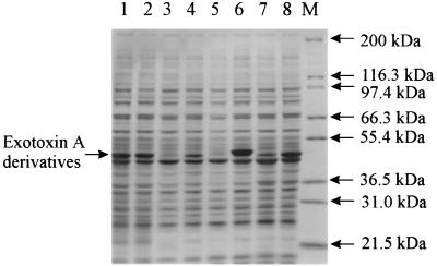
To confirm that amplification of tRNA genes resulted in increased accumulation of the corresponding tRNAs, we performed an RT-PCR analysis of total RNA isolated from BL21(λDE3) cells carrying either pACYC184 or pACYC-IRL10. Our analysis revealed that in order to obtain equal concentrations of PCR-amplified fragments corresponding to products of the ileX, argU, and leuW genes, three or four additional PCR cycles were needed for RNA from cells carrying pACYC184 than for RNA from cells carrying pACYC-IRL10 (data not shown). Furthermore, to confirm that the amplified genes encode functional tRNAs, we constructed three plasmids that encode a fragment of Pseudomonas exotoxin A, pETI10PA10, pETR10PA25, and pETL10PA32. The N-terminal regions of the proteins encoded by these plasmids contained stretches of 10 isolucine (codon ATA), arginine (codon AGA), and leucine (codon CTA) residues, respectively. Figure 2 shows data for expression of Pseudomonas exotoxin A derivatives encoded by these plasmids in BL21(λDE3) cells containing either plasmid pACYC184 or plasmid pACYC-IRL10. Production of exotoxin A derivatives encoded by plasmids pETI10PA10, pETR10PA25, and pETL10PA32 was more efficient in cells containing plasmid pACYC-IRL10 than in cells containing control plasmid pACYC184. Protein encoded by parent plasmid pETPA5 was produced with the same efficiency in cells containing pACYC184 and in cells containing pACYC-IRL10. These data confirm that amplification of the ileX, argU, and leuW genes increased the functional levels of the corresponding tRNAs in E. coli.
FIG. 2.
Effect of amplification of the ileX, argU, and leuW genes in E. coli BL21(λDE3) on production of different derivatives of Pseudomonas exotoxin A. BL21(λDE3) cells were cotransformed with a plasmid encoding a derivative of Pseudomonas exotoxin A (lanes 1 and 2, pETPA5; lanes 3 and 4, pETI10PA10; lanes 5 and 6, pETR10PA25; lanes 7 and 8, pETL10PA32) and with either pACYC184 (lanes 1, 3, 5, and 7) or pACYC-IRL10 (lanes 2, 4, 6, and 8). Cells were induced with IPTG and lysed, and cell proteins were separated by 4 to 20% gradient SDS–PAGE and visualized by Coomassie blue staining. Lane M contained the Mark12 wide-range protein standard from Novex.
Construction of plasmids encoding fragments of BoNTs and expression of these fragments in E. coli.
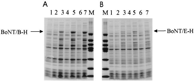
As described above, we constructed a set of plasmids encoding the light chains of BoNT/A, BoNT/B, BoNT/C, and BoNT/E, as well as the heavy chains of BoNT/B, BoNT/C, and BoNT/E. Fragments encoding the light and heavy chains of BoNTs were amplified from clostridial genomic DNA by using primers listed in Table 1. Amplified fragments were cloned into expression vectors to create BoNT fragment-encoding genes whose transcription was under control of the efficient bacteriophage T7 promoter, and the region around the start codon was also optimized to ensure efficient initiation of translation. To analyze expression of our recombinant genes, we introduced these plasmids into BL21(λDE3) cells that simultaneously were transformed with either pACYC184 or derivatives of this plasmid containing the ileX, argU, and leuW genes, and the proteins produced were analyzed by SDS-PAGE. As shown in Fig. 3 and 4, cells cotransformed with both a BoNT fragment-encoding plasmid and plasmid pACYC184 produced proteins of interest in such low quantities that they were not detectable on Coomassie blue-stained gels. Also, we were not able to detect substantial amounts of BoNT fragments in the cells containing plasmid pACYC-Arg34, pACYC-L10, or pACYC-RL5 instead of pACYC184. In contrast, production of recombinant BoNT fragments was substantially greater in cells cotransformed with either pACYC-Ile7, pACYC-IleArg10, or pACYC-IRL10 and BoNT fragment-encoding plasmids. Thus, amplification of the ileX gene plays a major role in increasing the production of BoNT fragments. Also, cells containing plasmid pACYC-IleArg10 or pACYC-IRL10 produced proteins of interest at slightly (up to twofold) higher levels than cells containing plasmid pACYC-Ile7 produced these proteins. This improved production effect was observed with cells that were grown for 1.5 h after induction of expression with IPTG but not in cells grown for 16 h after induction of expression when no significant accumulation of proteins of interest was observed (data not shown).
FIG. 3.
Effect of amplification of the ileX, argU, and leuW genes in E. coli BL21(λDE3) on production of light chains of BoNT/A (A), BoNT/B (B), BoNT/C (C), and BoNT/E (D). The expression of each protein was evaluated in the presence or absence of amplified ileX, argU, or leuW genes, as follows: lane 1, no amplification (pACYC184); lane 2, pACYC-Ile7; lane 3, pACYC-Arg34; lane 4, pACYC-L10; lane 5, pACYC-IleArg10; lane 6, pACYC-RL5; and lane 7, pACYC-IRL10. Cells were induced with IPTG and lysed, and the total cell proteins were separated by 4 to 20% gradient SDS–PAGE and visualized by Coomassie blue staining. The arrows indicate the locations of the various neurotoxin proteins. The molecular weight markers used (lane M) were phosphorylase b (molecular weight, 97,400), bovine serum albumin (66,200), glutamate dehydrogenate (55,000), ovalbumin (42,700), aldolase (40,000), carbonic anhydrase (31,000), soybean trypsin inhibitor (21,500), and lysozyme (14,400).
FIG. 4.
Effect of amplification of the ileX, argU, and leuW genes in E. coli BL21(λDE3) on production of heavy chains of BoNT/B (A) and BoNT/E (B). The expression of each protein was evaluated in the absence or presence of the amplified ileX, argU, or leuW genes, as follows: lane 1, no amplification (pACYC184); lane 2, pACYC-Ile7; lane 3, pACYC-Arg34; lane 4, pACYC-L10; lane 5, pACYC-RL5; lane 6, pACYC-IleArg10; and lane 7, pACYC-IRL10. Cells were induced with IPTG and processed for SDS-PAGE as described in the legend to Fig. 2. The arrows indicate the locations of the expressed proteins BoNT/B-H and BoNT/E-H. The molecular weight markers used (lane M) were the molecular weight markers described in the legend to Fig. 3.
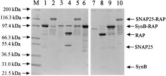
The identities of the proteins were confirmed with specific antibodies. Furthermore, to ensure the functional integrity of the toxin products as proteases, we carried out enzymatic activity tests as described above. Recombinants BoNT/A-L, BoNT/B-L, and BoNT/E-L were recovered from inclusion bodies by using the denaturation-renaturation procedure described above, and their enzymatic activities were tested. Figure 5 shows that BoNT/B-L was active in cleavage of the SynB-RAP fusion protein but was not active when the SNAP25-RAP fusion protein was the substrate. In contrast, BoNT/A-L (Fig. 6) and BoNT/E-L (data not shown) cleaved SNAP25-RAP and but did not cleave the SynB-RAP fusion protein. The activities of recombinant light chains were completely inhibited by EDTA. The specificities of BoNT/B-L and BoNT/E-L were also confirmed in vivo by using previously described systems (10, 13).
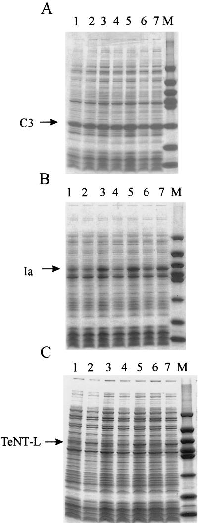
FIG. 5.
Effect of amplification of the ileX, argU, and leuW genes on production of the botulinum C3 protein (A), the iota toxin Ia protein (B), and the light chain of tetanus toxin (C) in BL21(λDE3) cells. The expression of each protein was evaluated in the absence or in the presence of amplified ileX, argU, or leuW genes, as follows: lane 1, no amplification (pACYC184); lane 2, pACYC-Arg34; lane 3, pACYC-Ile7; lane 4, pACYC-L10; lane 5, pACYC-IleArg10; lane 6, pACYC-RL5; and lane 7, pACYC-IRL10. Cells were induced with IPTG and processed for SDS-PAGE as described in the legend to Fig. 2. The arrows indicate the locations of proteins C3 and Ia and the light chain of tetanus toxin (TeNT-L). The molecular weight markers used (lane M) were the molecular weight markers described in the legend to Fig. 3.
FIG. 6.
Enzymatic activities of recombinant light chains of BoNT/A and BoNT/B. Substrate proteins SynB-RAP (lane 1) and SNAP25-RAP (lane 2) and light chains of BoNT/A (lane 3) and BoNT/B (lane 7) were included. Also included were a mixture of BoNT/A-L and SNAP25 in the absence (lane 4) and in the presence (lane 5) of EDTA (lane 5) and a mixture of BoNT/B-L and SynB-RAP in the absence (lane 8) and in the presence (lane 9) of EDTA. Lanes 6 and 10 contained a mixture of BoNT/A-L and SynB-RAP and a mixture of BoNT/B-L and SNAP25, respectively. Proteolytic activity was determined in the presence of 10 mM Tris-HCl (pH 6.8) and 1 mM ZnSO4 for 1.5 h at 37°C. Proteolytic products were separated by 4 to 20% gradient SDS-PAGE, and proteins were visualized with Coomassie blue as described in the text. The arrows on the left indicate the positions of molecular weight markers (lane M) obtained from the Mark12 wide-range protein standard (Novex), and the arrows on the right indicate the locations of substrate proteins and their products.
Construction and expression in E. coli of plasmids encoding proteins from different representatives of the genus Clostridium.
We hypothesized that amplification of tRNAs that recognize rarely used codons may be a useful strategy which has general applicability for improving the efficiency of heterologous expression in E. coli. To study this, we tested the applicability of this procedure for expression of other clostridial toxins. We used the C3 protein (16) from C. botulinum, the light and heavy chains of tetanus toxin from C. tetani, and the iota toxin Ia protein from C. perfringens (17) as prototypes. The corresponding sequences were amplified from the total DNAs of the corresponding microorganisms by using PCR and the specific primers listed in Table 1 and were placed under control of a bacteriphage T7 promoter with an efficient translation initiation site as described above. The resulting plasmids, pTSC3-7 (encoding the C3 protein), pETTeNT-L12Km and pETTeNT-H4Km (encoding the light and heavy chains of tetanus toxin, respectively), and pETiota11Km (encoding the iota toxin Ia protein), were introduced into BL21(λDE3) cells containing either pACYC184 or tRNA-encoding derivatives of this plasmid, and production of the corresponding recombinant proteins was analyzed by SDS-PAGE. Our analysis revealed that unlike plasmids encoding fragments of BoNTs, plasmids pTSC3-7, pETiota11Km, pETTeNT-L12Km (Fig. 5), and pETTeNT-H4Km (data not shown) gave relatively efficient production of recombinant proteins in E. coli cells that contained normal quantities of tRNA genes. Amplification of either ileX, argU, or leuW separately did not result in increased production of the C3 protein. Simultaneous amplification of the ileX and argU genes did slightly improve production of this protein. In the cases of the pETiota11Km, pETTeNT-L12Km (Fig. 5), and pETTeNT-H4Km (data not shown) plasmids, amplification of the ileX gene had a positive effect on production of recombinant proteins. Amplification of the argU gene in addition to amplification of the ileX gene also allowed us to improve production of the heavy chain of tetanus toxin encoded by pETTeNT-H4Km.
DISCUSSION
Low levels of expression of clostridial neurotoxins in traditional organisms such as E. coli may explain why our understanding of the mechanism of action of such an important class of toxins is lagging behind our understanding of the mechanism of action of other toxins. The fact that efficient expression of a tetanus toxin fragment was achieved after complete de novo synthesis of the coding sequence and adjustment of codons in the sequence on the basis of E. coli codon usage indicated the importance of the codons used in the sequence for efficient production of this protein (12). Whether this was fully attributable to codon usage or to other factors, such as the mRNA secondary structure, was not clear. Furthermore, this approach is expensive and requires a substantial amount of preliminary work before each protein can be expressed. In this study, we examined whether rarely used codons play a major role in decreasing the efficiency of production of clostridial neurotoxins in E. coli. By amplifying tRNA genes whose products recognize rarely used codons, we were able to significantly improve production of clostridial neurotoxin fragments in E. coli. We did this without changing the coding sequences of neurotoxin fragments, which confirmed that rarely used codons play a major role in efficient expression of clostridial neurotoxin-encoding genes in E. coli. Table 2 shows the frequencies of rarely used codons in the sequences and the effect of amplification of tRNA-encoding genes on the level of expression. The data show that there is a definite correlation between the frequency of a particular codon in the reading frame and the effect of amplification of the corresponding tRNA-encoding gene on the production of the corresponding protein. Indeed, of the three codons examined (ATA, AGA, and CTA), the ATA codon encoding isoleucine is the most frequently used codon in neurotoxin-encoding sequences, and amplification of the ileX gene has the most dramatic effect on the level of expression of this codon. The AGA codon is less prevalent than the ATA codon, and thus the effect of argU gene amplification is more modest. Nevertheless, there is not a strong correlation between the frequencies of rarely used codons in each gene and the effects of the corresponding tRNA amplification on the efficiency of expression. Indeed, although ATA occurs more frequently with the genes encoding the light chain of tetanus toxin (4.8%) and the iota toxin Ia protein (5.1%) than with the genes encoding the light chains of BoNT/A (3.8%) and BoNT/E (4.0%), amplification of the ileX gene had more dramatic effect on production of the last two proteins than on production of the first two. In addition, even though all of the clostridial genes examined in this study contain the CTA codon, expression of neither of them was improved by amplification of the leuW gene.
TABLE 2.
Codon usage in recombinant genes and effect of amplification of tRNA-encoding genes on expression in E. coli
| Plasmid | Codon usage (%)
|
Effect of tRNA gene amplification on expressiona
|
||||
|---|---|---|---|---|---|---|
| ATA | AGA | CTA | ileX (ATA) | argU (AGA) | leuW (CTA) | |
| PETBoNT/A-L22Km | 3.8 (19)b | 2.0 (10) | 1.0 (5) | ++ | − | − |
| PETBoNT/B-L10 | 6.2 (29) | 2.3 (11) | 0.2 (1) | ++ | + | − |
| pETBoNT/C-L20 | 5.9 (28) | 3.1 (15) | 0.8 (4) | ++ | − | − |
| pETBoNT/E-L31 | 4.0 (18) | 2.4 (11) | 1.5 (7) | ++ | + | − |
| pETBoNT/B-H18 | 6.4 (57) | 1.9 (17) | 1.0 (9) | ++ | + | − |
| pETBoNT/C-H14 | 5.0 (48) | 2.5 (24) | 0.9 (9) | ++ | + | − |
| pETBoNT/E-H10 | 4.4 (40) | 2.2 (20) | 0.3 (3) | ++ | + | − |
| pTSC3-7 | 2.4 (6) | 3.2 (8) | 0.8 (2) | +/− | +/− | − |
| pETiota11Km | 5.1 (23) | 1.7 (8) | 0.8 (4) | + | − | − |
| pETTeNT-L12Km | 4.8 (24) | 1.8 (9) | 1.4 (7) | + | − | − |
| pETTeNT-H4Km | 5.7 (53) | 1.8 (17) | 1.1 (11) | ++ | + | − |
| pETPA5 | 0 | 0 | 0 | − | − | − |
| pETI10PA10 | 2.5 (10) | 0 | 0 | ++ | − | − |
| pETR10PA25 | 0 | 2.5 (10) | 0 | − | ++ | − |
| pETL10PA32 | 0 | 0 | 2.5 (10) | − | − | ++ |
++, strong effect; +, moderate effect; −, no effect; +/−, weak effect.
The values in parentheses are numbers of codons in the coding sequence.
It is noteworthy that the disruptive effect of rarely used codons on translation efficiency depends not only on the prevalence of these codons but also on their relative locations in the gene. Indeed, in recombinant genes encoded by plasmids pETBoNT/E-L31 and pETR10PA25, the frequencies of the AGA codon are practically the same (2.4 and 2.5%, respectively). Nevertheless, amplification of the argU gene had a more pronounced effect on expression of the recombinant gene from plasmid pETR10PA25, in which all 10 AGA codons were clustered together, than on expression of the recombinant gene from plasmid pETBoNT/E-L31, in which 11 AGA codons were randomly spreaded throughout the coding sequence.
The lack of an effect of leuW gene amplification on expression of clostridial proteins suggests that the disruptive effect of rarely used codons becomes noticeable only when the total number and relative frequency of these codons in the gene are higher than certain minimum values. Our results for expression of exotoxin A derivatives encoded by plasmids pETI10PA10, pETR10PA25, and pETL10PA32 also suggest that the effective number of codons varies with each type of codon. Indeed, of the three types of codons tested, the ATA codon had the most dramatic effect on production of proteins in E. coli cells. A stretch of 10 of these codons was sufficient to completely inhibit production of an exotoxin A derivative in E. coli cells that contained normal level of tRNAs. When the same protein was encoded by a gene that contained 10 CTA codons instead of ATA codons, production of the protein was significantly enhanced in the same E. coli cells. Our results also demonstrate that amplification of tRNA-encoding genes for rare codons can be used for optimization of protein production and may be applicable to many other genes for production of proteins that have great commercial value.
ACKNOWLEDGMENTS
We thank Said Goueli and Josephine Grosh (Promega Corporation) for critical reviews of the manuscript and Uri Vertiev (Gamaleya Research Institute of Epidemiology and Microbiology, Moscow, Russia) for providing clostridial DNAs.
REFERENCES
- 1.Ausubel F M, Brent R, Kingston R E, Moore D D, Seidman J G, Smith J A, Struhl K. Current protocols in molecular biology. New York, N.Y: Greene Publishing Associates and Wiley-Interscience; 1987. [Google Scholar]
- 2.Binz T, Kurazono H, Popoff M R, Eklund M W, Sakaguchi G, Kozaki S, Krieglstein K, Henschen A, Gill D M, Niemann H. Nucleotide sequence of the gene encoding Clostridium botulinum neurotoxin type D. Nucleic Acids Res. 1990;18:5556. doi: 10.1093/nar/18.18.5556. [DOI] [PMC free article] [PubMed] [Google Scholar]
- 3.Blasi J, Chapman E R, Yamasaki S, Binz T, Niemann H, Jahn R. Botulinum neurotoxin C1 blocks neurotransmitter release by means of cleaving HPC-1/syntaxin. EMBO J. 1993;12:4821–4828. doi: 10.1002/j.1460-2075.1993.tb06171.x. [DOI] [PMC free article] [PubMed] [Google Scholar]
- 4.Carruthers A, Kiene K, Carruthers J. Botulinum A exotoxin use in clinical dermatology. J Am Acad Dermatol. 1996;34:788–797. doi: 10.1016/s0190-9622(96)90016-x. [DOI] [PubMed] [Google Scholar]
- 5.Del Tito B J, Jr, Ward J M, Hodgson J, Gershater C J, Edwards H, Wysocki L A, Watson F A, Sathe G, Kane J F. Effects of a minor isoleucyl tRNA on heterologous protein translation in Escherichia coli. J Bacteriol. 1995;177:7086–7091. doi: 10.1128/jb.177.24.7086-7091.1995. [DOI] [PMC free article] [PubMed] [Google Scholar]
- 6.East A K, Richardson P T, Allaway D, Collins M D, Roberts T A, Thompson D E. Sequence of the gene encoding type F neurotoxin of Clostridium botulinum. FEMS Microbiol Lett. 1992;75:225–230. doi: 10.1016/0378-1097(92)90408-g. [DOI] [PubMed] [Google Scholar]
- 7.Garcia G M, Mar P K, Mullin D A, Walker J R, Prather N E. The E. coli dnaY gene encodes an arginine transfer RNA. Cell. 1986;45:453–459. doi: 10.1016/0092-8674(86)90331-4. [DOI] [PubMed] [Google Scholar]
- 8.Goldman E, Rosenberg A H, Zubay G, Studier F W. Consecutive low-usage leucine codons block translation only when near the 5′ end of a message in Escherichia coli. J Mol Biol. 1995;245:467–473. doi: 10.1006/jmbi.1994.0038. [DOI] [PubMed] [Google Scholar]
- 9.Hauser D, Eklund M W, Kurazono H, Binz T, Niemann H, Gill D M, Boquet P, Popoff M R. Nucleotide sequence of Clostridium botulinum C1 neurotoxin. Nucleic Acids Res. 1990;18:4924. doi: 10.1093/nar/18.16.4924. [DOI] [PMC free article] [PubMed] [Google Scholar]
- 10.Hua S Y, Raciborska D A, Trimble W S, Charlton M P. Different VAMP/synaptobrevin complexes for spontaneous and evoked transmitter release at the crayfish neuromuscular junction. J Neurophysiol. 1998;80:3233–3246. doi: 10.1152/jn.1998.80.6.3233. [DOI] [PubMed] [Google Scholar]
- 11.Komine Y, Adachi T, Inokuchi H, Ozeki H. Genomic organization and physical mapping of the transfer RNA genes in Escherichia coli K12. J Mol Biol. 1990;212:579–598. doi: 10.1016/0022-2836(90)90224-A. [DOI] [PubMed] [Google Scholar]
- 12.Makoff A J, Oxer M D, Romanos M A, Fairweather N F, Ballantine S. Expression of tetanus toxin fragment C in E. coli: high level expression by removing rare codons. Nucleic Acids Res. 1989;17:10191–10202. doi: 10.1093/nar/17.24.10191. [DOI] [PMC free article] [PubMed] [Google Scholar]
- 13.Martin T F, Kowalchyk J A. Docked secretory vesicles undergo Ca2+-activated exocytosis in a cell-free system. J Biol Chem. 1997;272:14447–14453. doi: 10.1074/jbc.272.22.14447. [DOI] [PubMed] [Google Scholar]
- 14.Nakajima N, Ozeki H, Shimura Y. In vitro transcription of the supB-E tRNA operon of Escherichia coli. Characterization of transcription products. J Biol Chem. 1982;257:11113–11120. [PubMed] [Google Scholar]
- 15.Nakajima N, Ozeki H, Shimura Y. Organization and structure of an E. coli tRNA operon containing seven tRNA genes. Cell. 1981;23:239–249. doi: 10.1016/0092-8674(81)90288-9. [DOI] [PubMed] [Google Scholar]
- 16.Nemoto Y, Namba T, Kozaki S, Narumiya S. Clostridium botulinum C3 ADP-ribosyltransferase gene. Cloning, sequencing, and expression of a functional protein in Escherichia coli. J Biol Chem. 1991;266:19312–19329. [PubMed] [Google Scholar]
- 17.Perelle S, Gibert M, Boquet P, Popoff M R. Characterization of Clostridium perfringens iota-toxin genes and expression in Escherichia coli. Infect Immun. 1993;61:5147–5156. doi: 10.1128/iai.61.12.5147-5156.1993. . (Erratum, 63:4967, 1995.) [DOI] [PMC free article] [PubMed] [Google Scholar]
- 18.Poulet S, Hauser D, Quanz M, Niemann H, Popoff M R. Sequences of the botulinal neurotoxin E derived from Clostridium botulinum type E (strain Beluga) and Clostridium butyricum (strains ATCC 43181 and ATCC 43755) Biochem Biophys Res Commun. 1992;183:107–113. doi: 10.1016/0006-291x(92)91615-w. [DOI] [PubMed] [Google Scholar]
- 19.Rojiani M V, Jakubowski H, Goldman E. Relationship between protein synthesis and concentrations of charged and uncharged tRNATrp in Escherichia coli. Proc Natl Acad Sci USA. 1990;87:1511–1515. doi: 10.1073/pnas.87.4.1511. [DOI] [PMC free article] [PubMed] [Google Scholar]
- 20.Schiavo G, Benfenati F, Poulain B, Rossetto O, Polverino de Laureto P, DasGupta B R, Montecucco C. Tetanus and botulinum-B neurotoxins block neurotransmitter release by proteolytic cleavage of synaptobrevin. Nature. 1992;359:832–835. doi: 10.1038/359832a0. [DOI] [PubMed] [Google Scholar]
- 21.Schiavo G, Poulain B, Rossetto O, Benfenati F, Tauc L, Montecucco C. Tetanus toxin is a zinc protein and its inhibition of neurotransmitter release and protease activity depend on zinc. EMBO J. 1992;11:3577–3583. doi: 10.1002/j.1460-2075.1992.tb05441.x. [DOI] [PMC free article] [PubMed] [Google Scholar]
- 22.Schiavo G, Rossetto O, Catsicas S, Polverino de Laureto P, DasGupta B R, Benfenati F, Montecucco C. Identification of the nerve terminal targets of botulinum neurotoxin serotypes A, D, and E. J Biol Chem. 1993;268:23784–23787. [PubMed] [Google Scholar]
- 23.Schiavo G, Santucci A, Dasgupta B R, Mehta P P, Jontes J, Benfenati F, Wilson M C, Montecucco C. Botulinum neurotoxins serotypes A and E cleave SNAP-25 at distinct COOH-terminal peptide bonds. FEBS Lett. 1993;335:99–103. doi: 10.1016/0014-5793(93)80448-4. [DOI] [PubMed] [Google Scholar]
- 24.Scott A B. Botulinum toxin injection into extraocular muscles as an alternative to strabismus surgery. J Pediatr Ophthalmol Strabismus. 1980;17:21–25. doi: 10.3928/0191-3913-19800101-06. [DOI] [PubMed] [Google Scholar]
- 25.Scott A B, Kennedy R A, Stubbs H A. Botulinum A toxin injection as a treatment for blepharospasm. Arch Ophthalmol. 1985;103:347–350. doi: 10.1001/archopht.1985.01050030043017. [DOI] [PubMed] [Google Scholar]
- 26.Spanjaard R A, Chen K, Walker J R, van Duin J. Frameshift suppression at tandem AGA and AGG codons by cloned tRNA genes: assigning a codon to argU tRNA and T4 tRNA(Arg) Nucleic Acids Res. 1990;18:5031–5036. doi: 10.1093/nar/18.17.5031. [DOI] [PMC free article] [PubMed] [Google Scholar]
- 27.Strickland D K, Ashcom J D, Williams S, Burgess W H, Migliorini M, Argraves W S. Sequence identity between the alpha 2-macroglobulin receptor and low density lipoprotein receptor-related protein suggests that this molecule is a multifunctional receptor. J Biol Chem. 1990;265:17401–17404. [PubMed] [Google Scholar]
- 28.Strickland D K, Ashcom J D, Williams S, Battey F, Behre E, McTigue K, Battey J F, Argraves W S. Primary structure of alpha 2-macroglobulin receptor-associated protein. Human homologue of a Heymann nephritis antigen. J Biol Chem. 1991;266:13364–13369. [PubMed] [Google Scholar]
- 29.Studier F W, Moffatt B A. Use of bacteriophage T7 RNA polymerase to direct selective high-level expression of cloned genes. J Mol Biol. 1986;189:113–130. doi: 10.1016/0022-2836(86)90385-2. [DOI] [PubMed] [Google Scholar]
- 30.Thompson D E, Brehm J K, Oultram J D, Swinfield T J, Shone C C, Atkinson T, Melling J, Minton N P. The complete amino acid sequence of the Clostridium botulinum type A neurotoxin, deduced by nucleotide sequence analysis of the encoding gene. Eur J Biochem. 1990;189:73–81. doi: 10.1111/j.1432-1033.1990.tb15461.x. [DOI] [PubMed] [Google Scholar]
- 31.Thompson D E, Hutson R A, East A K, Allaway D, Collins M D, Richardson P T. Nucleotide sequence of the gene coding for Clostridium barati type F neurotoxin: comparison with other clostridial neurotoxins. FEMS Microbiol Lett. 1993;108:175–182. doi: 10.1111/j.1574-6968.1993.tb06095.x. [DOI] [PubMed] [Google Scholar]
- 32.Wahab S Z, Rowley K O, Holmes W M. Effects of tRNA(1Leu) overproduction in Escherichia coli. Mol Microbiol. 1993;7:253–263. doi: 10.1111/j.1365-2958.1993.tb01116.x. [DOI] [PubMed] [Google Scholar]
- 33.Zdanovsky A G, Chiron M, Pastan I, FitzGerald D J. Mechanism of action of Pseudomonas exotoxin. Identification of a rate-limiting step. J Biol Chem. 1993;268:21791–21799. [PubMed] [Google Scholar]


