

During the current COVID‐19 pandemic, dermatologists in countries with the highest incidences have noted an increase in consultations for chilblain‐like lesions. In the region of Murcia, an area in southeast Spain with a population of around 1.5 million, we and other dermatologists collected these findings over a period of 1 week. In most cases, the photographs were taken by the patients themselves and they consulted their general practitioners before being referred to us via our regional teledermatology platform. Patients were asked about fever, cough, shortness of breath and gastrointestinal symptoms. Only cases with acral erythematous or purplish papules/plaques and no previous history of chilblains or autoimmune disease were included (Fig. 1).
Figure 1.
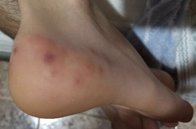
Purpuric oedematous plaques on the heel.
From 13 to 19 April 2020, we collected 41 cases. Mean age was 16 years (range 1–74 years) and 53% of patients were men. The most frequent location of the lesion was the feet alone (80%), followed by the hands and feet together (10%), the hands (7%) and the ears (2%). Of the 41 patients, 6 (14.6%) had extracutaneous symptoms, which preceded the skin lesions in half of the cases. All lesions resolved within days without treatment or only topical steroids for itch relief (Fig. 2).
Figure 2.
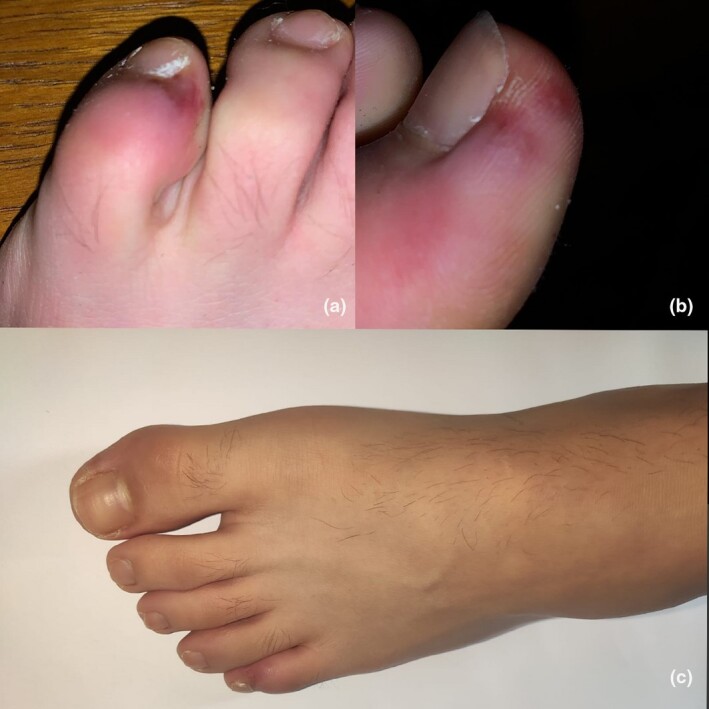
(a,b) Erythematous purpuric lesions associated with secondary blistering of the toes; (c) 14 days later, the lesions had resolved without treatment.
With the restrictions on access to diagnostic tests, we could only perform PCR in 19 cases, which were all negative. However, six patients (14.6%) were cohabiting with at least one person who had a confirmed diagnosis of COVID‐19.
We assess only patients who presented chilblains with no clear explanation for this given their medical history. Although infrequent, one known trigger for this type of lesion is viral infection,1, 2 and the development of this outbreak in spring, with temperatures exceeding 20 °C, just days after reaching the peak number of cases of COVID‐19 in Spain makes us suspect there may well be a connection between both events. Simple pernio is an exceptional finding in spring in this area, give the warm temperature.
Alramthan and Aldaraji2 recently published a report of two similar cases and Piccolo et al.3 also published a similar case series. Kolivras et al.4 described the histopathological findings in one case, which consisted of vacuolar degeneration of the basal layer with scattered necrotic keratinocytes and a dense superficial and deep perivascular and perieccrine lymphocytic infiltrate with no vasculitis.
In a pre‐COVID world, diagnosis of these patients would have been different. In the current situation, with limited PCR tests, our patients usually do not qualify for testing as they have mild, if any, symptoms. The 19 cases we did test by PCR were negative. Likewise, during lockdown, the population is advised against coming into hospitals and biopsies are not taken routinely. Thus, we can not confirm the relation between COVID‐19 and chilblain‐like lesions. However, the epidemiological background, the absence of other triggers and the fact that no lesions of this kind were diagnosed in our area in April 2019 should make clinicians consider the possibility of chilblains being a potential sign of COVID‐19 infection and act accordingly, as asymptomatic and paucisymptomatic patients can potentially transmit viral diseases, such as influenza.4, 5 Serological tests are needed to exclude recent SARS‐CoV‐2 infection in this context, where PCR results are usually negative.
Acknowledgement
We thank our fellow local dermatologists for their invaluable collaboration in this study.
Contributor Information
J. López‐Robles, Department of Dermatology Hospital Morales Meseguer Murcia Spain Department of Dermatology Hospital Virgen del Castillo Yecla, Murcia Spain.
de la I. Hera, Department of Dermatology Hospital de la Vega Lorenzo Guirao Cieza Spain.
J. Pardo‐Sánchez, Dermatology Service Complejo Hospitalario Universitario de Cartagena Murcia Spain
J. Ruiz‐Martínez, Department of Dermatology Hospital General Universitario Reina Sofia Murcia Spain Department of Dermatology Hospital Rafael Mendez Lorca, Murcia Spain.
E. Cutillas‐Marco, Department of Dermatology Hospital General Universitario Reina Sofia Murcia Spain
References
- Crowson AN, Magro CM. Idiopathic perniosis and its mimics: a clinical and histological study of 38 cases. Hum Pathol 1997; 28: 478–84. [DOI] [PubMed] [Google Scholar]
- Alramthan A, Aldaraji W. A case of COVID‐19 presenting in clinical picture resembling chilblains disease. First report from the Middle East. Clin Exp Dermatol 2020;45: 746–8.. [DOI] [PMC free article] [PubMed] [Google Scholar]
- Piccolo V, Neri I, Filippeschi C et al. Chilblain‐like lesions during COVID‐19 epidemic: a preliminary study on 63 patients. J Eur Acad Dermatol Venereol 2020;34: e291–3.. [DOI] [PMC free article] [PubMed] [Google Scholar]
- Kolivras A, Dehavay F, Delplace D et al. Coronavirus (COVID‐19) infection induced chilblains: a case report with histopathological findings. JAAD Case Reports 2020;6: 489–92.. [DOI] [PMC free article] [PubMed] [Google Scholar]
- Ip DK, Lau LL, Leung NH et al. Viral shedding and transmission potential of asymptomatic and paucisymptomatic influenza virus infections in the community. Clin Infect Dis 2017; 64: 736–42. [DOI] [PMC free article] [PubMed] [Google Scholar]