PLOS ONE logoLink to PLOS ONE
. 2022 Jun 22;17(6):e0269323. doi: 10.1371/journal.pone.0269323

Using deep-learning in fetal ultrasound analysis for diagnosis of cystic hygroma in the first trimester

Mark C Walker 1,2,3,4,5,6,*, Inbal Willner 1,5, Olivier X Miguel 2, Malia S Q Murphy 2, Darine El-Chaâr 1,2,4,5, Felipe Moretti 1,5, Alysha L J Dingwall Harvey 2, Ruth Rennicks White 2,5, Katherine A Muldoon 1,2, André M Carrington 7,8, Steven Hawken 2,4, Richard I Aviv 8,9,10
Editor: Ruxandra Stoean11
PMCID: PMC9216531  PMID: 35731736

Abstract

Objective

To develop and internally validate a deep-learning algorithm from fetal ultrasound images for the diagnosis of cystic hygromas in the first trimester.

Methods

All first trimester ultrasound scans with a diagnosis of a cystic hygroma between 11 and 14 weeks gestation at our tertiary care centre in Ontario, Canada were studied. Ultrasound scans with normal nuchal translucency were used as controls. The dataset was partitioned with 75% of images used for model training and 25% used for model validation. Images were analyzed using a DenseNet model and the accuracy of the trained model to correctly identify cases of cystic hygroma was assessed by calculating sensitivity, specificity, and the area under the receiver-operating characteristic (ROC) curve. Gradient class activation heat maps (Grad-CAM) were generated to assess model interpretability.

Results

The dataset included 289 sagittal fetal ultrasound images;129 cystic hygroma cases and 160 normal NT controls. Overall model accuracy was 93% (95% CI: 88–98%), sensitivity 92% (95% CI: 79–100%), specificity 94% (95% CI: 91–96%), and the area under the ROC curve 0.94 (95% CI: 0.89–1.0). Grad-CAM heat maps demonstrated that the model predictions were driven primarily by the fetal posterior cervical area.

Conclusions

Our findings demonstrate that deep-learning algorithms can achieve high accuracy in diagnostic interpretation of cystic hygroma in the first trimester, validated against expert clinical assessment.

Introduction

Artificial intelligence (AI) and machine learning models are increasingly being applied in clinical diagnostics, including medical imaging [1]. Deep-learning is a class of machine learning models inspired by artificial neural networks which can process large amounts of data to identify important features that are predictive of outcomes of interest. Importantly, model performance can be continuously and incrementally improved as data accrues. Deep-learning models perform well in pattern recognition, making them particularly useful in the interpretation of medical images [2]. A systematic review and meta-analysis of studies reporting on the diagnostic performance of deep-learning models in the identification of disease features from medical images found that they performed equivalently to trained clinicians, however, few studies make direct comparisons between deep-learning models and health care professionals [2].

AI has many potential uses in ultrasonography, including automating standardized plane detection, and extracting quantitative information about anatomical structure and function. Inter-operator heterogeneity in ultrasound image acquisition poses unique challenges for the use of AI in this space, and are further impeded by a lack of standardized imaging planes [3]. Still, AI has been successfully tested in a range of organ systems, and there is growing interest for its application in obstetric ultrasonography for fetal structure identification, automated measurement of fetal growth parameters, and for the diagnosis of congenital anomalies [4]. Uptake of AI applications into clinical practice is beginning, however, ongoing investigation is warranted to demonstrate the feasibility, validity and reliability of such tools.

Cystic hygroma is a congenital lymphangioma documented in approximately 1 in 800 pregnancies and 1 in 8 000 live births [5]. It is commonly associated with chromosomal abnormalities including Trisomy 21, Turner Syndrome, and anatomical malformations [68]. Cystic hygroma is diagnosed based on the assessment of nuchal translucency (NT) thickness between the fetal skin and the subcutaneous soft tissue at the neck and cervical spine [4]. Diagnosis is straightforward and possible through routine first- or second-trimester ultrasonography. Affected pregnancies require extensive antenatal and postpartum management which may include early cytogenetic testing for suspected aneuploidy, comprehensive assessment of fetal anatomy, and postpartum surgical intervention [8, 9]. Some studies have investigated automated and semi-automated systems for measuring NT thickness, however, no studies to date have investigated AI and deep-learning models to assess diagnoses associated with NT thickness, including cystic hygroma [10, 11].

Given the unique challenges in applying AI in fetal ultrasonography, cystic hygroma is an ideal condition to investigate the feasibility of using deep-learning models in the interpretation of ultrasound images. We sought to develop a deep-learning model that could analyze fetal ultrasound images and correctly identify cases of cystic hygroma compared to normal controls.

Methods

Study setting and design

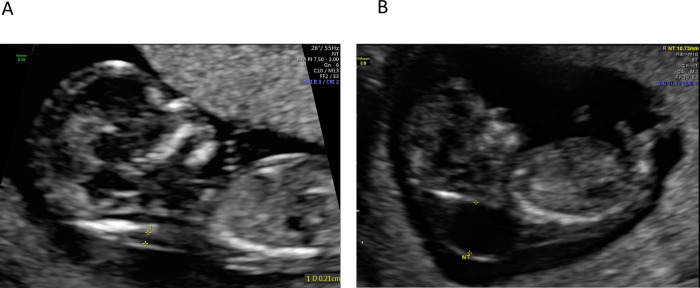
This was a retrospective study conducted at The Ottawa Hospital, a multi-site tertiary-care facility in Ottawa, Canada, with a catchment area of 1.3 million people. First trimester ultrasound images taken between March 2014 and March 2021 were retrieved from the institutional Picture Archiving and Communication System (PACS) and saved in Digital Imaging and Communications in Medicine (DICOM) format. Eligible images were those that included a mid-sagittal view of the fetus taken between 11 and 14 weeks gestation. Cases were identified if there was a final clinical diagnosis of a cystic hygroma in the ultrasound report (Fig 1A). A set of normal ultrasound images from NT screens were used for controls and were retrieved between March 2021 and June 2021 (Fig 1B). Cases and normal images were reviewed and verified by a clinical expert (IW). Patients were not contacted and patient consent was not required to access the images. Data were de-identified and fully anonymized for the model training. This study was reviewed and approved by the Ottawa Health Sciences Network Research Ethics Board (OHSN REB #20210079).

Fig 1.

Fig 1

Fetal ultrasound images of normal (A) and cystic hygroma (B) scans.

A 4-fold cross-validation (4CV) design was used, whereby the same deep-learning architecture was tested and trained four different times using randomly partitioned versions (folds) of the image dataset. For each fold, 75% of the dataset was used for model training, and 25% was used for model validation. The 4CV design was chosen instead of the more commonly used 10-fold cross-validation (10CV) design to optimize the performance of the deep-learning models within our small dataset. With 4CV, each prediction error affects the accuracy of the model by 1.4%, and the sensitivity by 3.1%. Had a 10CV approach been used, each prediction error would have affected model accuracy by 3.4% and the sensitivity by 7.8%.

Data preparation

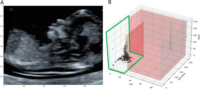
DICOM images included coloured annotations such as calipers, text, icons, profile traces (Fig 2A) and patient personal health information (PHI), which were removed prior to analysis. PHI was handled by cropping the images to remove the identifying information contained in the image borders. Coloured annotations were removed by first converting image data from the Red Green Blue (RGB) colour space to the Hue Saturation Value (HSV) colour space. Image pixels belonging to the grey ultrasound image were empirically identified and ranged from 0–27, 0–150 and 0–255 for the H, S and V values, respectively (Fig 2B).

Fig 2. Identifying image annotations on a normal NT scan.

Fig 2

(A) Image annotations included calipers, text, icons, and profile traces, all of which were removed prior to model training. (B) 3D Scatter Plot of HSV image data. Each point represents one image pixel and its associated HSV values. The red region highlights the range of values which do not belong to the grayscale ultrasound image. The area encircled in green shows pixel values that belong to the grayscale ultrasound image. Grayscale images had H, S and V values ranging from 0–27, 0–150 and 0–255, respectively.

Pixels outside of these ranges were determined to be part of the coloured annotations. Third, a binary mask of the image was created where annotation pixels were labelled ‘1’ and ultrasound image pixels were labelled ‘0’. The binary mask was then dilated with a 5x5 kernel to include contours surrounding the annotations. Last, the Navier-Stokes image infill method [12] was used to artificially reconstruct the ultrasound image without annotations (Fig 3).

Fig 3. Removal of image annotations on a scan with cystic hygroma diagnosis.

Fig 3

(A) Ultrasound image before annotations were removed. Yellow calipers (bottom middle) are visible, along with text annotations (top left). (B) The binary mask of the image which was generated to define the region of the image that need to be infilled (white pixels). (C) Result of the Navier-Stokes image infill method; all image annotations have been removed.

After DICOM images were cleaned, they were converted to grayscale (1 channel image). Intensities were standardized to a mean of zero and a standard deviation of one for better stability during the neural network training [13]. Finally, the images were resized to 256 x 256 pixels.

Neural network model

A DenseNet convolutional neural network (CNN) model architecture[14] was used to classify images as “normal” or those with “cystic hygroma”. Specifically, we used the DenseNet169 PyTorch model [15], with the input layer modified to have a shape of 256 x 256 x 1 pixels and the output layer modified to produce two possible outcome features: 1) normal and 2) cystic hygroma. DenseNet169 was chosen because of its advantages over the ResNet architecture as shown in a publication by Gao et al. (2017) demonstrating that DenseNet achieves high performance while requiring less computation [12]. On some image classification benchmarks DenseNet demonstrated better performance and efficiency than the ResNet model.

Model training

Model training was performed from scratch with random weights initialization for 1000 epochs (i.e., step-in optimization) using the cross-entropy loss function [16], the Adam optimizer [17, 18] (epsilon = 1e-8, beta1 = 0.9, beta2 = 0.999) and a batch size of 64. The available pretrained DenseNet models were trained on ImageNet. ImageNet and fetal ultrasound images are significantly different, therefore CNN models for fetal US applications should not be pretrained on ImageNet. We trained the architecture using our dataset of ultrasound images from The Ottawa Hospital with random weights initialization. The learning rate was set to 1e-2 and reduced by a factor of 0.72 at every 100 epochs (gamma = 0.72, step size = 100) using a learning rate step scheduler [19].

Data augmentation was used during model training to increase the generalizability of the CNN model. Augmentations included random horizontal flip of the ultrasound images (50% probability), random rotation in the range of [–15, 15] degrees, random translation (x±10% and y±30% of image size) and random shearing in the range of [-0.2, 0.2] degrees. These augmentations were performed dynamically at batch loading. Due to the randomness of these operations, a single training image underwent different augmentations at each epoch.

To address imbalance in the number of normal NT and cystic hygroma images in the training dataset, cystic hygroma images were randomly up sampled, with replacement, to match the number of normal NT images.

Model validation

For each epoch, the validation data were used to assess the total number of true and false positives, and true and false negatives and used to calculate accuracy, sensitivity, specificity and the area under the receiver-operating characteristic curve (AUC). For each fold, the performance metrics for the epoch with the highest level of accuracy were reported. The mean, standard deviation and 95% confidence intervals (CI) of the performance metrics across all folds were then computed.

Explainability

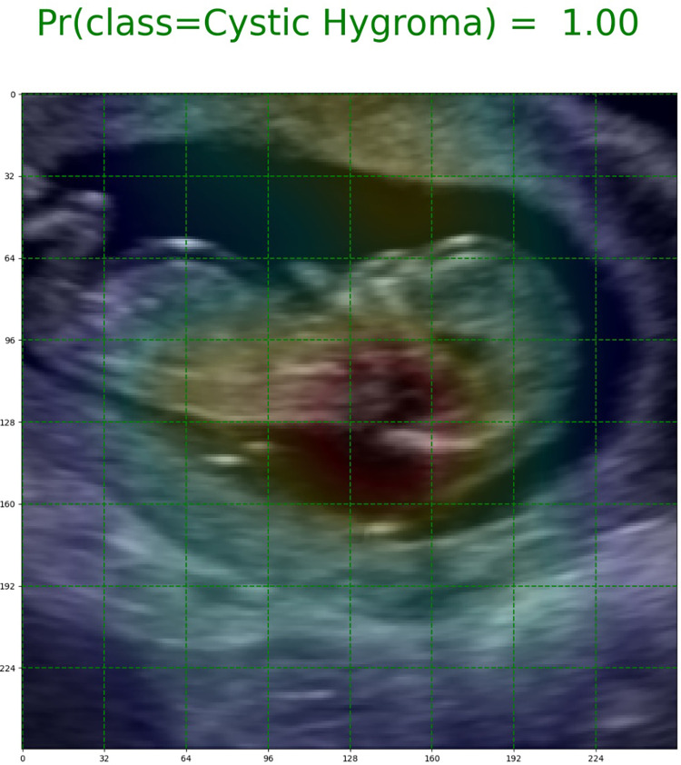
The Gradient-weighted Class Activation Mapping (Grad-CAM) method was used to improve the interpretability of our trained DenseNet models, and visually contextualize important features in the image data that were used for model predictions [20]. Grad-CAM is a widely used technique for the visual explanation of deep-learning algorithms [21, 22]. With this approach, heat maps were generated from 8x8 feature maps to highlight regions of each image that were the most important for model prediction (Fig 4).

Fig 4. Grad-CAM image of a cystic hygroma case.

Fig 4

The green gridlines indicate the size of the feature maps (8x8) used to generate the heat maps. The red highlights the region of the image that influenced the model’s prediction the most.

Results

The image dataset included 289 unique ultrasound images; 160 control images with normal NT measurements, and 129 cases with cystic hygroma. A total of 217 images were used for model training, and 72 images were used for model validation (Table 1).

Table 1. Partitioning of data across training and validation datasetsa.

Overall, n (%) Normal NT images, n (%) Cystic hygroma images, n (%)
Total dataset 289 (100%) 160 (100%) 129 (100%)
Training dataset 217 (75.1%) 120 (75%) 97 (75.2%) b
Validation dataset 72 (24.9%) 40 (25%) 32 (24.8%)

NT, nuchal translucency.

aColumn statistics are provided.

b97 original images; 23 cystic hygroma images were randomly resampled to reduce imbalance between the two groups in the training dataset, to produce in a final cystic hygroma training dataset of 120 images.

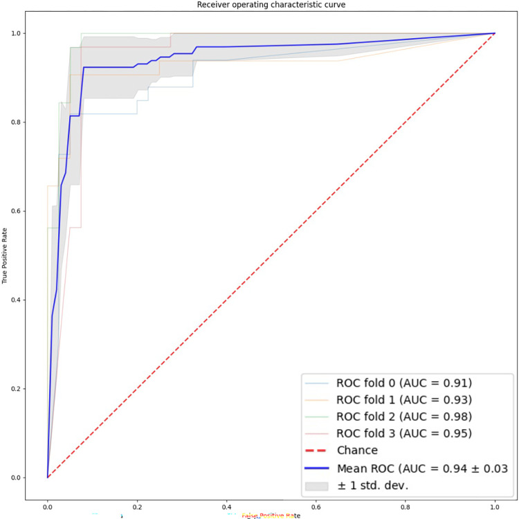
Table 2 shows the results for all 4 cross validation folds. All 4 models performed well in the validation set. The overall mean accuracy was 93% (95% CI: 88–98%), and the area under the receiver operating characteristic curve was 0.94 (95% CI: .89–1.0) (Fig 5). The sensitivity was 92% (95% CI: 79–100%) and the specificity was 94% (95% CI: 91–96%).

Table 2. 4-fold cross validation resultsa.

FOLD NUMBER OVERALL PERFORMANCE
Fold 0 Fold 1 Fold 2 Fold 3 Mean±SD 95% Confidence Interval
True positives, n 27 29 32 31 29.75±1.92 26.0–33.5
True negatives, n 38 38 37 37 37.50±0.50 36.5–38.5
False positives, n 2 2 3 3 2.50±0.50 1.5–3.5
False negatives, n 6 3 0 1 2.50±2.29 0.0–7.0
Accuracy 0.89 0.93 0.96 0.94 0.93±0.03 0.88–0.98
Sensitivity 0.82 0.91 1.00 0.97 0.92±0.07 0.79–1.0
Specificity 0.95 0.95 0.93 0.93 0.94±0.01 0.91–0.96
AUC 0.91 0.93 0.98 0.95 0.94±0.03 0.89–1.0

AUC, area under the curve; SD, standard deviation.

aDecision threshold in the receiver-operating characteristic (ROC) curve was set at 0.5 in the [0,1] range of predicted probability (or class membership).

Fig 5. Receiver operating characteristic plot summarizing performance of all four cross validation folds.

Fig 5

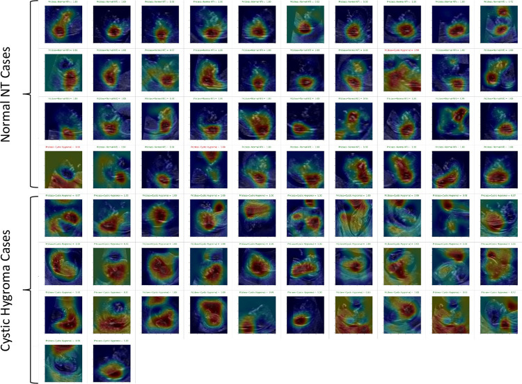
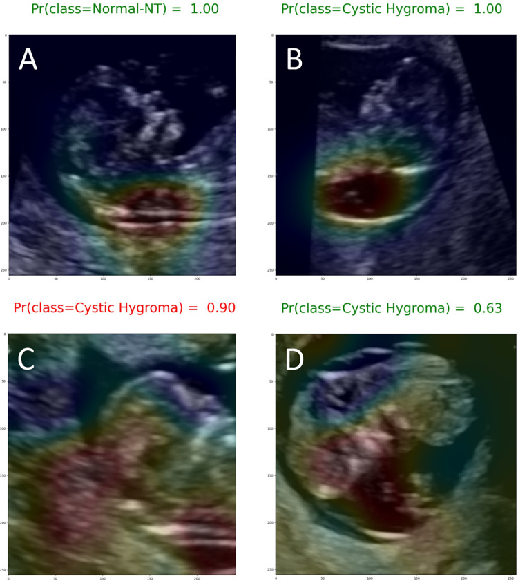
Most of the Grad-CAM heat maps highlighted the fetal head and neck (Fig 6). Although some heat maps specifically highlighted the posterior cervical region, in the area used for NT measurement (Fig 7A and 7B), poor localization did occur (Fig 7C and 7D).

Fig 6. Grad-CAM heat maps for the full validation set of Fold 2.

Fig 6

Top 4 rows are normal NT cases and bottom 4 rows are cystic hygroma cases. Red colours highlight regions of high importance and blue colours highlight regions of low or no importance. Therefore, a good model would have Grad-CAM heatmaps that highlight the head and neck area for both normal and cystic hygroma images.

Fig 7. Exemplary Grad-CAM heat maps.

Fig 7

(A) Normal NT case with good localization in which the model predicted the correct class with a high (1.00) output probability (true negative). (B) Cystic hygroma case with good localization in which the model predicted the correct class with a high (1.00) output probability (true positive). (C) Normal NT case showing poor localization in which the model predicted this class incorrectly with a 0.90 output probability (false positive). (D) Cystic hygroma case showing poor localization in which the model predicted the correct class, but with an output probability that suggests uncertainty (0.63) (true positive).

There were 10 false negatives and 10 false positives. The misclassified images were reviewed with a clinical expert (MW) and it was determined that misclassifications commonly happened when the fetus was close to the placental membrane leading the heat map to focus on another part of the brain.

Discussion

Our findings demonstrate the feasibility of using deep-learning models to interpret fetal ultrasound images and identify cystic hygroma diagnoses with high performance in a dataset of first trimester ultrasound scans. The model achieved excellent prediction of cystic hygroma with a sensitivity of 92% (95% CI: 79–100%) and specificity of 94% (95% CI: 91–96%). This study contributes to the literature on AI and medical diagnostics, and more specifically to the use of AI in fetal ultrasonography where there are scant data.

Ultrasound is critical in the observation of fetal growth and development, however, small fetal structures, involuntary fetal movements and poor image quality make neonatal image acquisition and interpretation challenging. Our study has shown that deep-learning and Grad-CAM heat maps can correctly identify the fetal head and neck region to identify abnormalities. There have been several investigations focusing on AI-based localization of standard planes in fetal ultrasonography, suggesting that AI-models perform as well as clinicians in obtaining reasonable planes for image capture and diagnosis [2327]. Building on this literature, cystic hygroma has not been evaluated yet it is an ideal fetal diagnostic condition to assess the accuracy of AI-based models because it is a clearly visible diagnosis to the trained expert.

More recently, others have sought to apply machine learning methods to the identification of fetal malformations, with promising results. Xie et al. developed and tested a CNN-based deep-learning model in a dataset of nearly 30,000 fetal brain images, including over 14,000 images with common central nervous system abnormalities [28]. Although, the final model was able to discriminate well between normal and abnormal images (sensitivity and specificity were 96.9% and 95.9%, respectively) in a hold out test set, the models were not trained to distinguish between specific brain abnormalities and did not have information on cystic hygroma diagnoses. In a large sample of 2D ultrasounds, fetal echocardiograms and anatomy scans from second trimester pregnancies, Arnaout et al. trained CNN models to accurately localize the fetal heart and detect complex congenital heart disease [29]. Their models achieved high sensitivity (95%, 95%CI, 84–99), and specificity (96%, 95%CI, 95–97) and were validated in several independent datasets. Finally, Baumgartner et al. [25] demonstrated the potential for such systems to operate in real-time. Their findings suggest that deep-learning models could be used “live” to guide sonographers with image capture and recognition during clinical practice. The model proposed by Baumgartner et al. was trained for detection of multiple fetal standard views in freehand 2D ultrasound data. They achieved a 91% recall (i.e., sensitivity) on the profile standard views which is close to the evaluation metrics obtained in our study. Although published data on deep-learning models for the identification of specific malformations are generally lacking, our findings, combined with those of others in this space demonstrate the feasibility of deep-learning models for supporting diagnostic decision-making.

Using the latest data augmentation and image dimensionality techniques, we have developed a deep-learning algorithm with very good predictive accuracy on a relatively small dataset. The strengths of this study include our use of the k-fold cross-validation experiment design. Although computationally intensive, k-fold cross-validation reduces model bias and variance, as most of the data are used for model training and validation. In addition, removal of calipers and text annotations from ultrasound images established a dataset free from the clinical bias on which to develop our models. Annotations on routinely collected ultrasound images have historically limited their utility for medical AI research [30]. Furthermore, our use of Grad-CAM heat maps enabled transparent reporting of how the deep-learning models performed on a case-by-case basis. With this approach, we were able to confirm excellent localization for most of the images used in our dataset. Class-discriminative visualization enables the user to understand where models fail (i.e., why the models predicted what they predicted) and can be used to inform downstream model enhancements. Additionally, all false negative and false positive images were reviewed and it was determined that misclassifications commonly occurred when the fetus was close to the placental membrane. Future work could collect more images where the fetus is close to membrane, up-sample the images that were error prone, and further train the model to incorporate this pattern.

Our study is not without limitations. First, as a single centre study, the sample size available for developing and validating deep-learning model was relatively small. However, use of data augmentation to increase the variability in our dataset, enrichment of cystic hygroma cases in our training set, and use of the k-fold cross validation experiment design are all well-accepted strategies to overcome the limitations of small datasets [31]. Second, although we removed all image annotations, we cannot discount the possibility that the blending and infill methods used to reconstitute the images influenced the deep-learning algorithm. However, the Grad-CAM heatmaps provide reassurance that fetal craniocervical regions were driving the deep-learning algorithm, and that the model appropriately places high importance on regions which are clinically relevant for diagnosis. Given the relatively low incidence of congenital anomalies such as cystic hygroma, a natural extension of this work will be to introduce our models to a larger, multi-centre dataset with more variability in the image parameters and greater feature variety specific to cystic hygroma.

Conclusions

In this proof-of-concept study, we demonstrate the potential for deep-learning to support early and reliable identification of cystic hygroma from first trimester ultrasound scans. We present a novel application of convolutional neural networks to automatically identify cases of cystic hygroma and localise the relevant fetal structures for clinical decision-making. With further development, including testing in a large multi-site dataset and external validation, our approach may be applied to a range of other fetal anomalies typically identified by ultrasonography.

Acknowledgments

The authors would like to acknowledge that this study took place on unceded Algonquin Anishinabe territory.

Data Availability

The ultrasound images used in this study are not currently available to upload to PLOS ONE. All images belong to The Ottawa Hospital. A formal request can be made to the IRB, who would then consult a privacy officer at The Ottawa Hospital. Requests can be sent to the corresponding author (Dr. Mark Walker) or the Ottawa Health Sciences Network Research Ethics Board (rebadministration@ohri.ca)/(http://www.ohri.ca/ohsn-reb/contacts.htm).

Funding Statement

This study was supported by a Canadian Institutes of Health Research Foundation Grant (FDN 148438)(MCW). The funding agency played no role in the study design, data collection and analysis, decision to publish, or preparation of the manuscript.

References

Decision Letter 0

Ruxandra Stoean

25 Mar 2022

PONE-D-21-40543Using deep-learning algorithms in fetal ultrasound analysis for diagnosis of cystic hygroma in the first trimesterPLOS ONE

Dear Dr. Walker,

Thank you for submitting your manuscript to PLOS ONE. After careful consideration, we feel that it has merit but does not fully meet PLOS ONE’s publication criteria as it currently stands. Therefore, we invite you to submit a revised version of the manuscript that addresses the points raised during the review process.

The positioning of the paper against the state of the art needs to be more thoroughly addressed. Also the experiments (setup + results) need further clarification. The choice for selecting the DenseNet169 model is not explained; at least another different architecture should be applied in comparison, in order to highlight DenseNet169 as being the better alternative. The interpretation of the visual part (human + GradCAM) is not detailed. The cases that were erroneously predicted should be discussed.

Please submit your revised manuscript by May 09 2022 11:59PM. If you will need more time than this to complete your revisions, please reply to this message or contact the journal office at plosone@plos.org. When you're ready to submit your revision, log on to https://www.editorialmanager.com/pone/ and select the 'Submissions Needing Revision' folder to locate your manuscript file.

Please include the following items when submitting your revised manuscript:

  • A rebuttal letter that responds to each point raised by the academic editor and reviewer(s). You should upload this letter as a separate file labeled 'Response to Reviewers'.

  • A marked-up copy of your manuscript that highlights changes made to the original version. You should upload this as a separate file labeled 'Revised Manuscript with Track Changes'.

  • An unmarked version of your revised paper without tracked changes. You should upload this as a separate file labeled 'Manuscript'.

If you would like to make changes to your financial disclosure, please include your updated statement in your cover letter. Guidelines for resubmitting your figure files are available below the reviewer comments at the end of this letter.

If applicable, we recommend that you deposit your laboratory protocols in protocols.io to enhance the reproducibility of your results. Protocols.io assigns your protocol its own identifier (DOI) so that it can be cited independently in the future. For instructions see: https://journals.plos.org/plosone/s/submission-guidelines#loc-laboratory-protocols. Additionally, PLOS ONE offers an option for publishing peer-reviewed Lab Protocol articles, which describe protocols hosted on protocols.io. Read more information on sharing protocols at https://plos.org/protocols?utm_medium=editorial-email&utm_source=authorletters&utm_campaign=protocols.

We look forward to receiving your revised manuscript.

Kind regards,

Ruxandra Stoean

Academic Editor

PLOS ONE

Journal Requirements:

When submitting your revision, we need you to address these additional requirements.

1. Please ensure that your manuscript meets PLOS ONE's style requirements, including those for file naming. The PLOS ONE style templates can be found at 

https://journals.plos.org/plosone/s/file?id=wjVg/PLOSOne_formatting_sample_main_body.pdf and 

https://journals.plos.org/plosone/s/file?id=ba62/PLOSOne_formatting_sample_title_authors_affiliations.pdf

2. Please provide additional details regarding participant consent. In the ethics statement in the Methods and online submission information, please ensure that you have specified (1) whether consent was informed and (2) what type you obtained (for instance, written or verbal, and if verbal, how it was documented and witnessed). If your study included minors, state whether you obtained consent from parents or guardians. If the need for consent was waived by the ethics committee, please include this information.

If you are reporting a retrospective study of medical records or archived samples, please ensure that you have discussed whether all data were fully anonymized before you accessed them and/or whether the IRB or ethics committee waived the requirement for informed consent. If patients provided informed written consent to have data from their medical records used in research, please include this information.

3. We note that the grant information you provided in the ‘Funding Information’ and ‘Financial Disclosure’ sections do not match. 

When you resubmit, please ensure that you provide the correct grant numbers for the awards you received for your study in the ‘Funding Information’ section.

4. Thank you for stating the following in the Acknowledgments Section of your manuscript: " This study was supported by a Canadian Institutes of Health Research Foundation Grant (FDN 148438). The funding agency was not involved in study design, analysis or interpretation of data. The agency was not involved writing of this manuscript or in the decision to submit the article for publication"

We note that you have provided funding information that is not currently declared in your Funding Statement. However, funding information should not appear in the Acknowledgments section or other areas of your manuscript. We will only publish funding information present in the Funding Statement section of the online submission form. 

Please remove any funding-related text from the manuscript and let us know how you would like to update your Funding Statement. Currently, your Funding Statement reads as follows: 

"This study was supported by a Canadian Institutes of Health Research Foundation Grant (FDN 148438)(MCW). The funding agency payed no role in the study design, data collection and analysis, decision to publish, or preparation of the manuscript."

Please include your amended statements within your cover letter; we will change the online submission form on your behalf.

5. We note that you have indicated that data from this study are available upon request. PLOS only allows data to be available upon request if there are legal or ethical restrictions on sharing data publicly. For more information on unacceptable data access restrictions, please see http://journals.plos.org/plosone/s/data-availability#loc-unacceptable-data-access-restrictions. 

In your revised cover letter, please address the following prompts:

a) If there are ethical or legal restrictions on sharing a de-identified data set, please explain them in detail (e.g., data contain potentially sensitive information, data are owned by a third-party organization, etc.) and who has imposed them (e.g., an ethics committee). Please also provide contact information for a data access committee, ethics committee, or other institutional body to which data requests may be sent.

b) If there are no restrictions, please upload the minimal anonymized data set necessary to replicate your study findings as either Supporting Information files or to a stable, public repository and provide us with the relevant URLs, DOIs, or accession numbers. For a list of acceptable repositories, please see http://journals.plos.org/plosone/s/data-availability#loc-recommended-repositories.

We will update your Data Availability statement on your behalf to reflect the information you provide.

6. PLOS requires an ORCID iD for the corresponding author in Editorial Manager on papers submitted after December 6th, 2016. Please ensure that you have an ORCID iD and that it is validated in Editorial Manager. To do this, go to ‘Update my Information’ (in the upper left-hand corner of the main menu), and click on the Fetch/Validate link next to the ORCID field. This will take you to the ORCID site and allow you to create a new iD or authenticate a pre-existing iD in Editorial Manager. Please see the following video for instructions on linking an ORCID iD to your Editorial Manager account: https://www.youtube.com/watch?v=_xcclfuvtxQ

[Note: HTML markup is below. Please do not edit.]

Reviewers' comments:

Reviewer's Responses to Questions

Comments to the Author

1. Is the manuscript technically sound, and do the data support the conclusions?

The manuscript must describe a technically sound piece of scientific research with data that supports the conclusions. Experiments must have been conducted rigorously, with appropriate controls, replication, and sample sizes. The conclusions must be drawn appropriately based on the data presented.

Reviewer #1: Partly

Reviewer #2: No

Reviewer #3: Yes

**********

2. Has the statistical analysis been performed appropriately and rigorously?

Reviewer #1: No

Reviewer #2: No

Reviewer #3: Yes

**********

3. Have the authors made all data underlying the findings in their manuscript fully available?

The PLOS Data policy requires authors to make all data underlying the findings described in their manuscript fully available without restriction, with rare exception (please refer to the Data Availability Statement in the manuscript PDF file). The data should be provided as part of the manuscript or its supporting information, or deposited to a public repository. For example, in addition to summary statistics, the data points behind means, medians and variance measures should be available. If there are restrictions on publicly sharing data—e.g. participant privacy or use of data from a third party—those must be specified.

Reviewer #1: No

Reviewer #2: No

Reviewer #3: No

**********

4. Is the manuscript presented in an intelligible fashion and written in standard English?

PLOS ONE does not copyedit accepted manuscripts, so the language in submitted articles must be clear, correct, and unambiguous. Any typographical or grammatical errors should be corrected at revision, so please note any specific errors here.

Reviewer #1: Yes

Reviewer #2: Yes

Reviewer #3: Yes

**********

5. Review Comments to the Author

Please use the space provided to explain your answers to the questions above. You may also include additional comments for the author, including concerns about dual publication, research ethics, or publication ethics. (Please upload your review as an attachment if it exceeds 20,000 characters)

Reviewer #1: The proposed work deals with a DenseNet architecture for Cystic hygroma diagnosis in fetal ultrasound images. The paper is technically intermediate; I have the following issues that need to be resolved before acceptance.

1) The title of the paper is "Using Deep Learning Algorithms ...", but, the authors used only one DenseNet169 architecture. The name should be changed.

2) If there are other related works, then cite and describe them, what is the novelty of your work compared to such works.

3) In the Study Setup and Design section, it is mentioned that a 4-fold cross-validation was used instead of a 10-fold cross-validation. For clarity, show learning curves to better describe the behavior of the model.

4) On line 97, it written that “four different deep learning models were tested and trained…”, but only the DenseNet169 architecture was used in the work. This needs to be clarified.

5) Explain why, among all known architectures, the DenseNet169 architecture was used, give arguments for such a choice. How were the hyper-parameters chosen?

6) It is necessary to clarify the details of the architecture. So the DenseNet169 model was trained from scratch? Have the architecture weights been fine-tuned? From which layer?

7) References used in the discussion section should be cited first in Introduction. The discussion of the results should be clarified.

Reviewer #2: In the Model Training, the authors did not describe the detials of method of training the CNN model by using the utrasound image, as well as did not describe the structure of the CNN. If the authors just applied the CNN model ,which is trained by other peoples, to classify images as “normal” or those with “cystic hygroma”, I think this way is not suitable for the medical image, because the medical images are different from nature image. The author should build the network by themselves, including make the image label for the model training.

Reviewer #3: This manuscript described a study used DenseNet to classify fetal ultrasound image for diagnosis of cystic hygroma. The proposed method for the application is reasonable and the results were promising and expected. In this study, the authors also used Grad-CAM to demonstrate the explainability of deep learning model. This manuscript was well organized.

I have three concerns to this manuscript:

1. If the diagnosis of cystic hygroma is easy for human eyes, what is the effect of having such a model?

2. From the Fig. 5, the red color regions in normal and cystic hygroma seem at same location (head and neck). Thus the authors should explain how to interpret the difference between normal and cystic hygroma.

3. The number of error cases in the study is small. The authors should discuss what happened in these cases.

**********

6. PLOS authors have the option to publish the peer review history of their article (what does this mean?). If published, this will include your full peer review and any attached files.

If you choose “no”, your identity will remain anonymous but your review may still be made public.

Do you want your identity to be public for this peer review? For information about this choice, including consent withdrawal, please see our Privacy Policy.

Reviewer #1: No

Reviewer #2: No

Reviewer #3: No

[NOTE: If reviewer comments were submitted as an attachment file, they will be attached to this email and accessible via the submission site. Please log into your account, locate the manuscript record, and check for the action link "View Attachments". If this link does not appear, there are no attachment files.]

While revising your submission, please upload your figure files to the Preflight Analysis and Conversion Engine (PACE) digital diagnostic tool, https://pacev2.apexcovantage.com/. PACE helps ensure that figures meet PLOS requirements. To use PACE, you must first register as a user. Registration is free. Then, login and navigate to the UPLOAD tab, where you will find detailed instructions on how to use the tool. If you encounter any issues or have any questions when using PACE, please email PLOS at figures@plos.org. Please note that Supporting Information files do not need this step.

PLoS One. 2022 Jun 22;17(6):e0269323. doi: 10.1371/journal.pone.0269323.r002

Author response to Decision Letter 0


8 May 2022

Response to Reviewers

General comments:

The positioning of the paper against the state of the art needs to be more thoroughly addressed. Also the experiments (setup + results) need further clarification. The choice for selecting the DenseNet169 model is not explained; at least another different architecture should be applied in comparison, in order to highlight DenseNet169 as being the better alternative. The interpretation of the visual part (human + GradCAM) is not detailed. The cases that were erroneously predicted should be discussed

• Thank you for raising these points for clarification. We have clarified that we did not conduct a comparison paper on multiple architectures. Our study was designed to assess if DenseNet169 is an appropriate architecture, rather than how it compares to other architectures. We chose DenseNet169 because it has more connections than other architectures (i.e. ResNet).

• Interpretation of the visual part (human +GradCam) is not detailed – We have expanded on this issue. All cystic hygroma images had been diagnosed in ultrasound reports and each image was abstracted and confirmed by a clinical expert independent review (IW). The following text has been added:

o ‘Cases were images if there was a final clinical diagnosis of a cystic hygroma in the ultrasound report. A set of normal ultrasound images from NT screens were used for controls and were retrieved between March 2021 and June 2021. All images were reviewed and verified by a clinical expert (IW).’(Methods, Study setting and design)

• There were 10 false negatives and 10 false positives. The misclassified images were reviewed with an independent clinical expert (MCW) and it was determined that misclassifications commonly happened when the fetus was close to the placental membrane leading the heat map to focus on another part of the anatomy. To improve this, future work could collect more images that have fetus close to membrane and upsample the images that were error prone. The following text has been added:

o ‘There were 10 false negatives and 10 false positives. The misclassified images were reviewed with a clinical expert (MW) and it was determined that misclassifications commonly happened when the fetus was close to the placental membrane leading the heat map to focus on another part of the brain.’(Results)

1. Please ensure that your manuscript meets PLOS ONE's style requirements, including those for file naming. The PLOS ONE style templates can be found at

https://journals.plos.org/plosone/s/file?id=wjVg/PLOSOne_formatting_sample_main_body.pdf and

https://journals.plos.org/plosone/s/file?id=ba62/PLOSOne_formatting_sample_title_authors_affiliations.pdf

• Thank you. The manuscript has been reviewed and formatted with PLOS ONE style requirements

2. Please provide additional details regarding participant consent. In the ethics statement in the Methods and online submission information, please ensure that you have specified (1) whether consent was informed and (2) what type you obtained (for instance, written or verbal, and if verbal, how it was documented and witnessed). If your study included minors, state whether you obtained consent from parents or guardians. If the need for consent was waived by the ethics committee, please include this information.

• Thank you for raising these points for clarification. This study was a retrospective study of ultrasound images and consent was waived. We have included the following text in the manuscript:

o ‘Patients were not contacted, and patient consent was not required to access the images. Data were de-identified and fully anonymized for the model training. This study was reviewed and approved by the Ottawa Health Sciences Network Research Ethics Board (OHSN REB #20210079).’ (Methods)

3. If you are reporting a retrospective study of medical records or archived samples, please ensure that you have discussed whether all data were fully anonymized before you accessed them and/or whether the IRB or ethics committee waived the requirement for informed consent. If patients provided informed written consent to have data from their medical records used in research, please include this information.

• Thank you for raising these points for clarification. This study was a retrospective study of ultrasound images and all images were fully anonymized prior to model training. We have included the following text in the manuscript:

o ‘Patients were not contacted, and patient consent was no required to access the images. Data were de-identified and fully anonymized for the model training. This study was reviewed and approved by the Ottawa Health Sciences Network Research Ethics Board (OHSN REB #20210079).’ (Methods)

3. We note that the grant information you provided in the ‘Funding Information’ and ‘Financial Disclosure’ sections do not match. When you resubmit, please ensure that you provide the correct grant numbers for the awards you received for your study in the ‘Funding Information’ section.

• Thank you for raising these points for clarification. All information on funding has been removed from the manuscript and is only included in the online submission form.

4. Thank you for stating the following in the Acknowledgments Section of your manuscript: " This study was supported by a Canadian Institutes of Health Research Foundation Grant (FDN 148438). The funding agency was not involved in study design, analysis or interpretation of data. The agency was not involved writing of this manuscript or in the decision to submit the article for publication". We note that you have provided funding information that is not currently declared in your Funding Statement. However, funding information should not appear in the Acknowledgments section or other areas of your manuscript. We will only publish funding information present in the Funding Statement section of the online submission form.

• Thank you for raising these points for clarification. All information on funding has been removed from the manuscript and is only included in the online submission form.

5. We note that you have indicated that data from this study are available upon request. PLOS only allows data to be available upon request if there are legal or ethical restrictions on sharing data publicly. For more information on unacceptable data access restrictions, please see http://journals.plos.org/plosone/s/data-availability#loc-unacceptable-data-access-restrictions.

In your revised cover letter, please address the following prompts:

a) If there are ethical or legal restrictions on sharing a de-identified data set, please explain them in detail (e.g., data contain potentially sensitive information, data are owned by a third-party organization, etc.) and who has imposed them (e.g., an ethics committee). Please also provide contact information for a data access committee, ethics committee, or other institutional body to which data requests may be sent.

• Thank you for raising this point. The ultrasound images are not currently available to upload to PLOS ONE. All images belong to The Ottawa Hospital. A formal request can be made to the IRB, who would then consult a privacy officer at The Ottawa Hospital. Requests can be sent to the corresponding author (Dr. Mark Walker) or the Ottawa Health Sciences Network Research Ethics Board (rebadministration@ohri.ca). We have also included this information in the cover letter

b) If there are no restrictions, please upload the minimal anonymized data set necessary to replicate your study findings as either Supporting Information files or to a stable, public repository and provide us with the relevant URLs, DOIs, or accession numbers. For a list of acceptable repositories, please see http://journals.plos.org/plosone/s/data-availability#loc-recommended-repositories.

We will update your Data Availability statement on your behalf to reflect the information you provide.

• Thank you for updating the data availability statement on our behalf.

6. PLOS requires an ORCID iD for the corresponding author in Editorial Manager on papers submitted after December 6th, 2016. Please ensure that you have an ORCID iD and that it is validated in Editorial Manager.

• Thank you for raising this point. The ORCID ID has been added for the corresponding author.

Reviewer #1: The proposed work deals with a DenseNet architecture for Cystic hygroma diagnosis in fetal ultrasound images. The paper is technically intermediate; I have the following issues that need to be resolved before acceptance.

1) The title of the paper is "Using Deep Learning Algorithms ...", but, the authors used only one DenseNet169 architecture. The name should be changed.

• Thank you for highlighting this point. We have changed the title to ‘Using deep-learning in fetal ultrasound analysis for diagnosis of cystic hygroma in the first trimester’.

2) If there are other related works, then cite and describe them, what is the novelty of your work compared to such works.

• Thank you for highlighting this point. We have expanded the discussion section to highlight other related works and emphasize the novelty of our project. Briefly, the main novelty of this study is the application of deep-learning and explainable AI techniques specifically for cystic hygroma diagnosis. Other works in the field used similar technical methods (deep-learning + GradCAM) but with different conditions (i.e. medical diagnoses not cystic hygroma). The references below have been emphasized in the manuscript:

o Baumgartner CF, Kamnitsas K, Matthew J, Fletcher TP, Smith S, Koch LM, Kainz B, Rueckert D. SonoNet: Real-Time Detection and Localisation of Fetal Standard Scan Planes in Freehand Ultrasound. IEEE Transactions on Medical Imaging. 2017;36(11):2204-2215. doi:10.1109/TMI.2017.2712367

o Xie HN, Wang N, He M, Zhang LH, Cai HM, Xian JB, Lin MF, Zheng J, Yang YZ. Using deep‐learning algorithms to classify fetal brain ultrasound images as normal or abnormal. Ultrasound in Obstetrics & Gynecology. 2020;56(4):579-587. doi:10.1002/uog.21967

o Arnaout R, Curran L, Zhao Y, Levine JC, Chinn E, Moon-Grady AJ. An ensemble of neural networks provides expert-level prenatal detection of complex congenital heart disease. Nature Medicine. 2021;27(5):882-891. doi:10.1038/s41591-021-01342-5

3) In the Study Setup and Design section, it is mentioned that a 4-fold cross-validation was used instead of a 10-fold cross-validation. For clarity, show learning curves to better describe the behavior of the model.

• Thank you for raising these points for clarification. We conducted a 4-fold cross-validation. The 4-fold cross-validation (4CV) design was chosen instead of the more commonly used 10-fold cross-validation (10CV) design to optimize the performance of the deep-learning models within our small dataset. With 4CV, each prediction error affects the accuracy of the model by 1.4%, and the sensitivity by 3.1%. Had a 10CV approach been used, each prediction error would have affected model accuracy by 3.4% and the sensitivity by 7.8%.

4) On line 97, it written that “four different deep learning models were tested and trained…”, but only the DenseNet169 architecture was used in the work. This needs to be clarified.

• Thank you for raising this point. We conducted a 4-fold cross-validation where every fold in the cross-validation experiment trains one DenseNet169 architecture. Therefore, each fold produces 1 trained model yielding a total of 4 different DenseNet169 models (i.e. same architecture, trained on 4 folds, producing 4 different models).

• The following text has been added:

o ‘A 4-fold cross-validation (4CV) design was used, whereby the same deep-learning architecture was tested and trained four different times using randomly partitioned versions (folds) of the image dataset. For each fold, 75% of the dataset was used for model training, and 25% was used for model validation. The 4CV design was chosen instead of the more commonly used 10-fold cross-validation (10CV) design to optimize the performance of the deep-learning models within our small dataset. With 4CV, each prediction error affects the accuracy of the model by 1.4%, and the sensitivity by 3.1%. Had a 10CV approach been used, each prediction error would have affected model accuracy by 3.4% and the sensitivity by 7.8%.’ (Methods, Study setting and design)

5) Explain why, among all known architectures, the DenseNet169 architecture was used, give arguments for such a choice. How were the hyper-parameters chosen?

• Thank you for raising these points for clarification. We have expanded on this in the methods section. The following text has been added:

o ‘DenseNet169 was chosen because of its advantages over the ResNet architecture as shown in [12]. On some image classification benchmarks DenseNet demonstrated better performance and efficiency than the ResNet model.’ (Methods, Neural Network Model)

o ‘Default hyperparameters were chosen and no hyperparameters tuning was conducted. Batch size was chosen to maximize graphic processing units (GPU) memory usage. Learning rate (lr) scheduler parameters were chosen to cover lr range of 0.01 to 0.0005. Large lr (0.01) are good for initialization of the model but will not converge as well as smaller lr (0.0005). Therefore, a decreasing lr is used to get the best of both large and small lrs.’ (Methods, Model Training)

6) It is necessary to clarify the details of the architecture. So, the DenseNet169 model was trained from scratch? Have the architecture weights been fine-tuned? From which layer?

• Thank you for raising these points for clarification. Yes, the DenseNet169 was trained from scratch and did not use pretrained weights. Pretrained DenseNet169 architecture was not available for grayscale images and potentially would not have worked appropriately with medical ultrasound images. We trained the architecture using our dataset of ultrasound images from The Ottawa Hospital with random weights initialization.

• The following text has been added to the manuscript:

o ‘Pretrained DenseNet169 architecture was not available for grayscale images and potentially would not have worked appropriately with medical ultrasound images. We trained the architecture using our dataset of ultrasound images from The Ottawa Hospital with random weights initialization.’(Methods, Model Training)

7) References used in the discussion section should be cited first in Introduction. The discussion of the results should be clarified.

• Thank you for highlighting this point. We have adjusted to introduction to include the references.

Reviewer #2: In the Model Training, the authors did not describe the details of method of training the CNN model by using the ultrasound image, as well as did not describe the structure of the CNN. If the authors just applied the CNN model ,which is trained by other peoples, to classify images as “normal” or those with “cystic hygroma”, I think this way is not suitable for the medical image, because the medical images are different from nature image. The author should build the network by themselves, including make the image label for the model training.

• Thank you for raising these points for consideration. This is an important issue that was raised with Reviewer 1 as well. We have expanded this section in detail and including more information on the method for training the CNN model and choice of architecture. Please see the comments to Reviewer 1 in Questions 3,4,5 and 6

• We have referenced the full description of the architecture of the DenseNet for readers who want to know more. See comments to reviewer 1 question 5 for explanation of our decision to use DenseNet.

• We used off-the-shelves DenseNet169 model trained from scratch. No pretrained weights and no fine-tuning was done. In other words, we built the dataset (made the image label) and trained the model ourselves (trained from scratch). We did not use a model trained by other people (ex: pretrained on ImageNet dataset) for two main reasons: 1) Models pretrained with ultrasound images were not readily available; 2) Medical images such as ultrasound are significantly different from natural images (eg. RGB vs grayscale, ultrasound artefacts different than natural image artefacts etc.) therefore models pretrained with natural images could be unsuitable.

Reviewer #3: This manuscript described a study used DenseNet to classify fetal ultrasound image for diagnosis of cystic hygroma. The proposed method for the application is reasonable and the results were promising and expected. In this study, the authors also used Grad-CAM to demonstrate the explainability of deep learning model. This manuscript was well organized. I have three concerns to this manuscript:

1. If the diagnosis of cystic hygroma is easy for human eyes, what is the effect of having such a model?

• Thank you for raising these points for clarification. Through fetal ultrasound, cystic hygroma is an easily identifiable congenital anomaly for trained experts. We began our pilot study with a fetal anomaly with a clearly visible diagnosis to assess the feasibility of using deep learning with ultrasounds before moving on to more challenging diagnoses. The following text has been included in the manuscript:

o ‘There have been several investigations focusing on AI-based localization of standard planes in fetal ultrasonography, suggesting that AI-models perform as well as clinicians in obtaining reasonable planes for image capture and diagnosis,[21–25]. Building on this literature, cystic hygroma has not been evaluated yet it is an ideal fetal diagnostic condition to assess the accuracy of AI-based models because it is a clearly visible diagnosis to the trained expert.’(Discussion)

2. From the Fig. 5, the red color regions in normal and cystic hygroma seem at same location (head and neck). Thus, the authors should explain how to interpret the difference between normal and cystic hygroma.

• Thank you for raising these points for clarification. The head and neck area are the correct area to distinguish cystic hygroma from normal. In figure 5, the GradCAM confirmed that the convolutional neural network was giving attention to the correct anatomical location for cystic hygroma ascertainment. Therefore, a good model would have GradCAM heatmaps that highlight the head and neck area for both normal and cystic hygroma images since a normal image is the absence of the disease.

• The following text has been added:

o ‘Fig 5. Grad-CAM heat maps for the full validation set of Fold 2. Top 4 rows are normal NT cases and bottom 4 rows are cystic hygroma cases. Red colours highlight regions of high importance and blue colours highlight regions of low or no importance. Therefore, a good model would have GradCAM heatmaps that highlight the head and neck area for both normal and cystic hygroma images.’ (Results, Figure 5).

• The following text has been added to clarify how to interpret the difference between normal and cystic hygroma

o ‘Cystic hygroma is characterized by increased nuchal translucency (i.e. fluid under the skin behind the fetal neck) with visible septations. Diagnosis is straightforward and possible through routine first- or second-trimester ultrasonography. Affected pregnancies require extensive antenatal and postpartum management which may include early cytogenetic testing for suspected aneuploidy, comprehensive assessment of fetal anatomy, and postpartum surgical intervention.’ (Introduction)

3. The number of error cases in the study is small. The authors should discuss what happened in these cases.

• Thank you for raising this point. In this pilot study there were 10 false negative images and 10 false positives. Each image was reviewed by a clinical expert (MW) and it was noted that misclassifications often happened when the fetus was close to the placental membrane leading the heat map to focus away from the head and neck region and on to another part of the body To improve this, future work could collect more images where the fetus is close to membrane, up-sample the images that were error prone, and further train the model to incorporate this pattern.

• The following text has been added to the manuscript:

o ‘There were 10 false negatives and 10 false positives. The misclassified images were reviewed with a clinical expert (MCW) and it was determined that misclassifications commonly happened when the fetus was close to the placental membrane leading the heat map to focus on another part of the body.’(Results, Figure 6).

o ‘Class-discriminative visualization enables the user to understand where models fail (i.e., why the models predicted what they predicted) and can be used to inform downstream model enhancements. Additionally, all false negative and false positive images were reviewed and it was determined that misclassifications commonly occurred when the fetus was close to the placental membrane. Future work could collect more images where the fetus is close to membrane, up-sample the images that were error prone, and further train the model to incorporate this pattern.’ (Discussion)

Attachment

Submitted filename: Response_to_reviewers_AI_Cystic_hygroma_28mar22.docx

Decision Letter 1

Ruxandra Stoean

19 May 2022

Using deep-learning in fetal ultrasound analysis for diagnosis of cystic hygroma in the first trimester

PONE-D-21-40543R1

Dear Dr. Walker,

We’re pleased to inform you that your manuscript has been judged scientifically suitable for publication and will be formally accepted for publication once it meets all outstanding technical requirements.

Within one week, you’ll receive an e-mail detailing the required amendments. When these have been addressed, you’ll receive a formal acceptance letter and your manuscript will be scheduled for publication.

An invoice for payment will follow shortly after the formal acceptance. To ensure an efficient process, please log into Editorial Manager at http://www.editorialmanager.com/pone/, click the 'Update My Information' link at the top of the page, and double check that your user information is up-to-date. If you have any billing related questions, please contact our Author Billing department directly at authorbilling@plos.org.

If your institution or institutions have a press office, please notify them about your upcoming paper to help maximize its impact. If they’ll be preparing press materials, please inform our press team as soon as possible -- no later than 48 hours after receiving the formal acceptance. Your manuscript will remain under strict press embargo until 2 pm Eastern Time on the date of publication. For more information, please contact onepress@plos.org.

Kind regards,

Ruxandra Stoean

Academic Editor

PLOS ONE

Additional Editor Comments (optional):

Reviewers' comments:

Reviewer's Responses to Questions

Comments to the Author

1. If the authors have adequately addressed your comments raised in a previous round of review and you feel that this manuscript is now acceptable for publication, you may indicate that here to bypass the “Comments to the Author” section, enter your conflict of interest statement in the “Confidential to Editor” section, and submit your "Accept" recommendation.

Reviewer #1: All comments have been addressed

Reviewer #3: All comments have been addressed

**********

2. Is the manuscript technically sound, and do the data support the conclusions?

The manuscript must describe a technically sound piece of scientific research with data that supports the conclusions. Experiments must have been conducted rigorously, with appropriate controls, replication, and sample sizes. The conclusions must be drawn appropriately based on the data presented.

Reviewer #1: Yes

Reviewer #3: Yes

**********

3. Has the statistical analysis been performed appropriately and rigorously?

Reviewer #1: Yes

Reviewer #3: Yes

**********

4. Have the authors made all data underlying the findings in their manuscript fully available?

The PLOS Data policy requires authors to make all data underlying the findings described in their manuscript fully available without restriction, with rare exception (please refer to the Data Availability Statement in the manuscript PDF file). The data should be provided as part of the manuscript or its supporting information, or deposited to a public repository. For example, in addition to summary statistics, the data points behind means, medians and variance measures should be available. If there are restrictions on publicly sharing data—e.g. participant privacy or use of data from a third party—those must be specified.

Reviewer #1: Yes

Reviewer #3: No

**********

5. Is the manuscript presented in an intelligible fashion and written in standard English?

PLOS ONE does not copyedit accepted manuscripts, so the language in submitted articles must be clear, correct, and unambiguous. Any typographical or grammatical errors should be corrected at revision, so please note any specific errors here.

Reviewer #1: Yes

Reviewer #3: Yes

**********

6. Review Comments to the Author

Please use the space provided to explain your answers to the questions above. You may also include additional comments for the author, including concerns about dual publication, research ethics, or publication ethics. (Please upload your review as an attachment if it exceeds 20,000 characters)

Reviewer #1: (No Response)

Reviewer #3: The authors had addressed my concerns and the manuscript had been revised according to review commnets. I have no further comments.

**********

7. PLOS authors have the option to publish the peer review history of their article (what does this mean?). If published, this will include your full peer review and any attached files.

If you choose “no”, your identity will remain anonymous but your review may still be made public.

Do you want your identity to be public for this peer review? For information about this choice, including consent withdrawal, please see our Privacy Policy.

Reviewer #1: No

Reviewer #3: No

Acceptance letter

Ruxandra Stoean

26 May 2022

PONE-D-21-40543R1

Using deep-learning in fetal ultrasound analysis for diagnosis of cystic hygroma in the first trimester

Dear Dr. Walker:

I'm pleased to inform you that your manuscript has been deemed suitable for publication in PLOS ONE. Congratulations! Your manuscript is now with our production department.

If your institution or institutions have a press office, please let them know about your upcoming paper now to help maximize its impact. If they'll be preparing press materials, please inform our press team within the next 48 hours. Your manuscript will remain under strict press embargo until 2 pm Eastern Time on the date of publication. For more information please contact onepress@plos.org.

If we can help with anything else, please email us at plosone@plos.org.

Thank you for submitting your work to PLOS ONE and supporting open access.

Kind regards,

PLOS ONE Editorial Office Staff

on behalf of

Dr. Ruxandra Stoean

Academic Editor

PLOS ONE

Associated Data

    This section collects any data citations, data availability statements, or supplementary materials included in this article.

    Supplementary Materials

    Attachment

    Submitted filename: Response_to_reviewers_AI_Cystic_hygroma_28mar22.docx

    Data Availability Statement

    The ultrasound images used in this study are not currently available to upload to PLOS ONE. All images belong to The Ottawa Hospital. A formal request can be made to the IRB, who would then consult a privacy officer at The Ottawa Hospital. Requests can be sent to the corresponding author (Dr. Mark Walker) or the Ottawa Health Sciences Network Research Ethics Board (rebadministration@ohri.ca)/(http://www.ohri.ca/ohsn-reb/contacts.htm).


    Articles from PLoS ONE are provided here courtesy of PLOS

    RESOURCES