

Abstract
Background:
Isolated unstable Lisfranc ligament injuries in elite athletes are associated with a lengthy period of rehabilitation and prolonged absence from competition.
Purpose:
To assess the efficacy of a knotless, interosseous suture button system for repairing isolated unstable ligamentous Lisfranc injuries and its capacity to allow accelerated rehabilitation with earlier weightbearing and return-to-play times in elite athletes.
Study Design:
Case series; Level of evidence, 4.
Methods:
The authors retrospectively reviewed data from a prospectively compiled database for elite athletes treated by a single surgeon. All included patients had clinical and magnetic resonance imaging evidence of an unstable isolated complete ligamentous Lisfranc injury requiring surgical reduction and stabilization. All patients underwent surgery using a knotless interosseous suture button to achieve stabilization, followed by a standardized postoperative regimen involving full weightbearing at 4 weeks, and all had a minimum postoperative follow-up of 2 years.
Results:
Included were 12 patients: 7 National Rugby League (NRL) players, 2 professional dancers, 1 Olympic gymnast, 1 professional wakeboarder, and 1 professional NRL referee. The mean age of the patients was 21.1 years (range, 16-34 years). Ten patients underwent acute surgical stabilization within 3 weeks of the injury, and 2 patients sustained chronic isolated Lisfranc instability that was initially treated nonoperatively. All athletes were able to return to full weightbearing by 4 weeks postoperatively, successfully returned to training by 9 to 12 weeks, and returned to full competition by 12 to 16 weeks. No major complications were reported.
Conclusion:
Knotless interosseous suture button stabilization was a reliable treatment option for both acute and chronic isolated ligamentous Lisfranc injuries in these elite athletes. This technique does not require hardware removal, allows early weightbearing with accelerated rehabilitation, and may shorten the return-to-play interval.
Keywords: Lisfranc, sports, outcome studies, rugby, sports trauma
Injuries to the foot account for more than 15% of all athletic injuries, with midfoot sprains being sustained by approximately 4% of collegiate American football players per year. 25,26 Lisfranc injuries in the sporting population tend to be primarily ligamentous, in comparison with the bony trauma sustained during high-energy Lisfranc injuries from motor vehicle accidents and falls from height. 2,13,22,29 The typical mechanism of injury in athletes involves an axial force being applied during plantarflexion with slight rotation. 13,22,29 Lisfranc injuries in sport can be associated with prolonged return-to-play (RTP) time frames, which can be detrimental to an elite athlete’s career. A recent case series on the management of ligamentous Lisfranc injuries in elite athletes found that the mean RTP was 24.1 weeks for screw fixation. 15
Up to 20% of Lisfranc joint injuries are overlooked or misdiagnosed, particularly in the sporting population, because they are ligamentous and have subtle findings on imaging. 16,24,32,40 Inadequate recognition, even when initial radiographs appear normal, can delay treatment and result in substantial deformity and dysfunction and potentially be career-ending for athletes. Recent advances in magnetic resonance imaging (MRI) of the Lisfranc joint complex have allowed better understanding of the complex system of ligamentous structures involved. 6 The Lisfranc ligament extends from the medial base of the second metatarsal to the lateral joint surface of the medial cuneiform and is composed of plantar, interosseous, and dorsal components. 6,20 The current study concerns complete tears of this complex, which involve all 3 of these ligamentous structures.
MRI is often crucial to pinpoint the ligamentous pathology, with specialist musculoskeletal radiologists able to identify and diagnose purely ligamentous injuries. 24 A recent systematic review found that direct assessment of ligamentous Lisfranc injuries is best achieved by MRI over conventional radiography, ultrasonography, or computed tomography. 33 This encourages the utilization of MRI, particularly in elite athletes who would benefit from prompt diagnosis and management. In Australia, elite athletes are managed by team physicians and physical therapists, and therefore, MRI is often ordered over other conventional orthopaedic imaging modalities to shorten the diagnostic time frame.
Nonoperative treatment is indicated in stable, nondisplaced Lisfranc ligament sprains. 29 Where displacement and instability of the Lisfranc joint occurs, precise anatomical reduction is imperative for optimal long-term outcomes. 15,17,23,28,29 Reduction and fixation can be achieved through percutaneous pinning, screw fixation, suture button fixation, bridge plating, and primary arthrodesis. 14,34 In this case series, we report the use of a knotless suture button device made up an oval and circular button connected by 4 strands of No. 2 ultra-high-molecular-weight polyethylene and polyester wire (Knotless TightRope; Arthrex). 3 Although limited to small case series, several studies utilizing suture buttons have reported satisfactory results, achieving stable fixation of the Lisfranc joint and allowing early return to weightbearing while eliminating further surgery to remove the hardware. 4,7 –12,19,37
The objective of this study was to assess the efficacy of this knotless interosseous suture button device to repair purely ligamentous Lisfranc injuries in elite athletes. We hypothesized that this device would allow for an accelerated and predictable rehabilitation protocol resulting in earlier RTP as well as avoiding a second operation for hardware removal.
Methods
Local institutional ethics approval was obtained for the study protocol. Data were reviewed retrospectively from a prospectively compiled database of patients treated by the primary author (M.S.) between January 2017 and January 2020 for ligamentous Lisfranc injuries. All patients in this study were high-level athletes and had been referred to the primary author by a qualified sports medicine team physician. Typically, weightbearing radiographs are the initial investigation for a patient evaluated with a midfoot injury, and if indeterminate, MRI can be utilized to better visualize soft tissue damage. Given the population of professional athletes in this series and the importance of a prompt definitive diagnosis, team physicians referred all patients in this series to a specialist musculoskeletal radiologist for an MRI as the primary investigation.
In all included patients, MRI demonstrated a complete Lisfranc ligament tear involving the dorsal, interosseous, and plantar components (Figure 1). There were no cases of associated intercuneiform ligamentous involvement, and patients with associated fractures or bony ligamentous avulsions were excluded. Two patients had chronic Lisfranc joint injuries that had been managed nonoperatively elsewhere. At the time of referral, all patients were unable to play their respective sport because of instability. On examination, provocative maneuvers were used to assess instability of the Lisfranc joint directly. Tests included the piano key test, heel-raise maneuver, and evaluation of the patients’ ability to walk on their toes.
Figure 1.

(A) Conventional radiograph showing diastasis of the first and second metatarsal bases. (B) Magnetic resonance imaging scan showing a complete Lisfranc ligament tear (arrow).
Included in the study were patients who were elite athletes, demonstrating clinical instability of the midfoot on examination, with an MRI-confirmed isolated complete tear of the Lisfranc ligament and no involvement of the intercuneiform ligaments. Elite athletes were defined as professional athletes playing their respective sport at the national level or higher. In addition, patients were required to have a minimum of 2 years of postoperative follow-up. Patients were excluded if they had an associated bony injury.
Patient demographics were recorded, including age, sex, and type of sport played, alongside the MRI features reported. All study patients underwent identical surgical procedures and postoperative protocols. All patients were reviewed at 2 weeks, 6 weeks, 3 months, 6 months, 1 year, and 2 years, and we recorded the time to return to weightbearing, training, and playing as well as any complications that may have arisen.
Operative Technique
A 3-cm dorsal longitudinal incision was made over the lateral aspect of the second tarsometatarsal joint before carefully dissecting over to the Lisfranc joint. The extensor digitorum brevis muscle was retracted, and the neurovascular bundle was identified and protected with blunt retractors (Figure 2). The Lisfranc ligament was adequately exposed and its stability assessed using a freer dissector to confirm the complete tear of the dorsal, interosseous, and plantar components. At this point, intercuneiform stability was also assessed. None of the patients in this series demonstrated intercuneiform instability, which was consistent with the MRI findings. Any debris hindering reduction within the joint was removed (Figure 2).
Figure 2.

Dorsal view of the left foot. Lisfranc joint exposure. Freer elevator within the unstable Lisfranc joint, demonstrating an obvious gap. The extensor digitorum brevis muscle and neurovascular bundle are under the Ragnell retractor on the right.
For the 2 chronic cases included in this case series, the joint between the base of the second metatarsal and the medial cuneiform was scarified using a curette to stimulate a scar response postoperatively. The Lisfranc ligament was reduced to its anatomic position under direct visualization, and the joint was stabilized with a reduction clamp. The freer dissector was used to confirm that there was no gapping between the medial cuneiform and the medial aspect of the base of the second metatarsal.
Similarly to the technique reported in the paper by Cottom et al, 11 a guide wire was passed under fluoroscopy from the lateral proximal flare of the base of the second metatarsal to the plantar medial portion of the medial cuneiform. A 3.7-mm drill bit was then used to drill the suture button bone tunnel. The interosseous suture button was advanced through the bone tunnel from the base of the second metatarsal to the medial cuneiform. A 2012 study reported the risk of soft tissue interposition between the medial button of the interosseous suture button and the bony cortex, leading to subsequent soft tissue necrosis, loosening of the suture button, and potentially rediastasis. 35 The authors recommend the use of a medial incision to ensure that no soft tissue is trapped between the button and the bony cortex. 35 We also use a small medial incision (<1 cm) to avoid entrapment of any soft tissues, especially the tibialis anterior tendon between the button and the cortex of the medial cuneiform. This technique also avoids excessive use of the image intensifier intraoperatively to evaluate the placement of the button.
The lateral circular button was then advanced to the lateral bone surface of the base of the second metatarsal by pulling on the lateral threads of the suture button. Once the circular button was lying flat on the bone surface, adequate tension was applied by pulling on the lateral threads to compress the Lisfranc joint between the 2 buttons (Figure 3). The reduction clamp was removed, and the reduction and stability of the Lisfranc joint were assessed using the freer dissector. After routine closure of the incisions, the patient’s foot was placed in a short-leg posterior plaster slab.
Figure 3.

Dorsal view of the left foot. The Lisfranc joint was reduced and the ligament anatomically restored with tightened round button (arrow) in the lateral aspect of the base of the second metatarsal. Note the small Kocher-Langenbeck retractor protecting the neurovascular bundle on the right.
Postoperative Rehabilitation
Postoperatively, patients were nonweightbearing for the first 2 weeks in a posterior splint. At 2 weeks, the patient started partial weightbearing in a controlled ankle motion walker boot and progressed to full weightbearing in the boot after 4 weeks. Chemical thromboprophylaxis was maintained until full weightbearing to reduce the risk of deep vein thrombosis. 36 At 6 to 8 weeks, the patient could transition to an athletic shoe and begin progressive low-impact activity and training. By 9 to 12 weeks, if there was no clinical instability, the patient could tolerate heel raises and single-leg hopping, and if there was radiographic evidence of preserved alignment and stability in the affected joint, the patient could progressively return to full training and playing. Postoperative anteroposterior radiographs were performed to assess the reduction and position of the knotless suture button (Figure 4).
Figure 4.
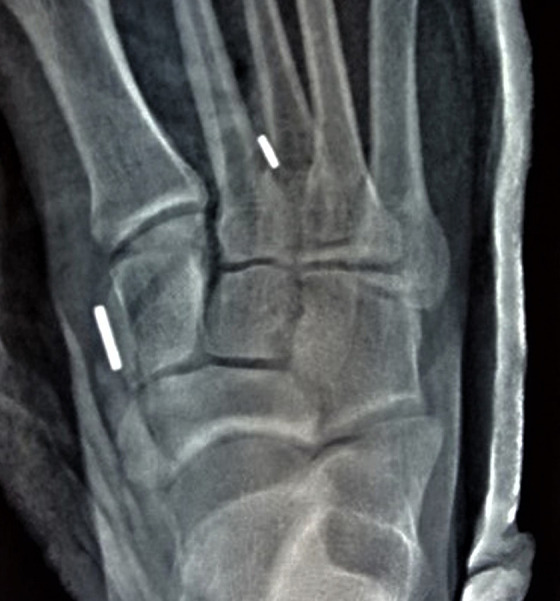
Postoperative anteroposterior radiograph showing the restored anatomic alignment of the Lisfranc joint. The large button is seated on the medial cuneiform, and the small button is on the second metatarsal.
Results
Overall, 12 athletes who underwent fixation of a ligamentous Lisfranc injury using the knotless interosseous suture button met the inclusion and exclusion criteria. The mean age of the patients was 21.1 years (range, 16-34 years), with a mean follow-up period of 42 months. As presented in Table 1, the 12 cases included 7 professional National Rugby League (NRL) players, 2 professional dancers, 1 Olympic gymnast, 1 professional wakeboarder, and 1 professional NRL referee. The NRL referee was included as referees also follow strict training schedules and run up to 15 km a game; therefore, they have similar expected outcomes to the other athletes.
Table 1.
Patient, Injury, and RTP Details a
| Time Frame, wk | ||||||||
|---|---|---|---|---|---|---|---|---|
| Patient No. | Age, y | Sex | Sport | Injury Details | Injury to Fixation | WB | RTT | RTP |
| 1 | 18 | M | NRL | FT ligamentous | 2 | 4 | 10 | 14 |
| 2 | 18 | M | NRL | FT ligamentous | 2 | 4 | 10 | 14 |
| 3 | 16 | M | NRL | FT ligamentous | 1 | 4 | 9 | 12 |
| 4 | 20 | M | Wakeboarding | FT ligamentous | 2 | 4 | 10 | 12 |
| 5 | 34 | M | NRL referee | FT ligamentous | 2 | 4 | 12 | 14 |
| 6 | 21 | M | NRL | FT ligamentous | 1 | 4 | 10 | N/A |
| 7 | 25 | F | WNRL | FT ligamentous | 2 | 4 | 12 | N/A |
| 8 | 25 | M | NRL | FT ligamentous | 1 | 4 | 10 | N/A |
| 9 | 17 | M | Olympic gymnast | FT ligamentous | 2 | 4 | 12 | 14 |
| 10 | 21 | F | Dancer | FT ligamentous | 2 | 4 | 12 | 14 |
| 11 | 22 | M | NRL | Chronic FT ligamentous | 20 | 4 | 10 | N/A |
| 12 | 16 | F | Dancer | Chronic FT ligamentous | 27 | 4 | 12 | 16 |
a F, female; FT, full thickness; M, male; N/A, not available (return to play occurred during the off-season); NRL, National Rugby League; NRLW, Women’s National Rugby League; RTP, return to play; RTT, return to training; WB, weightbearing.
Ten patients underwent surgery within 3 weeks of the injury. Regarding the 2 patients with chronic Lisfranc joint injuries, 1 was an NRL player who had gross instability of the midfoot with pushoff, and the other was a professional ballerina who was experiencing considerable instability en pointe. Both were unable to perform the heel-raise maneuver effectively because of “giving way” or a sensation of weakness in the midfoot. All patients had isolated complete ligamentous Lisfranc joint injuries, which were reported on MRI scans by a specialized musculoskeletal radiologist. There were no associated intercuneiform injuries in any of the patients included in this series.
All patients started full weightbearing at 4 weeks, started training by 9 to 12 weeks, and had returned to full competition by 12 to 16 weeks. Four of the athletes in this case series were fit to return to competition during their off-season, and as a result, RTP time frames could not be accurately calculated for these cases. All patients were fit to return to competition between 2 and 4 weeks from their return-to-training date, and all were able to return to their preinjury level of competition.
No major short-term complications (eg, wound dehiscence, surgical-site infection, implant failure, or loss of reduction) were observed at follow-up. One patient reported mild irritation from the medial button at the 3-month follow-up, but it was not severe enough to have it removed. Another patient reported paresthesia on the dorsum of the foot that had resolved by the 6-week follow-up.
Discussion
To our knowledge, this is the first case series reporting the use of a knotless interosseous suture button device to repair ligamentous Lisfranc injuries in elite athletes. It is a guide to both treating surgeons and team physicians on the potential accelerated RTP for athletes with purely ligamentous Lisfranc injuries when using this operative technique. All 12 patients who met the inclusion and exclusion criteria were full weightbearing at 4 weeks, were training by 9 to 12 weeks, and had returned to full competition by 12 to 16 weeks. All patients returned to their preinjury professional level, with no major complications or device failure observed in the 2-year follow-up.
The use of interosseous suture button stabilization for Lisfranc joint injuries is not new. Several studies, from as early as 2008, have reported the successful stabilization of Lisfranc injuries using the Arthrex Mini-TightRope system. 4,5,7,10,11,19 This study reports a technique of drilling from the base of the second metatarsal to the medial cuneiform. This places the oval button over the medial cuneiform, instead of the second metatarsal. This technique has been previously reported by Cottom et al 11 when using the Mini-TightRope. The alternative technique of drilling from the medial cuneiform to the second metatarsal has been reported in several previous case series. 4,5,8,9 The author believes that the technique described by Cottom et al 11 is technically less challenging and minimizes the intraoperative use of an image intensifier. In terms of surgical technique, the central difference with our study is that we used a larger 3.7-mm knotless interosseous suture button instead of the mini (2.7-mm) interosseous suture button.
The use of the larger suture button has also been recently reported by Cho et al 8 and Chun et al, 9 both in 2021. Both drilled a 3.5-mm tunnel in the opposite direction of that described in our series, while also reporting the use of a knotted suture button. The knotless system utilized in our case series eliminates the need for tying a suture onto the button. This avoids the possible risk of knot irritation to soft tissue and suture abscess, which has been reported in the suture button fixation of syndesmosis injuries. 18,35,39 Additionally, Cho et al 8 discuss the concern of potential irritation caused by the large oval button when seated on the second metatarsal. The technique reported in our series reduces the risk of this irritation, as the large button is placed on the medial cuneiform.
Cottom et al 10 also reported using an additional intercuneiform screw. They reported the subsequent removal of the intercuneiform screw in 9 of their patients, secondary to loosening or residual pain. Jain et al 19 also reported using an additional intercuneiform suture button. No intercuneiform joint instability was observed preoperatively in our patients, and on the most recent follow-up, there were no concerns relating to this.
Our study also differs from these previous case series using suture button stabilization in that we focused on a larger group of 12 elite athletes, had a minimum follow-up of 2 years, and report an accelerated rehabilitation protocol. We report a return to full weightbearing at 4 weeks postoperatively, compared with 12 weeks in Cho et al 8 and 6 weeks in several other case series using suture button stabilization. 4,14,19,37 Jain et al 19 and Charlton et al, 7 who also used a suture button stabilization system in elite athletes, reported returns to competition of 21 weeks and 26 weeks, respectively. It appears that the larger suture button device used in this series safely allowed earlier weightbearing than previous studies, allowing patients to return to competition much earlier, at 12 to 16 weeks. There were no problems encountered in our study with this accelerated postoperative protocol, and all athletes were able to return to full competition level. Similar to previous studies using suture button devices, 4,5,7,9,11,14,19 we did not encounter any wound problems, device loosening, failure of implants, recurrent instabilities, or loss of reduction in our patient group.
The conventional method of fixation for Lisfranc injuries is open reduction and screw fixation or bridge plating. The main advantages of using the suture button technique are (1) the capacity to return to weightbearing significantly earlier and allow for more predictable and aggressive rehabilitation, (2) the elimination of a second operation and associated costs to remove the hardware, (3) less joint disruption and articular surface damage caused by screw fixation, and (4) a less rigid fixation that allows physiologic motion and flexibility of the Lisfranc joint. 1,4,7,11,21,30,31,37
A recent study by Deol et al 15 reported the repair of Lisfranc injuries in elite athletes using conventional screw fixation. Comparably with our cohort, 7 of the 17 elite athletes in their case series experienced purely ligamentous injuries. Five of these patients were managed using a single Lisfranc screw, and 2 required an additional tarsometatarsal screw. 15 With screw fixation, these patients were nonweightbearing postoperatively for a total of 8 weeks, compared with only 4 weeks in our cohort. Moreover, with screw fixation, hardware failure can occur once weightbearing is initiated, so patients in this study underwent a second procedure for removal of hardware at 16 weeks, further delaying their rehabilitation. 15 After the second procedure, the athletes were allowed to gradually return to training at 19 weeks. Excluding a player who retired because of the injury, the average return to training for this group was 19.3 weeks, and the average return to competition was 24.1 weeks. 15 Similarly, in Mora et al, 27 all 33 recreational athletes underwent removal of hardware 6 months postoperatively and returned to unrestricted sporting activities by 8 months. In a third study also reporting the use of screw fixation by Vosbikian et al, 38 the active patients wanting optimal motion without risk for hardware failure underwent a second operation to remove hardware at a mean of 6.9 months after the initial operation. Using our technique with the knotless interosseous suture button fixation, athletes were able to return to full training in as early as 9 weeks, compared with the average of 19 weeks for the elite athletes who underwent screw fixation for purely ligamentous Lisfranc injuries in Deol et al.15 More importantly, a second procedure was not required to remove the implant. In reporting this, the authors do note the higher initial implant cost with the suture button device compared with the screw fixation.
Limitations
We acknowledge the limitations of this study, which include a small sample size, lack of functional outcome scoring, lack of a treatment comparison group, inclusion of only isolated Lisfranc injuries with no associated intercuneiform injuries or fractures, and lack of long-term postoperative imaging to demonstrate maintenance of reduction. In addition, all study patients were highly motivated professional athletes with regular contact with specialist team doctors and physical therapists, and therefore, results may not be equal in lower-level athletes.
Conclusion
Knotless interosseous suture button stabilization is a reliable treatment option for both acute and chronic isolated ligamentous Lisfranc injuries in elite athletes. This technique does not require hardware removal, allows early weightbearing with accelerated rehabilitation, and may shorten the RTP interval. Further research with higher-level comparative studies is needed to assess the outcomes of these implants.
Footnotes
Final revision submitted March 6, 2022; accepted March 23, 2022.
The authors declared that they have no conflicts of interest in the authorship and publication of this contribution. AOSSM checks author disclosures against the Open Payments Database (OPD). AOSSM has not conducted an independent investigation on the OPD and disclaims any liability or responsibility relating thereto.
Ethical approval for this study was obtained from St. Vincent’s Clinic.
References
- 1. Alberta FG, Aronow MS, Barrero M, et al. Ligamentous Lisfranc joint injuries: a biomechanical comparison of dorsal plate and transarticular screw fixation. Foot Ankle Int. 2005;26(6):462–473. [DOI] [PubMed] [Google Scholar]
- 2. Attia AK, Mahmoud K, Alhammoud A, d’Hooghe P, Farber D. Return to play after low-energy Lisfranc injuries in high-demand individuals: a systematic review and meta-analysis of athletes and active military personnel. Orthop J Sports Med. 2021;9(3):2325967120988158. [DOI] [PMC free article] [PubMed] [Google Scholar]
- 3. Baravarian B, Geffen D. Lisfranc Tightrope. Foot Ankle Spec. 2009;2(5):249–250. [DOI] [PubMed] [Google Scholar]
- 4. Brin YS, Nyska M, Kish B. Lisfranc injury repair with the TightRope™ device: a short-term case series. Foot Ankle Int. 2010;31(7):624–627. [DOI] [PubMed] [Google Scholar]
- 5. Cardile C, Cazzaniga C, Manzini B, Marasco R, Ragni P. Lisfranc injuries in adolescents: a case report and literature review. Foot (Edinb). 2021;47:101812. [DOI] [PubMed] [Google Scholar]
- 6. Castro M, Melão L, Canella C, et al. Lisfranc joint ligamentous complex: MRI with anatomic correlation in cadavers. AJR Am J Roentgenol. 2010;195(6):W447. [DOI] [PubMed] [Google Scholar]
- 7. Charlton T, Boe C, Thordarson DB. Suture button fixation treatment of chronic Lisfranc injury in professional dancers and high-level athletes. J Dance Med Sci. 2015;19(4):135–139. [DOI] [PubMed] [Google Scholar]
- 8. Cho J, Kim J, Min TH, et al. Suture button vs conventional screw fixation for isolated Lisfranc ligament injuries. Foot Ankle Int. 2021;42(5):598–608. [DOI] [PubMed] [Google Scholar]
- 9. Chun DI, Kim J, Min TH, et al. Fixation of isolated Lisfranc ligament injury with the TightRope™: a technical report. Orthop Traumatol Surg Res. 2021;107(6):102940. [DOI] [PubMed] [Google Scholar]
- 10. Cottom JM, Graney CT, Sisovsky C. Treatment of Lisfranc injuries using interosseous suture button: a retrospective review of 84 cases with a minimum 3-year follow-up. J Foot Ankle Surg. 2020;59(6):1139–1143. [DOI] [PubMed] [Google Scholar]
- 11. Cottom JM, Hyer CF, Berlet GC. Treatment of Lisfranc fracture dislocations with an interosseous suture button technique: a review of 3 cases. J Foot Ankle Surg. 2008;47(3):250–258. [DOI] [PubMed] [Google Scholar]
- 12. Crates JM, Barber FA, Sanders EJ. Subtle Lisfranc subluxation: results of operative and nonoperative treatment. J Foot Ankle Surg. 2015;54(3):350–355. [DOI] [PubMed] [Google Scholar]
- 13. Curtis MJ, Myerson M, Szura B. Tarsometatarsal joint injuries in the athlete. Am J Sports Med. 1993;21(4):497–502. [DOI] [PubMed] [Google Scholar]
- 14. Delman C, Patel M, Campbell M, Kreulen C, Giza E. Flexible fixation technique for Lisfranc injuries. Foot Ankle Int. 2019;40(11):1338–1345. [DOI] [PubMed] [Google Scholar]
- 15. Deol RS, Roche A, Calder JDF. Return to training and playing after acute Lisfranc injuries in elite professional soccer and rugby players. Am J Sports Med. 2016;44(1):166–170. [DOI] [PubMed] [Google Scholar]
- 16. Englanoff G, Anglin D, Hutson HR. Lisfranc fracture-dislocation: a frequently missed diagnosis in the emergency department. Ann Emerg Med. 1995;26(2):229–233. [DOI] [PubMed] [Google Scholar]
- 17. Hardcastle P, Reschauer R, Kutscha-Lissberg E, Schoffmann W. Injuries to the tarsometatarsal joint. Incidence, classification and treatment. J Bone Joint Surg Br. 1982;64-B(3):349–356. [DOI] [PubMed] [Google Scholar]
- 18. Hodgson P, Thomas R. Avoiding suture knot prominence with suture button along distal fibula: technical tip. Foot Ankle Int. 2011;32(9):908–909. [DOI] [PubMed] [Google Scholar]
- 19. Jain K, Drampalos E, Clough TM. Results of suture button fixation with targeting device aid for displaced ligamentous Lisfranc injuries in the elite athlete. Foot (Edinb). 2017;30:43–46. [DOI] [PubMed] [Google Scholar]
- 20. Johnson A, Hill K, Ward J, Ficke J. Anatomy of the Lisfranc ligament. Foot Ankle Spec. 2008;1(1):19–23. [DOI] [PubMed] [Google Scholar]
- 21. Klitzman R, Zhao H, Zhang LQ, Strohmeyer G, Vora A. Suture-button versus screw fixation of the syndesmosis: a biomechanical analysis. Foot Ankle Int. 2010;31(1):69–75. [DOI] [PubMed] [Google Scholar]
- 22. Lewis JS, Anderson RB. Lisfranc injuries in the athlete. Foot Ankle Int. 2016;37(12):1374–1380. [DOI] [PubMed] [Google Scholar]
- 23. Curtis M, Myerson M, Szura B. Tarsometatarsal joint injuries in the athlete. Am J Sports Med. 1993;21(4):497–502. [DOI] [PubMed] [Google Scholar]
- 24. McBryde JAM, Hoffman JL. Injuries to the foot and ankle in athletes. South Med J. 2004;97(8):738–741. [DOI] [PubMed] [Google Scholar]
- 25. McHale KJ, Rozell JC, Milby AH, Carey JL, Sennett BJ. Outcomes of Lisfranc injuries in the National Football League. Am J Sports Med. 2016;44(7):1810–1817. [DOI] [PubMed] [Google Scholar]
- 26. Meyer SA, Callaghan JJ, Albright JP, Crowley ET, Powell JW. Midfoot sprains in collegiate football players. Am J Sports Med. 1994;22(3):392–401. [DOI] [PubMed] [Google Scholar]
- 27. Mora AD, Kao M, Alfred T, et al. Return to sports and physical activities after open reduction and internal fixation of Lisfranc injuries in recreational athletes. Foot Ankle Int. 2018;39(7):801–807. [DOI] [PubMed] [Google Scholar]
- 28. Myerson SM, Cerrato AR. Current management of tarsometatarsal injuries in the athlete. J Bone Joint Surg. 2008;90(11):2522–2533. [PubMed] [Google Scholar]
- 29. Nunley JA, Vertullo CJ. Classification, investigation, and management of midfoot sprains: Lisfranc injuries in the athlete. Am J Sports Med. 2002;30(6):871–878. [DOI] [PubMed] [Google Scholar]
- 30. Panchbhavi VK, Vallurupalli S, Yang J, Andersen CR. Screw fixation compared with suture-button fixation of isolated Lisfranc ligament injuries. J Bone Joint Surg Am. 2009;91(5):1143–1148. [DOI] [PubMed] [Google Scholar]
- 31. Pelt CE, Bachus KN, Vance RE, Beals TC. A biomechanical analysis of a tensioned suture device in the fixation of the ligamentous Lisfranc injury. Foot Ankle Int. 2011;32(4):422–431. [DOI] [PubMed] [Google Scholar]
- 32. Sherief TI, Mucci B, Greiss M. Lisfranc injury: how frequently does it get missed? And how can we improve? Injury. 2006;38(7):856–860. [DOI] [PubMed] [Google Scholar]
- 33. Sripanich Y, Weinberg MW, Krähenbühl N, et al. Imaging in Lisfranc injury: a systematic literature review. Skeletal Radiol. 2020;49(1):31–53. [DOI] [PubMed] [Google Scholar]
- 34. Stavlas P, Roberts CS, Xypnitos FN, Giannoudis PV. The role of reduction and internal fixation of Lisfranc fracture-dislocations: a systematic review of the literature. Int Orthop. 2010;34(8):1083–1091. [DOI] [PMC free article] [PubMed] [Google Scholar]
- 35. Storey P, Gadd RJ, Blundell C, Davies MB. Complications of suture button ankle syndesmosis stabilization with modifications of surgical technique. Foot Ankle Int. 2012;33(9):717–721. [DOI] [PubMed] [Google Scholar]
- 36. Sullivan M, Eusebio ID, Haigh K, et al. Prevalence of deep vein thrombosis in low-risk patients after elective foot and ankle surgery. Foot Ankle Int. 2019;40(3):330–335. [DOI] [PubMed] [Google Scholar]
- 37. Tzatzairis T, Firth G, Parker L. Adolescent Lisfranc injury treated with TightRope™: a case report and review of literature. World J Orthop. 2019;10(2):115–122. [DOI] [PMC free article] [PubMed] [Google Scholar]
- 38. Vosbikian M, O’Neil JT, Piper C, Huang R, Raikin SM. Outcomes after percutaneous reduction and fixation of low-energy Lisfranc injuries. Foot Ankle Int. 2017;38(7):710–715. [DOI] [PubMed] [Google Scholar]
- 39. Willmott HJS, Singh B, David LA. Outcome and complications of treatment of ankle diastasis with tightrope fixation. Injury. 2009;40(11):1204–1206. [DOI] [PubMed] [Google Scholar]
- 40. Yan A, Chen SR, Ma X, Shi Z, Hogan M. Updates on Lisfranc complex injuries. Foot Ankle Orthop. 2021;6(1):2473011420982275. [DOI] [PMC free article] [PubMed] [Google Scholar]