Abstract
Formulation of cationic liposomes is a key factor that determine the gene knockdown efficiency by cationic liposomes/siRNA complexes (siRNA lipoplexes). Here, to determine the optimal combination of cationic lipid and phospholipid in cationic liposomes for in vitro and in vivo gene knockdown using siRNA lipoplexes, three types of cationic lipid were used, namely 1,2-dioleoyl-3-trimethylammonium-propane (DOTAP), dimethyldioctadecylammonium bromide (DDAB) and 11-[(1,3-bis(dodecanoyloxy)-2-((dodecanoyloxy)methyl)propan-2-yl)amino]-N,N,N-trimethyl-11-oxoundecan-1-aminium bromide (TC-1-12). Thereafter, 30 types of cationic liposome composed of each cationic lipid with phosphatidylcholine or phosphatidylethanolamine containing saturated or unsaturated dialkyl chains (C14, C16, or C18) were prepared. The inclusion of phosphatidylethanolamine containing unsaturated and long dialkyl chains with DOTAP- or DDAB-based cationic liposomes induced strong luciferase gene knockdown in human breast cancer MCF-7-Luc cells stably expressing luciferase gene. Furthermore, the inclusion of phosphatidylcholine or phosphatidylethanolamine containing saturated and short dialkyl chains or unsaturated and long dialkyl chains into TC-1-12-based cationic liposomes resulted in high gene knockdown efficacy. When cationic liposomes composed of DDAB/1,2-dioleoyl-sn-glycero-3-phosphoethanolamine (DOPE), TC-1-12/DOPE and TC-1-12/1-palmitoyl-2-oleoyl-sn-glycero-3-phosphoethanolamine were used, significant gene knockdown occurred in the lungs of mice following systemic injection of siRNA lipoplexes. Overall, the present findings indicated that optimal phospholipids in cationic liposome for in vitro and in vivo siRNA transfection were affected by the types of cationic lipid used.
Keywords: cationic liposome, siRNA delivery, phospholipid, lung, gene knockdown
Introduction
Small interfering (si)RNA is a promising tool for inhibiting specific gene expression (1). Cationic liposomes have attracted attention for siRNA delivery to cells (2,3) as siRNA/cationic liposome complexes (siRNA lipoplexes) are efficiently delivered into cells and notably suppress target gene expression (2). To prepare cationic liposomes, liposomal formulations are used to combine cationic and neutral helper lipids, such as phospholipid or cholesterol (Chol), to increase transfection efficiency and stability (4). However, the structures of cationic lipid and phospholipid, such as head group and alkyl chains, affect the transfection efficiency of siRNA lipoplexes (5,6). Therefore, discovering an optimal combination of cationic and neutral helper lipid for liposomal formulations is key for achieving efficient siRNA transfection.
The most common cationic lipids for siRNA delivery with cationic liposomes are 1,2-dioleoyl-3-trimethylammonium-propane (DOTAP) (7–10), 1,2-di-O-octadecenyl-3-trimethylammonium-propane (10–12) and dimethyldioctadecylammonium bromide (DDAB) (9,13) which contain a quaternary ammonium group in the head group. 1,2-Dioleoyl-sn-glycero-3-phosphoethanolamine (DOPE), which has a small head group, is unsaturated and has long dialkyl chains; it is often used as a neutral helper lipid to prepare cationic liposome (7–12) because DOPE destabilizes siRNA lipoplexes in endosomes by inducing a change in conformation at acidic pH when incorporated into the liposomal formulation (14).
Previously, we selected 6 types of cationic Chol derivative and 11 types of cationic lipid with dialkyl or trialkyl chains to prepare 17 types of cationic liposome composed of cationic lipid with DOPE for siRNA delivery and evaluate gene knockdown efficacy (5). Among these cationic liposomes, those composed of dialkyl (DDAB) or trialkyl cationic lipid [11-((1,3-bis(dodecanoyloxy)-2-((dodecanoyloxy)methyl)propan-2-yl)amino)-N,N,N-trimethyl-11-oxoundecan-1-aminium bromide; TC-1-12] with DOPE notably suppressed targeted mRNA expression in the mouse lung following systemic injection of siRNA lipoplexes. TC-1-12 is a novel cationic lipid developed by our laboratory that exhibits high siRNA transfection efficiency both in vitro and in vivo (5,6). However, to the best of our knowledge, few reports have been published regarding the effect of phospholipids in cationic liposomal formulations on gene knockdown efficacy using siRNA lipoplexes (15,16).
To determine the effect of phospholipids in cationic liposomes on gene knockdown activity of siRNA lipoplexes, three types of cationic lipid (DOTAP, DDAB and TC-1-12) were selected to prepare 30 types of cationic liposome composed of cationic lipid with phosphatidylcholine or phosphatidylethanolamine containing saturated or unsaturated dialkyl chains (C14, C16 or C18) and in vitro and in vivo gene knockdown efficacy was evaluated to find optimal phospholipids in cationic liposome for siRNA transfection.
Materials and methods
Materials
DDAB (cat. no. DC-1-18) and TC-1-12 were obtained from Sogo Pharmaceutical Co., Ltd. DOTAP (cat. no. 890895C) was obtained from Avanti Polar Lipids, Inc. 1,2-Distearoyl-sn-glycero-3-phosphocholine (DSPC; cat. no. MC-8080), 1,2-dipalmitoyl-sn-glycero-3-phosphocholine (DPPC; cat. no. MC-6060), 1,2-dimyristoyl-sn-glycero-3-phosphocholine (DMPC; cat. no. MC-4040), 1,2-dioleoyl-sn-glycero-3-phosphocholine (DOPC; cat. no. MC-8181), 1-palmitoyl-2-oleoyl-sn-glycero-3-phosphocholine (POPC; cat. no. MC-6081), 1,2-distearoyl-sn-glycero-3-phosphoethanolamine (DSPE; cat. no. ME-8080), 1,2-dipalmitoyl-sn-glycero-3-phosphoethanolamine (DPPE; cat. no. ME-6060), 1,2-dimyristoyl-sn-glycero-3-phosphoethanolamine (DMPE; cat. no. ME-4040), DOPE (cat. no. ME-8181) and 1-palmitoyl-2-oleoyl-sn-glycero-3-phosphoethanolamine (POPE; cat. no. ME-6081; all COATSOME®) were obtained from NOF Corporation. All other chemicals were of the highest grade available.
siRNAs
siRNA sequences of firefly luciferase (Luc siRNA), non-silencing siRNA [negative control (Cont) siRNA] and cyanine 5 (Cy5)-conjugated Cont siRNA (Cy5-siRNA) were designed as reported previously (17,18) and synthesized by Sigma-Aldrich (Merck KGaA). siRNA sequences were as follows: Luc siRNA passenger strand, 5′-CCGUGGUGUUCGUGUCUAAGA-3′ and guide strand, 5′-UUAGACACGAACACCACGGUA-3′ and Cont siRNA passenger strand, 5′-GUACCGCACGUCAUUCGUAUC-3′ and guide strand, 5′-UACGAAUGACGUGCGGUACGU-3′. For Cy5-siRNA, Cy5 dye was conjugated at the 5′-end of the passenger strand of Cont siRNA. The siRNA sequences of mouse Tie2 and luciferase siRNA (Cont 2 siRNA), which served as a negative control of Tie2 siRNA, were designed as reported previously (5,19) and synthesized by Japan Bio Services Co., Ltd. The siRNA sequences were as follows: Tie2 passenger strand, 5′-CcAuCaUuUgCcCaGaUaU-3′ and guide strand, 5′-aUaUcUgGgCaAaUgAuGg-3′ and Cont 2 siRNA passenger strand, 5′-AuCaCgUaCgCgGaAuAcUuCgA-3′ and guide strand, 5′-uCgAaGuAuUcCgCgUaCgUgAu-3′. Lowercase letters represent 2′-O-methyl-modified nucleotides.
Preparation of cationic liposomes and siRNA lipoplexes
The cationic liposomes were prepared from cationic lipid and phospholipid at a molar ratio of 1:1. To prepare cationic liposomes, cationic lipid and phospholipid were dissolved in chloroform or chloroform/methanol (9:1, v/v). Chloroform and methanol were evaporated under vacuum on a rotary evaporator at 60°C to obtain a thin film. The thin film was hydrated with water at 60°C by vortex mixing for 30 sec at 3,000 rpm and sonicated in a bath-type sonicator (Bransonic® 2510J-MTH, 42 kHz; Branson UL Trasonics Co.) for 5–10 min at 60°C.
To prepare siRNA lipoplexes, liposome was added to 50 pmol siRNA at a charge ratio (+:-) of 4:1, vortexed for 10 sec at 3,000 rpm and room temperature and left at room temperature for 15 min, as previously described (9,20). The charge ratio represents molar ratio of cationic lipid in cationic liposomes to siRNA phosphate.
The particle size, particle size distribution [polydispersity index (PDI)] and ζ-potential of cationic liposomes and siRNA lipoplexes were measured using a light-scattering photometer (cat. no. ELS-Z2; Otsuka Electronics Co., Ltd.), as previously reported (21).
Free siRNA levels in siRNA lipoplexes
siRNA lipoplexes were prepared at charge ratios (+:-) of 1:1-4:1. The amount of free siRNA in siRNA lipoplexes were measured using exclusion assay with SYBR® Green I Nucleic Acid Gel Stain (Takara Bio Inc.) and calculated based on the standard curves of free siRNA as previously reported (21).
Cell culture
Human breast cancer MCF-7 cells stably expressing firefly luciferase (MCF-7-Luc), constructed by transfection of plasmid pcDNA3 containing firefly luciferase (hLuc) gene (GenBank no. AY535007.1) from plasmid psiCHECK2 (Promega Corporation) were donated by Dr Kenji Yamato (University of Tsukuba, Tsukuba, Japan). MCF-7-Luc cells were cultured in RPMI-1640 medium supplemented with 10% heat-inactivated fetal bovine serum (FBS) and 1.2 mg/ml G418 sulfate (geneticin, Wako Pure Chemical Industries, Ltd.) at 37°C in a humidified atmosphere with 5% CO2.
Gene knockdown in vitro using siRNA lipoplexes
MCF-7-Luc cells were seeded in 6-well culture plates at a density of 3×10⁵ cells/well at 37°C. After 24 h, each siRNA lipoplex with 50 pmol Cont siRNA or Luc siRNA was diluted in 1 ml RPMI-1640 medium supplemented with 10% FBS and added to the cells (final concentration, 50 nM). At 48 h post-transfection, luciferase activity was measured as counts per second (cps)/µg protein using PicaGene MelioraStar-LT Luminescence Reagent (Toyo Ink Co. Ltd.) and BCA reagent (Pierce™ BCA Protein Assay kit; Thermo Fisher Scientific, Inc.) as reported previously (21). Luciferase activity (%) was calculated relative to that of untransfected cells.
Cytotoxicity of siRNA lipoplexes
Each siRNA lipoplex sample with 5 pmol Cont siRNA was diluted in 100 µl RPMI-1640 medium supplemented with 10% FBS and added to MCF-7-Luc cells at 50% confluency in 96-well plates (final concentration, 50 nM). Following 24 h incubation at 37°C, cell viability was measured via WST-8 assay (Dojindo Laboratories, Inc.), as previously reported (21). WST-8 substrate was incubated with cells at 37°C for 60 min.
Biodistribution of siRNA following intravenous injection of siRNA lipoplexes into mice
Ethical approval for this study was obtained from the Institutional Animal Care and Use Committee of Hoshi University (approval no. P21-039). A total of 8 female BALB/c mice (weight, 18–20 g; age, 8 weeks; Sankyo Labo Service Corporation, Inc.) were housed at 24°C and 55% humidity under 12/12-h light/dark cycle (lights on at 8:00 a.m.) with food and water ad libitum. siRNA lipoplexes with 20 µg Cy5-siRNA were administered intravenously to mice via the lateral tail vein (n=1/siRNA lipoplex). At 1 h post-injection of siRNA lipoplexes, mice were sacrificed via cervical dislocation; death was confirmed by cessation of heartbeat. Tissue (lung, heart, liver, spleen, and kidney) was analyzed by Cy5 fluorescence imaging using NightOWL LB981 NC100 system (Berthold Technologies GmbH & Co. KG), as previously described (21). The images were analyzed using IndiGo2 software (version 2.0.1.0) provided with the in vivo imaging system (Berthold Technologies). Following fluorescence imaging, tissue samples were frozen on dry ice and sliced into 16 µm sections. The localization of Cy5-siRNA was examined using a fluorescent microscope (Eclipse TS100-F; Nikon Corporation) with optical filter Cy5 HQ (excitation, 620/60 nm; dichroic mirror, 660 nm; emission, 700/75 nm; Nikon Corporation).
Agglutination assay
Erythrocyte suspension was prepared from whole blood of female BALB/c mice (age, 8 weeks; Sankyo Labo Service Corporation, Inc.), as previously described (13). siRNA lipoplexes with 2 µg siRNA were added to 100 µl 2% (v/v) erythrocyte suspension. Following incubation for 15 min at 37°C, the sample was placed on a glass plate and agglutination was observed with light microscope at 100× magnification.
Expression of Tie2 mRNA in the lung following systemic injection of siRNA lipoplexes
siRNA lipoplexes with 20 µg Cont 2 or Tie2 siRNA were administered intravenously to 8-week-old female BALB/c mice via the lateral tail veins (n=3-4/siRNA lipoplex). No siRNA lipoplexes caused mouse death following systemic injection. The lung was excised at 48 h post-injection of siRNA lipoplexes and total RNA was isolated using Isogen II (Nippon Gene Co., Ltd.). cDNA was synthesized from total RNA using PrimeScript™ RT Master Mix (Takara Bio Inc.) according to the manufacturer's protocol and quantitative PCR was performed using a Roche Light Cycler 96 system with FastStart Essential DNA Probes Master (Roche Diagnostics GmbH) and TaqMan Gene expression assays [Tie2, cat. no. Mm00443243_m1 and phosphatase and tensin homolog (PTEN), cat. no. Mm00477208_m1; both Applied Biosystems; Thermo Fisher Scientific, Inc.; primer sequences not available]. The thermocycling conditions were as follows: Initial denaturation at 95°C for 600 sec, followed by 45 cycles of denaturation at 95°C for 10 sec and primer annealing and extension at 60°C for 30 sec (two-step amplification). The expression levels of Tie2 mRNA were normalized to PTEN in each sample as reported previously (19) and analyzed using the comparative Cq (2−ΔΔCq) method (22).
Statistical analysis
Data are presented as the mean + SD of triplicate assessments. The statistical significance was determined by unpaired Student's t test or one-way ANOVA followed by Tukey's post hoc test using GraphPad Prism version 4.0 (GraphPad Software, Inc.). P<0.05 was considered to indicate a statistically significant difference.
Results
Characterization of cationic liposomes and siRNA lipoplexes
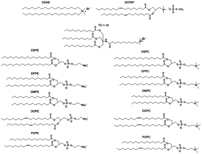
The present study aimed to determine whether alkyl chain length, saturation and the head group of phospholipids in cationic liposomes affect gene knockdown efficacy following treatment with siRNA lipoplexes. DOTAP and DDAB were used as dialkyl cationic lipids, TC-1-12 was used as a trialkyl cationic lipid and DSPE, DPPE, DMPE, POPE, DOPE, DSPC, DPPC, DMPC, POPC and DOPC were used as phospholipids (Fig. 1). Cationic liposomes were prepared from cationic lipid/phospholipid at a molar ratio of 1:1 (Table I, Table II, Table III).
Figure 1.
Structure of cationic lipids and phospholipids. DDAB, dimethyldioctadecylammonium bromide; DOTAP, 1,2-dioleoyl-3-trimethylammonium-propane methyl sulfate salt; TC-1-12, 11-[(1,3-bis(dodecanoyloxy)-2-((dodecanoyloxy)methyl)propan-2-yl)amino]-N,N,N-trimethyl-11-oxoundecan-1-aminium bromide; DSPC, 1,2-distearoyl-sn-glycero-3-phosphocholine; DPPC, 1,2-dipalmitoyl-sn-glycero-3-phosphocholine; DMPC, 1,2-dimyristoyl-sn-glycero-3-phosphocholine; DOPC, 1,2-dioleoyl-sn-glycero-3-phosphocholine; POPC, 1-palmitoyl-2-oleoyl-sn-glycero-3-phosphocholine; DSPE, 1,2-distearoyl-sn-glycero-3-phosphoethanolamine; DPPE, 1,2-dipalmitoyl-sn-glycero-3-phosphoethanolamine; DMPE, 1,2-dimyristoyl-sn-glycero-3-phosphoethanolamine; DOPE, 1,2-dioleoyl-sn-glycero-3-phosphoethanolamine; POPE, 1-palmitoyl-2-oleoyl-sn-glycero-3-phosphoethanolamine.
Table I.
Particle size and ζ-potential of DDAB-based cationic liposomes and small interfering RNA lipoplexes.
| Liposome | Lipoplexb | |||||
|---|---|---|---|---|---|---|
|
|
|
|||||
| Liposome | Sizea, nm | PDI | ζ-potentiala, mV | Sizea, nm | PDI | ζ-potentiala, mV |
| LP-DDAB/DSPE | 101.9±0.9 | 0.24±0.01 | 46.9±1.8 | 152.1±3.2 | 0.24±0.01 | 38.7±1.4 |
| LP-DDAB/DPPE | 108.1±1.5 | 0.22±0.01 | 44.7±0.3 | 209.2±5.5 | 0.29±0.01 | 40.2±1.4 |
| LP-DDAB/DMPE | 111.2±2.6 | 0.23±0.01 | 47.3±0.3 | 167.6±1.4 | 0.18±0.01 | 43.8±2.1 |
| LP-DDAB/DOPE | 103.0±1.3 | 0.21±0.01 | 52.2±1.2 | 181.0±4.7 | 0.17±0.02 | 47.1±1.1 |
| LP-DDAB/POPE | 99.6±1.0 | 0.21±0.01 | 46.2±0.7 | 190.2±2.6 | 0.21±0.01 | 42.3±1.7 |
| LP-DDAB/DSPC | 132.4±2.0 | 0.23±0.01 | 45.9±0.7 | Aggregation | ND | ND |
| LP-DDAB/DPPC | 123.1±1.4 | 0.23±0.01 | 47.8±1.7 | 149.7±0.6 | 0.20±0.01 | 41.9±3.0 |
| LP-DDAB/DMPC | 106.7±2.3 | 0.23±0.01 | 45.9±0.2 | 158.1±5.8 | 0.20±0.01 | 37.5±2.0 |
| LP-DDAB/DOPC | 120.0±1.3 | 0.27±0.00 | 45.1±1.8 | 169.3±2.6 | 0.21±0.00 | 40.2±1.8 |
| LP-DDAB/POPC | 110.2±5.4 | 0.20±0.06 | 45.5±4.8 | 178.3±17.7 | 0.17±0.07 | 35.7±0.2 |
Aggregation indicates particle size >1 µm.
In water.
Charge ratio (+:-) of cationic lipid to siRNA phosphate, 4:1. Data are presented as the mean ± SD (n=3). PDI, polydispersity index; ND, not determined; DDAB, dimethyldioctadecylammonium bromide; DSPC, 1,2-distearoyl-sn-glycero-3-phosphocholine; DPPC, 1,2-dipalmitoyl-sn-glycero-3-phosphocholine; DMPC, 1,2-dimyristoyl-sn-glycero-3-phosphocholine; DOPC, 1,2-dioleoyl-sn-glycero-3-phosphocholine; POPC, 1-palmitoyl-2-oleoyl-sn-glycero-3-phosphocholine; DSPE, 1,2-distearoyl-sn-glycero-3-phosphoethanolamine; DPPE, 1,2-dipalmitoyl-sn-glycero-3-phosphoethanolamine; DMPE, 1,2-dimyristoyl-sn-glycero-3-phosphoethanolamine; DOPE, 1,2-dioleoyl-sn-glycero-3-phosphoethanolamine; POPE, 1-palmitoyl-2-oleoyl-sn-glycero-3-phosphoethanolamine; LP, liposome.
Table II.
Particle size and ζ-potential of DOTAP-based cationic liposomes and small interfering RNA lipoplexes.
| Liposome | Lipoplexb | |||||
|---|---|---|---|---|---|---|
|
|
|
|||||
| Liposome | Sizea, nm | PDI | ζ-potentiala, mV | Sizea, nm | PDI | ζ-potentiala, mV |
| LP-DOTAP/DSPE | 111.8±2.0 | 0.23±0.01 | 50.3±0.5 | 216.3±27.3 | 0.19±0.07 | 48.7±2.7 |
| LP-DOTAP/DPPE | 116.0±0.9 | 0.20±0.01 | 48.3±0.7 | 163.7±6.5 | 0.21±0.01 | 41.1±0.7 |
| LP-DOTAP/DMPE | 113.7±1.8 | 0.28±0.01 | 52.4±2.2 | 184.0±5.6 | 0.20±0.01 | 47.8±2.2 |
| LP-DOTAP/DOPE | 109.2±1.0 | 0.21±0.01 | 53.7±1.6 | 179.1±5.7 | 0.16±0.01 | 42.8±1.5 |
| LP-DOTAP/POPE | 111.7±1.4 | 0.24±0.02 | 49.6±1.0 | 175.8±6.5 | 0.21±0.01 | 40.3±0.4 |
| LP-DOTAP/DSPC | 93.3±0.8 | 0.22±0.01 | 48.9±1.4 | 228.7±1.5 | 0.26±0.00 | 32.2±0.7 |
| LP-DOTAP/DPPC | 112.8±0.8 | 0.26±0.00 | 46.7±0.7 | 200.8±2.8 | 0.23±0.00 | 26.6±2.8 |
| LP-DOTAP/DMPC | 107.2±1.0 | 0.25±0.00 | 46.3±2.7 | 240.5±1.9 | 0.28±0.02 | 31.3±1.1 |
| LP-DOTAP/DOPC | 102.2±0.9 | 0.24±0.01 | 48.6±3.0 | 227.3±4.1 | 0.25±0.00 | 35.2±0.1 |
| LP-DOTAP/POPC | 115.5±2.0 | 0.26±0.01 | 46.0±1.6 | 233.1±3.0 | 0.28±0.01 | 31.3±0.6 |
In water.
Charge ratio (+:-) of cationic lipid to siRNA phosphate, 4:1. Data are presented as the mean ± SD (n=3). DOTAP, 1,2-dioleoyl-3-trimethylammonium-propane methyl sulfate salt; PDI, polydispersity index; DSPC, 1,2-distearoyl-sn-glycero-3-phosphocholine; DPPC, 1,2-dipalmitoyl-sn-glycero-3-phosphocholine; DMPC, 1,2-dimyristoyl-sn-glycero-3-phosphocholine; DOPC, 1,2-dioleoyl-sn-glycero-3-phosphocholine; POPC, 1-palmitoyl-2-oleoyl-sn-glycero-3-phosphocholine; DSPE, 1,2-distearoyl-sn-glycero-3-phosphoethanolamine; DPPE, 1,2-dipalmitoyl-sn-glycero-3-phosphoethanolamine; DMPE, 1,2-dimyristoyl-sn-glycero-3-phosphoethanolamine; DOPE, 1,2-dioleoyl-sn-glycero-3-phosphoethanolamine; POPE, 1-palmitoyl-2-oleoyl-sn-glycero-3-phosphoethanolamine; LP, liposome.
Table III.
Particle size and ζ-potential of TC-1-12-based cationic liposomes and small interfering RNA lipoplexes.
| Liposome | Lipoplexb | |||||
|---|---|---|---|---|---|---|
|
|
|
|||||
| Liposome | Sizea, nm | PDI | ζ-potentiala, mV | Sizea, nm | PDI | ζ-potentiala, mV |
| LP-TC-1-12/DSPE | 195.1±23.6 | 0.17±0.08 | 39.9±0.8 | 192.9±0.3 | 0.13±0.01 | 38.3±0.9 |
| LP-TC-1-12/DPPE | 111.7±1.0 | 0.24±0.01 | 40.3±1.2 | Aggregation | N.D. | N.D. |
| LP-TC-1-12/DMPE | 104.2±0.9 | 0.28±0.02 | 45.1±2.2 | Aggregation | N.D. | N.D. |
| LP-TC-1-12/DOPE | 122.5±0.9 | 0.24±0.00 | 41.6±0.8 | 179.6±3.7 | 0.21±0.01 | 43.4±1.5 |
| LP-TC-1-12/POPE | 104.0±1.7 | 0.25±0.01 | 40.3±1.3 | 182.5±3.2 | 0.24±0.01 | 32.6±4.0 |
| LP-TC-1-12/DSPC | 101.5±8.1 | 0.25±0.04 | 42.0±1.0 | 781.1±187.3 | 0.34±0.07 | 40.1±0.5 |
| LP-TC-1-12/DPPC | 110.2±2.0 | 0.28±0.01 | 43.4±0.4 | 485.2±28.0 | 0.25±0.01 | 40.0±1.0 |
| LP-TC-1-12/DMPC | 122.5±1.6 | 0.28±0.01 | 44.8±1.0 | 426.1±5.4 | 0.20±0.00 | 35.5±0.8 |
| LP-TC-1-12/DOPC | 138.9±2.8 | 0.27±0.02 | 41.5±1.0 | 272.4±13.5 | 0.23±0.05 | 37.0±1.2 |
| LP-TC-1-12/POPC | 113.8±1.2 | 0.25±0.01 | 42.7±3.1 | 380.9±21.4 | 0.17±0.01 | 31.3±1.3 |
Aggregation indicates particle size >1 µm.
In water.
Charge ratio (+:-) of cationic lipid to siRNA phosphate, 4:1. Data are presented as the mean ± SD (n=3). TC-1-12, 11-[(1,3-bis(dodecanoyloxy)-2-((dodecanoyloxy)methyl)propan-2-yl)amino]-N,N,N-trimethyl-11-oxoundecan-1-aminium bromide; PDI, polydispersity index; ND, not determined; DSPC, 1,2-distearoyl-sn-glycero-3-phosphocholine; DPPC, 1,2-dipalmitoyl-sn-glycero-3-phosphocholine; DMPC, 1,2-dimyristoyl-sn-glycero-3-phosphocholine; DOPC, 1,2-dioleoyl-sn-glycero-3-phosphocholine; POPC, 1-palmitoyl-2-oleoyl-sn-glycero-3-phosphocholine; DSPE, 1,2-distearoyl-sn-glycero-3-phosphoethanolamine; DPPE, 1,2-dipalmitoyl-sn-glycero-3-phosphoethanolamine; DMPE, 1,2-dimyristoyl-sn-glycero-3-phosphoethanolamine; DOPE, 1,2-dioleoyl-sn-glycero-3-phosphoethanolamine; POPE, 1-palmitoyl-2-oleoyl-sn-glycero-3-phosphoethanolamine; LP, liposome.
The size of cationic liposomes was 93–195 nm (PDI, 0.17-0.28) and ζ-potential was +40–54 mV (Table I, Table II, Table III). Our previous study reported that the optimal charge ratio (+:-) to prepare the siRNA lipoplexes was 4:1 for LP-DOTAP/DOPE, LP-DDAB/DOPE and LP-TC-1-12/DOPE (9,20). Therefore, subsequent experiments used charge ratio (+:-) of 4:1 to prepare siRNA lipoplexes. LP-DDAB/DSPC, LP-TC-1-12/DPPE and LP-TC-1-12/DMPE lipoplexes were aggregated (size, >1 µm) when liposomes were mixed with siRNA (Tables I and III). When TC-1-12 was combined with phosphatidylcholine to prepare cationic liposomes, lipoplex size increased to 270–780 nm (PDI, 0.17-0.34). However, the lipoplex sizes except for the combination of TC-1-12/phosphatidylcholine were 150–240 nm (PDI, 0.13-0.29) and ζ-potential was +31–49 mV.
Binding of siRNA to cationic liposomes
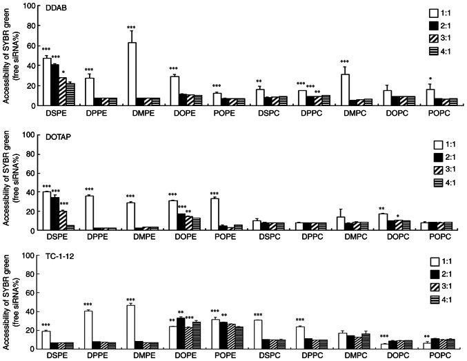
The binding of siRNA to each cationic liposome was assessed by exclusion assay with SYBR Green I. The addition of cationic liposomes to siRNA beyond charge ratios of 2:1-3:1 was found to markedly decrease the fluorescence of SYBR® Green I in the DDAB-, DOTAP-, and TC-1-12-based liposomes (Fig. 2). These results suggested that most DDAB-, DOTAP- and TC-1-12-based cationic liposomes exhibited greatest binding to siRNA at a charge ratio of 4:1.
Figure 2.
Effect of phospholipids in cationic liposomes on binding with siRNA. siRNA lipoplexes were formed at charge ratios (+:-) of 1:1-4:1 and used in an exclusion assay with SYBR® Green I Nucleic Acid Gel Stain. The amount of siRNA available to interact with the SYBR® Green I is expressed as a percentage of free siRNA without cationic liposome. Data are presented as the mean + SD (n=3). *P<0.05, **P<0.01, ***P<0.001 vs. 4:1. si, small interfering; DDAB, dimethyldioctadecylammonium bromide; DOTAP, 1,2-dioleoyl-3-trimethylammonium-propane methyl sulfate salt; TC-1-12, 11-[(1,3-bis(dodecanoyloxy)-2-((dodecanoyloxy)methyl)propan-2-yl)amino]-N,N,N-trimethyl-11-oxoundecan-1-aminium bromide; DSPC, 1,2-distearoyl-sn-glycero-3-phosphocholine; DPPC, 1,2-dipalmitoyl-sn-glycero-3-phosphocholine; DMPC, 1,2-dimyristoyl-sn-glycero-3-phosphocholine; DOPC, 1,2-dioleoyl-sn-glycero-3-phosphocholine; POPC, 1-palmitoyl-2-oleoyl-sn-glycero-3-phosphocholine; DSPE, 1,2-distearoyl-sn-glycero-3-phosphoethanolamine; DPPE, 1,2-dipalmitoyl-sn-glycero-3-phosphoethanolamine; DMPE, 1,2-dimyristoyl-sn-glycero-3-phosphoethanolamine; DOPE, 1,2-dioleoyl-sn-glycero-3-phosphoethanolamine; POPE, 1-palmitoyl-2-oleoyl-sn-glycero-3-phosphoethanolamine.
Effect of phospholipids in cationic liposomes on in vitro gene knockdown efficacy
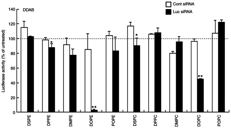
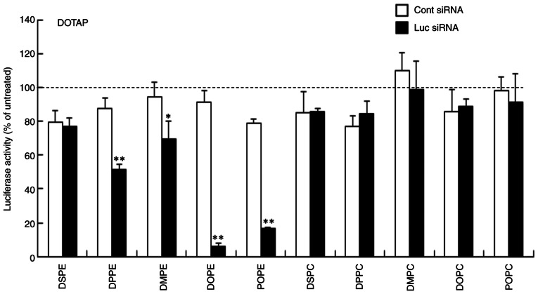
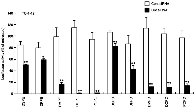
Previously, we reported that inclusion of DOPE in cationic liposomal formulations induces strong gene knockdown activity in cells; however, cationic liposomes with higher Chol content display decreased gene knockdown activity (6). To determine the effect of phospholipid in cationic liposomes on gene knockdown using siRNA lipoplexes, each siRNA lipoplex with Luc siRNA was transfected into MCF-7-Luc cells and gene knockdown efficacy was assessed by assaying luciferase activity. In DDAB- and DOTAP-based cationic liposomes, LP-DDAB/DOPE, LP-DOTAP/DOPE and LP-DOTAP/POPE lipoplexes caused strong gene knockdown efficacy (>80% knockdown compared with untreated cells; Figs. 3 and 4). LP-DDAB/DOPC and LP-DOTAP/DPPE lipoplexes exhibited moderate gene knockdown (56 and 49% knockdown, respectively, compared with untreated cells), and LP-DDAB/DPPE, LP-DDAB/DSPC, and LP-DOTAP/DMPE lipoplexes with Luc siRNA showed slightly gene knockdown compared with those with Cont siRNA. In TC-1-12-based liposomes, LP-TC-1-12/DMPE, LP-TC-1-12/DOPE, LP-TC-1-12/POPE, LP-TC-1-12/DMPC, LP-TC-1-12/DOPC and LP-TC-1-12/POPC lipoplexes had high gene knockdown efficacy (>80% knockdown compared with untreated cells; Fig. 5). LP-TC-1-12/DSPE, LP-TC-1-12/DPPE and LP-TC-1-12/DPPC lipoplexes exhibited moderate gene knockdown (41–57% knockdown compared with untreated cells), and LP-TC-1-12/DSPC lipoplexes with Luc siRNA showed slightly gene knockdown compared with those with Cont siRNA.
Figure 3.
Effect of phospholipids in DDAB-based cationic liposomes on gene knockdown using siRNA lipoplexes following transfection into MCF-7-Luc cells. siRNA lipoplexes with Cont or Luc siRNA were added to MCF-7-Luc cells at 50 nM and luciferase assay was performed 48 h post-transfection. Data are presented as the mean + SD (n=3). *P<0.05, **P<0.01 vs. Cont siRNA. Cont, control; Luc, luciferase; si, small interfering; DDAB, dimethyldioctadecylammonium bromide; DSPC, 1,2-distearoyl-sn-glycero-3-phosphocholine; DPPC, 1,2-dipalmitoyl-sn-glycero-3-phosphocholine; DMPC, 1,2-dimyristoyl-sn-glycero-3-phosphocholine; DOPC, 1,2-dioleoyl-sn-glycero-3-phosphocholine; POPC, 1-palmitoyl-2-oleoyl-sn-glycero-3-phosphocholine; DSPE, 1,2-distearoyl-sn-glycero-3-phosphoethanolamine; DPPE, 1,2-dipalmitoyl-sn-glycero-3-phosphoethanolamine; DMPE, 1,2-dimyristoyl-sn-glycero-3-phosphoethanolamine; DOPE, 1,2-dioleoyl-sn-glycero-3-phosphoethanolamine; POPE, 1-palmitoyl-2-oleoyl-sn-glycero-3-phosphoethanolamine.
Figure 4.
Effect of phospholipids in DOTAP-based cationic liposomes on gene knockdown using siRNA lipoplexes following transfection into MCF-7-Luc cells. siRNA lipoplexes with Cont or Luc siRNA were added to MCF-7-Luc cells at 50 nM and luciferase assay was performed 48 h post-transfection. Data are presented as the mean + SD (n=3). *P<0.05, **P<0.01 vs. Cont siRNA. Cont, control; Luc, luciferase; si, small interfering; DOTAP, 1,2-dioleoyl-3-trimethylammonium-propane methyl sulfate salt; DSPC, 1,2-distearoyl-sn-glycero-3-phosphocholine; DPPC, 1,2-dipalmitoyl-sn-glycero-3-phosphocholine; DMPC, 1,2-dimyristoyl-sn-glycero-3-phosphocholine; DOPC, 1,2-dioleoyl-sn-glycero-3-phosphocholine; POPC, 1-palmitoyl-2-oleoyl-sn-glycero-3-phosphocholine; DSPE, 1,2-distearoyl-sn-glycero-3-phosphoethanolamine; DPPE, 1,2-dipalmitoyl-sn-glycero-3-phosphoethanolamine; DMPE, 1,2-dimyristoyl-sn-glycero-3-phosphoethanolamine; DOPE, 1,2-dioleoyl-sn-glycero-3-phosphoethanolamine; POPE, 1-palmitoyl-2-oleoyl-sn-glycero-3-phosphoethanolamine.
Figure 5.
Effect of phospholipids in TC-1-12-based cationic liposomes on gene knockdown using siRNA lipoplexes following transfection into MCF-7-Luc cells. siRNA lipoplexes with Cont or Luc siRNA were added to MCF-7-Luc cells at 50 nM and luciferase assay was performed 48 h post-transfection. Data are presented as the mean + SD (n=3). *P<0.05, **P<0.01 vs. Cont siRNA. Cont, control; Luc, luciferase; si, small interfering; TC-1-12, 11-[(1,3-bis(dodecanoyloxy)-2-((dodecanoyloxy)methyl)propan-2-yl)amino]-N,N,N-trimethyl-11-oxoundecan-1-aminium bromide; DSPC, 1,2-distearoyl-sn-glycero-3-phosphocholine; DPPC, 1,2-dipalmitoyl-sn-glycero-3-phosphocholine; DMPC, 1,2-dimyristoyl-sn-glycero-3-phosphocholine; DOPC, 1,2-dioleoyl-sn-glycero-3-phosphocholine; POPC, 1-palmitoyl-2-oleoyl-sn-glycero-3-phosphocholine; DSPE, 1,2-distearoyl-sn-glycero-3-phosphoethanolamine; DPPE, 1,2-dipalmitoyl-sn-glycero-3-phosphoethanolamine; DMPE, 1,2-dimyristoyl-sn-glycero-3-phosphoethanolamine; DOPE, 1,2-dioleoyl-sn-glycero-3-phosphoethanolamine; POPE, 1-palmitoyl-2-oleoyl-sn-glycero-3-phosphoethanolamine.
Cytotoxicity induced by siRNA lipoplexes
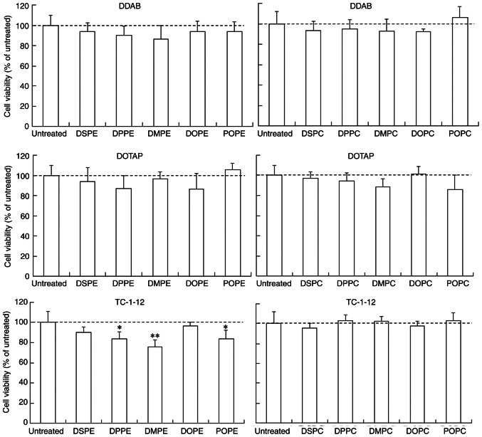
The effect of phospholipids in cationic liposomes on cytotoxicity was evaluated in MCF-7-Luc cells at 24 h post-transfection of siRNA lipoplexes. LP-TC-1-12/DPPE, LP-TC-1-12/DMPE and LP-TC-1-12/POPE lipoplexes showed slight cytotoxicity (75–84% cell viability), while the other lipoplexes did not exhibit cytotoxicity (>87% cell viability relative to untreated cells; Fig. 6). However, the addition of LP-TC-1-12/DPPE, LP-TC-1-12/DMPE and LP-TC-1-12/POPE without siRNA into the cells did not induce cytotoxic effects (100–105% cell viability; data not shown). These findings suggested that cationic liposomes composed of DDAB, DOTAP or TC-1-12 can be used for efficient siRNA transduction into cells with minimal toxicity.
Figure 6.
Effect of phospholipids in cationic liposomes on cell viability at 24 h post-transfection of siRNA lipoplexes into MCF-7-Luc cells. siRNA lipoplexes were added to MCF-7-Luc cells at 50 nM. Data are presented as the mean + SD (n=4-6). *P<0.05, **P<0.01 vs. untreated. si, small interfering; DDAB, dimethyldioctadecylammonium bromide; DOTAP, 1,2-dioleoyl-3-trimethylammonium-propane methyl sulfate salt; TC-1-12, 11-[(1,3-bis(dodecanoyloxy)-2-((dodecanoyloxy)methyl)propan-2-yl)amino]-N,N,N-trimethyl-11-oxoundecan-1-aminium bromide; DSPC, 1,2-distearoyl-sn-glycero-3-phosphocholine; DPPC, 1,2-dipalmitoyl-sn-glycero-3-phosphocholine; DMPC, 1,2-dimyristoyl-sn-glycero-3-phosphocholine; DOPC, 1,2-dioleoyl-sn-glycero-3-phosphocholine; POPC, 1-palmitoyl-2-oleoyl-sn-glycero-3-phosphocholine; DSPE, 1,2-distearoyl-sn-glycero-3-phosphoethanolamine; DPPE, 1,2-dipalmitoyl-sn-glycero-3-phosphoethanolamine; DMPE, 1,2-dimyristoyl-sn-glycero-3-phosphoethanolamine; DOPE, 1,2-dioleoyl-sn-glycero-3-phosphoethanolamine; POPE, 1-palmitoyl-2-oleoyl-sn-glycero-3-phosphoethanolamine.
Interaction between erythrocytes and siRNA lipoplexes
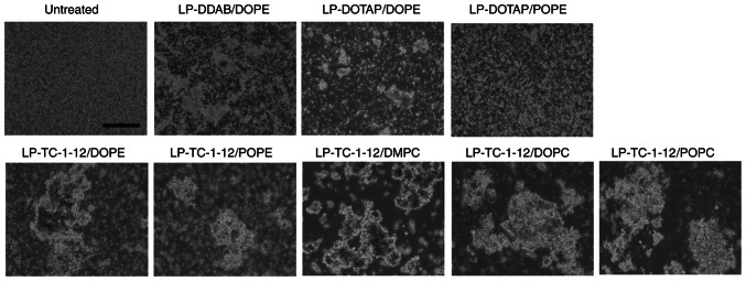
Positively charged lipoplexes cause agglutination via electrostatic interaction with negatively charged erythrocytes (23); these agglutinates are effectively trapped by highly extended lung capillaries (24). Therefore, to determine the effect of phospholipids in cationic liposomes on agglutination of siRNA lipoplexes with erythrocytes, siRNA lipoplexes were added to erythrocyte suspensions. LP-DDAB/DOPE, LP-DOTAP/DOPE, LP-DOTAP/POPE, LP-TC-1-12/DOPE, LP-TC-1-12/POPE, LP-TC-1-12/DMPC, LP-TC-1-12/DOPC and LP-TC-1-12/POPC were used as their lipoplexes exhibited high gene knockdown activity in cells (Table IV) and relatively small size (180–430 nm). All siRNA lipoplexes exhibited agglutination after mixing with the erythrocyte suspension, regardless of the type of phospholipid in the liposomal formulation (Fig. 7). In particular, TC-1-12-based siRNA lipoplexes formed large aggregates following mixing with erythrocyte suspension.
Table IV.
Summary of in vitro gene knockdown efficacy following treatment with siRNA lipoplexes.
| Cationic lipid | |||
|---|---|---|---|
|
|
|||
| Phospholipid | DDAB | DOTAP | TC-1-12 |
| DSPE | - | - | + |
| DPPE | - | + | - |
| DMPE | - | - | +++ |
| DOPE | +++ | +++ | +++ |
| POPE | - | +++ | +++ |
| DSPC | - | - | - |
| DPPC | - | - | ++ |
| DMPC | - | - | +++ |
| DOPC | ++ | - | +++ |
| POPC | - | - | +++ |
+++, >70; ++, 50–70; +, 30–49; -, <30% knockdown vs. control siRNA. DDAB, dimethyldioctadecylammonium bromide; DOTAP, 1,2-dioleoyl-3-trimethylammonium-propane methyl sulfate salt; TC-1-12, 11-[(1,3-bis(dodecanoyloxy)-2-((dodecanoyloxy)methyl)propan-2-yl)amino]-N,N,N-trimethyl-11-oxoundecan-1-aminium bromide; DSPC, 1,2-distearoyl-sn-glycero-3-phosphocholine; DPPC, 1,2-dipalmitoyl-sn-glycero-3-phosphocholine; DMPC, 1,2-dimyristoyl-sn-glycero-3-phosphocholine; DOPC, 1,2-dioleoyl-sn-glycero-3-phosphocholine; POPC, 1-palmitoyl-2-oleoyl-sn-glycero-3-phosphocholine; DSPE, 1,2-distearoyl-sn-glycero-3-phosphoethanolamine; DPPE, 1,2-dipalmitoyl-sn-glycero-3-phosphoethanolamine; DMPE, 1,2-dimyristoyl-sn-glycero-3-phosphoeth anolamine; DOPE, 1,2-dioleoyl-sn-glycero-3-phosphoethanolamine; POPE, 1-palmitoyl-2-oleoyl-sn-glycero-3-phosphoethanolamine; si, small interfering.
Figure 7.
Effect of phospholipids in cationic liposomes on agglutination of siRNA lipoplexes with erythrocytes. Lipoplexes with 2 µg siRNA were incubated with erythrocyte suspension. Scale bar, 200 µm. si, small interfering; DDAB, dimethyldioctadecylammonium bromide; DOTAP, 1,2-dioleoyl-3-trimethylammonium-propane methyl sulfate salt; TC-1-12, 11-[(1,3-bis(dodecanoyloxy)-2-((dodecanoyloxy)methyl)propan-2-yl)amino]-N,N,N-trimethyl-11-oxoundecan-1-aminium bromide; DMPC, 1,2-dimyristoyl-sn-glycero-3-phosphocholine; DOPC, 1,2-dioleoyl-sn-glycero-3-phosphocholine; POPC, 1-palmitoyl-2-oleoyl-sn-glycero-3-phosphocholine; DOPE, 1,2-dioleoyl-sn-glycero-3-phosphoethanolamine; POPE, 1-palmitoyl-2-oleoyl-sn-glycero-3-phosphoethanolamine.
Biodistribution of siRNA following systemic injection of siRNA lipoplexes
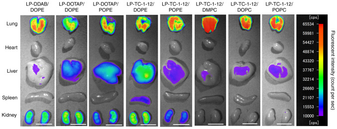
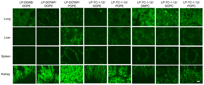
The present study investigated the effect of phospholipids in liposomal formulation on biodistribution of siRNA by ex vivo imaging at 1 h after systemic injection of lipoplexes with Cy5-siRNA. All siRNA lipoplexes caused siRNA accumulation in the lung (Figs. 8 and 9). In particular, LP-TC-1-12/DMPC, LP-TC-1-12/DOPC and LP-TC-1-12/POPC lipoplexes exhibited high siRNA accumulation in the lung, indicating that relatively large siRNA lipoplexes (270–430 nm) may form large agglutinations with blood components, resulting in efficient entrapment in lung capillaries. These results indicated that siRNA accumulation in the lung following systemic injection of siRNA lipoplexes may be affected by the size of siRNA lipoplexes, rather than the type of cationic lipid or phospholipid in cationic liposomes. In all the lipoplexes, the injected siRNAs were detected slightly in the liver and spleen, but not in heart. In the kidney, siRNA lipoplexes containing phosphatidylethanolamine showed high accumulation compared with the lipoplexes containing phosphatidylcholine.
Figure 8.
Effect of phospholipid in cationic liposomes on biodistribution of siRNA in mice at 1 h after systemic injection of siRNA lipoplexes. siRNA lipoplexes with 20 µg Cy5-siRNA were administered intravenously to mice. Cy5 fluorescence imaging of tissue was performed 1 h post-injection. Fluorescence intensity is illustrated using a color-coded scale (red, maximum; purple, minimum). Scale bar, 1 cm. si, small interfering; Cy5, cyanine 5; DDAB, dimethyldioctadecylammonium bromide; DOTAP, 1,2-dioleoyl-3-trimethylammonium-propane methyl sulfate salt; TC-1-12, 11-[(1,3-bis(dodecanoyloxy)-2-((dodecanoyloxy)methyl)propan-2-yl)amino]-N,N,N-trimethyl-11-oxoundecan-1-aminium bromide; DMPC, 1,2-dimyristoyl-sn-glycero-3-phosphocholine; DOPC, 1,2-dioleoyl-sn-glycero-3-phosphocholine; POPC, 1-palmitoyl-2-oleoyl-sn-glycero-3-phosphocholine; DOPE, 1,2-dioleoyl-sn-glycero-3-phosphoethanolamine; POPE, 1-palmitoyl-2-oleoyl-sn-glycero-3-phosphoethanolamine.
Figure 9.
Effect of phospholipids in cationic liposomes on biodistribution of siRNA in mice 1 h following intravenous injection of siRNA lipoplexes. siRNA lipoplex with 20 µg Cy5-siRNA was administered intravenously to mice. At 1 h post-injection, tissue was frozen and sliced to observe localization of Cy5-siRNA (green) using a fluorescent microscope. Scale bar, 100 µm. si, small interfering; Cy5, cyanine 5; DDAB, dimethyldioctadecylammonium bromide; DOTAP, 1,2-dioleoyl-3-trimethylammonium-propane methyl sulfate salt; TC-1-12, 11-[(1,3-bis(dodecanoyloxy)-2-((dodecanoyloxy)methyl)propan-2-yl)amino]-N,N,N-trimethyl-11-oxoundecan-1-aminium bromide; DMPC, 1,2-dimyristoyl-sn-glycero-3-phosphocholine; DOPC, 1,2-dioleoyl-sn-glycero-3-phosphocholine; POPC, 1-palmitoyl-2-oleoyl-sn-glycero-3-phosphocholine; DOPE, 1,2-dioleoyl-sn-glycero-3-phosphoethanolamine; POPE, 1-palmitoyl-2-oleoyl-sn-glycero-3-phosphoethanolamine.
Gene knockdown in the lung following systemic injection of siRNA lipoplexes
Tie2 gene is expressed in vascular endothelium (25,26) and has previously been used to evaluate gene knockdown efficacy of siRNA lipoplexes in the lung (19). The present study evaluated the effect of phospholipid in cationic liposomes on gene knockdown of Tie2 mRNA in pulmonary vascular endothelium at 48 h after single systemic injection of Tie2 siRNA lipoplexes into mice (Fig. 10). LP-DDAB/DOPE, LP-DOTAP/POPE, LP-TC-1-12/DOPE, LP-TC-1-12/POPE and LP-TC-1-12/DOPC were selected for evaluation of knockdown efficacy as their lipoplexes exhibited high gene knockdown activity (Fig. 3, Fig. 4, Fig. 5; Table IV) and maintained small size (180–270 nm). LP-DOTAP/DOPE was excluded from the evaluation as injection of LP-DOTAP/DOPE lipoplexes with 50 µg Tie2 siRNA has previously been shown not to induce gene knockdown of Tie2 mRNA in the lung (6). Systemic injection of LP-DDAB/DOPE, LP-TC-1-12/DOPE and LP-TC-1-12/POPE lipoplexes with Tie2 siRNA significantly suppressed Tie2 mRNA level (~50, 46 and 33% knockdown, respectively, compared with Cont 2 siRNA). Combined with the aforementioned results, these data indicated that accumulation of these siRNA lipoplexes in the lung did not decrease gene knockdown activity by agglutination with erythrocytes. However, LP-DOTAP/POPE and LP-TC-1-12/DOPC lipoplexes with Tie2 siRNA did not significantly suppress Tie2 mRNA levels in the lung.
Figure 10.
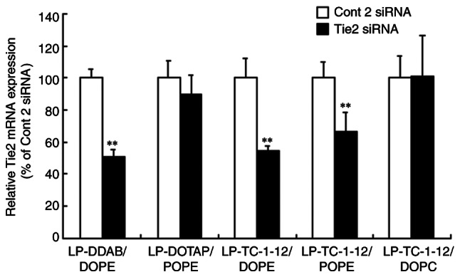
Effect of phospholipids in cationic liposomes on knockdown of Tie2 mRNA in the lung following systemic injection of Tie2 siRNA lipoplexes into mice. Tie2 mRNA levels in the lung were normalized to those of PTEN at 48 h after systemic administration of siRNA lipoplex with 20 µg Cont 2 or Tie2 siRNA. Tie2 expression (%) was calculated relative to that of mice treated with Cont 2 siRNA. Data are presented as the mean + SD (n=3-4). **P<0.01 vs. Cont 2 siRNA. si, small interfering; Cont, control; DDAB, dimethyldioctadecylammonium bromide; DOTAP, 1,2-dioleoyl-3-trimethylammonium-propane methyl sulfate salt; TC-1-12, 11-[(1,3-bis(dodecanoyloxy)-2-((dodecanoyloxy)methyl)propan-2-yl)amino]-N,N,N-trimethyl-11-oxoundecan-1-aminium bromide; DOPC, 1,2-dioleoyl-sn-glycero-3-phosphocholine; DOPE, 1,2-dioleoyl-sn-glycero-3-phosphoethanolamine; POPE, 1-palmitoyl-2-oleoyl-sn-glycero-3-phosphoethanolamine.
Discussion
Phosphatidylcholine and phosphatidylethanolamine are phospholipids in the cell membrane of most prokaryotes that are used as neutral helper lipids to prepare liposomes (15). Cationic liposomes composed of dialkyl cationic lipids with neutral helper lipids, such as DOPE, have been evaluated as carriers for siRNA delivery (5,6). The present study determined the effect of phospholipids in cationic liposomes on gene knockdown using siRNA lipoplexes. Following in vitro transfection, inclusion of phosphatidylcholines in DOTAP or DDAB-based cationic liposomes did not induce high gene knockdown by siRNA lipoplexes, although LP-DDAB/DOPC lipoplexes exhibited moderate gene knockdown activity. This may be because phosphatidylcholine has a larger head group than phosphatidylethanolamine (15). Du et al (27) reported that DOPC promotes stable laminar structure, limiting escape of cationic liposomes composed of DOTAP and DOPC from the endosome. However, DOPE promotes formation of inverted hexagonal lipid structures and cationic liposomes composed of DOTAP and DOPE exhibit improved transfection efficiency by destabilizing the endosomal membrane compared with those composed of DOTAP and DOPC (27). Such finding suggests that difference in headgroup structure between phosphatidylethanolamine and phosphatidylcholine may affect efficiency of siRNA transfection by cationic liposomes. In the present study, only combination with DOPE in DDAB-based cationic liposomes induced high gene knockdown activity. However, combination with DOPE or POPE in DOTAP-based cationic liposomes induced high gene knockdown in cells, indicating that saturation of dialkyl chains of cationic lipid and phospholipids may affect transfection efficiency following treatment with siRNA lipoplexes. The inclusion of phosphatidylethanolamine containing unsaturated and long dialkyl chains in DDAB or DOTAP-based liposomes induced strong gene knockdown in cells. By contrast, in TC-1-12-based liposomes, inclusion of phosphatidylcholine or phosphatidylethanolamine containing saturated and short or unsaturated and long dialkyl chains caused high gene knockdown by TC-1-12-based cationic liposomes. DDAB and DOTAP are cationic lipids with long anchors (C18 dialkyl chains), while TC-1-12 is cationic lipid with short anchor (C12 trialkyl chains). Thus, the difference in alkyl chain length of cationic lipids may affect the optimal combination of cationic lipid with phospholipids for gene knockdown activity. The phase transition temperatures (Tm) of phosphatidylcholine, DSPC, DPPC, DMPC, POPC and DOPC are 55, 41, 24, −2 and −17°C, respectively, and those of phosphatidylethanolamine, DSPE, DPPE, DMPE, POPE and DOPE are 74, 63, 50, 25 and −16°C, respectively (28). Phospholipids with unsaturated and long (POPE, DOPE, POPC and DOPC) or saturated and short dialkyl chains (DMPC and DMPE) have relatively low Tm and high membrane fluidity, indicating that high transfection activity by TC-1-12-based liposomes may be inversely associated with phospholipid Tm. Koulov et al (29) reported that cationic lipids with trialkyl chains promote greater vesicle fusion compared with those with structurally associated dialkyl or monoalkyl chains. Such findings indicate that TC-1-12-based cationic liposomes may be effective as vectors for siRNA delivery and exhibit fusogenic activity. Based on the present results, in vitro gene silencing activity was markedly affected by the type of phospholipid in cationic liposomes. The present study used MCF-7-Luc cells for evaluation of in vitro transfection efficiency by lipoplexes with Luc siRNA; however, further experiments are required to evaluate gene knockdown efficacy in other cancer cell lines using lipoplexes with Luc siRNAs targeting different sequences in luciferase gene.
In vivo, inclusion of phosphatidylethanolamine containing unsaturated and long dialkyl chains into DDAB or TC-1-12-based liposomes induced significant gene knockdown in the lung. Here, combination of DOTAP with phospholipid did not induce high in vivo gene knockdown. However, inclusion of Chol in DOTAP-based cationic liposomes was previously reported to increase gene knockdown in the lung (6), indicating that combination of DOTAP and Chol may be suitable for in vivo transfection. By contrast, DDAB-based cationic liposomes showed high in vivo gene knockdown efficacy when DDAB was combined with DOPE. Our recent study reported that cationic liposomes composed of DDAB and Chol induce high gene knockdown efficiency of Tie2 mRNA (>70%) when lipoplexes with 20 µg Tie2 siRNA are systemically injected into mice (13). DOTAP has unsaturated dialkyl chains (Tm, −12°C) (30) while DDAB has saturated dialkyl chains (Tm, 43°C) (31), indicating that combination of DOTAP and phospholipid may be unstable in blood circulation (37°C) following systemic injection, resulting in poor gene knockdown. Although Tm value of TC-1-12 is unknown, it was hypothesized that this value is low because of the short alkyl chains. Nonetheless, TC-1-12-based liposomes induced significant gene knockdown when combined with DOPE or POPE. It is unclear why TC-1-12 exhibited high in vivo transfection efficiency when combined with phosphatidylethanolamine containing unsaturated and long dialkyl chains. TC-1-12 may affect liposome membrane stability via its trialkyl chains (29). Further study is needed to evaluate the mechanism underlying in vivo gene knockdown by TC-1-12-based cationic liposomes. Among the present liposomal formulations, LP-DDAB/DOPE, LP-TC-1-12/DOPE and LP-TC-1-12/POPE are potential vectors for delivering siRNA into the lung. The present study evaluated mRNA levels in the lung following injection of siRNA lipoplexes; however, future experiments should also evaluate changes of protein levels in the lung.
In conclusion, the present study determined the effect of phospholipids in cationic liposomes on gene knockdown in breast cancer cells and mouse lung using siRNA lipoplexes. Differences in the structure of head groups and alkyl chains in phospholipids may have affected gene knockdown efficacy of siRNA lipoplexes. Overall, the present study provided information regarding optimal combination of cationic lipid and phospholipid for efficient siRNA delivery by cationic liposome.
Acknowledgements
The authors would like to thank Ms Ayaka Uchida and Ms Mayuko Miyauchi (Department of Molecular Pharmaceutics, Hoshi University, Tokyo, Japan) for assisting with in vitro gene knockdown using siRNA lipoplexes.
Funding Statement
Funding: No funding was received.
Availability of data and materials
The datasets used and/or analyzed in the current study are available from the corresponding author upon reasonable request.
Authors' contributions
YH conceived and designed the study and wrote the manuscript. MT, ST, KT, AS, NI, RY and KO performed the experiments. YH and MT confirm the authenticity of all the raw data. All authors have read and approved the final manuscript.
Ethics approval and consent to participate
The present study was approved by Institutional Animal Care and Use Committee of Hoshi University (approval no. P21-039).
Patient consent for publication
Not applicable.
Competing interests
The authors declare that they have no competing interests.
References
- 1.Wilson RC, Doudna JA. Molecular mechanisms of RNA interference. Annu Rev Biophys. 2013;42:217–239. doi: 10.1146/annurev-biophys-083012-130404. [DOI] [PMC free article] [PubMed] [Google Scholar]
- 2.Zhang S, Zhi D, Huang L. Lipid-based vectors for siRNA delivery. J Drug Target. 2012;20:724–735. doi: 10.3109/1061186X.2012.719232. [DOI] [PMC free article] [PubMed] [Google Scholar]
- 3.Zatsepin TS, Kotelevtsev YV, Koteliansky V. Lipid nanoparticles for targeted siRNA delivery-going from bench to bedside. Int J Nanomedicine. 2016;11:3077–3086. doi: 10.2147/IJN.S106625. [DOI] [PMC free article] [PubMed] [Google Scholar]
- 4.Barba AA, Bochicchio S, Dalmoro A, Lamberti G. Lipid delivery systems for nucleic-acid-based-drugs: From production to clinical applications. Pharmaceutics. 2019;11:360. doi: 10.3390/pharmaceutics11080360. [DOI] [PMC free article] [PubMed] [Google Scholar]
- 5.Hattori Y, Nakamura M, Takeuchi N, Tamaki K, Shimizu S, Yoshiike Y, Taguchi M, Ohno H, Ozaki K, Onishi H. Effect of cationic lipid in cationic liposomes on siRNA delivery into the lung by intravenous injection of cationic lipoplex. J Drug Target. 2019;27:217–227. doi: 10.1080/1061186X.2018.1502775. [DOI] [PubMed] [Google Scholar]
- 6.Hattori Y, Tamaki K, Ozaki KI, Kawano K, Onishi H. Optimized combination of cationic lipids and neutral helper lipids in cationic liposomes for siRNA delivery into the lung by intravenous injection of siRNA lipoplexes. J Drug Deliv Sci Technol. 2019;52:1042–1050. doi: 10.1016/j.jddst.2019.06.016. [DOI] [Google Scholar]
- 7.Taetz S, Bochot A, Surace C, Arpicco S, Renoir JM, Schaefer UF, Marsaud V, Kerdine-Roemer S, Lehr CM, Fattal E. Hyaluronic acid-modified DOTAP/DOPE liposomes for the targeted delivery of anti-telomerase siRNA to CD44-expressing lung cancer cells. Oligonucleotides. 2009;19:103–116. doi: 10.1089/oli.2008.0168. [DOI] [PubMed] [Google Scholar]
- 8.Dakwar GR, Braeckmans K, Ceelen W, De Smedt SC, Remaut K. Exploring the HYDRAtion method for loading siRNA on liposomes: The interplay between stability and biological activity in human undiluted ascites fluid. Drug Deliv Transl Res. 2017;7:241–251. doi: 10.1007/s13346-016-0329-4. [DOI] [PubMed] [Google Scholar]
- 9.Hattori Y, Nakamura A, Arai S, Kawano K, Maitani Y, Yonemochi E. siRNA delivery to lung-metastasized tumor by systemic injection with cationic liposomes. J Liposome Res. 2015;25:279–286. doi: 10.3109/08982104.2014.992024. [DOI] [PubMed] [Google Scholar]
- 10.Song H, Hart SL, Du Z. Assembly strategy of liposome and polymer systems for siRNA delivery. Int J Pharm. 2021;592:120033. doi: 10.1016/j.ijpharm.2020.120033. [DOI] [PubMed] [Google Scholar]
- 11.Kudsiova L, Welser K, Campbell F, Mohammadi A, Dawson N, Cui L, Hailes HC, Lawrence MJ, Tabor AB. Delivery of siRNA using ternary complexes containing branched cationic peptides: The role of peptide sequence, branching and targeting. Mol Biosyst. 2016;12:934–951. doi: 10.1039/C5MB00754B. [DOI] [PubMed] [Google Scholar]
- 12.Tagalakis AD, He L, Saraiva L, Gustafsson KT, Hart SL. Receptor-targeted liposome-peptide nanocomplexes for siRNA delivery. Biomaterials. 2011;32:6302–6315. doi: 10.1016/j.biomaterials.2011.05.022. [DOI] [PubMed] [Google Scholar]
- 13.Hattori Y, Saito H, Oku T, Ozaki K. Effects of sterol derivatives in cationic liposomes on biodistribution and gene-knockdown in the lungs of mice systemically injected with siRNA lipoplexes. Mol Med Rep. 2021;24:598. doi: 10.3892/mmr.2021.12237. [DOI] [PMC free article] [PubMed] [Google Scholar]
- 14.Rehman Z, Zuhorn IS, Hoekstra D. How cationic lipids transfer nucleic acids into cells and across cellular membranes: Recent advances. J Control Release. 2013;166:46–56. doi: 10.1016/j.jconrel.2012.12.014. [DOI] [PubMed] [Google Scholar]
- 15.Drescher S, van Hoogevest P. The phospholipid research center: Current research in phospholipids and their use in drug delivery. Pharmaceutics. 2020;12:1235. doi: 10.3390/pharmaceutics12121235. [DOI] [PMC free article] [PubMed] [Google Scholar]
- 16.Xue HY, Guo P, Wen WC, Wong HL. Lipid-based nanocarriers for RNA delivery. Curr Pharm Des. 2015;21:3140–3147. doi: 10.2174/1381612821666150531164540. [DOI] [PMC free article] [PubMed] [Google Scholar]
- 17.Hattori Y, Nakamura T, Ohno H, Fujii N, Maitani Y. siRNA delivery into tumor cells by lipid-based nanoparticles composed of hydroxyethylated cholesteryl triamine. Int J Pharm. 2013;443:221–229. doi: 10.1016/j.ijpharm.2012.12.017. [DOI] [PubMed] [Google Scholar]
- 18.Hattori Y, Kikuchi T, Nakamura M, Ozaki KI, Onishi H. Therapeutic effects of protein kinase N3 small interfering RNA and doxorubicin combination therapy on liver and lung metastases. Oncol Lett. 2017;14:5157–5166. doi: 10.3892/ol.2017.6830. [DOI] [PMC free article] [PubMed] [Google Scholar]
- 19.Fehring V, Schaeper U, Ahrens K, Santel A, Keil O, Eisermann M, Giese K, Kaufmann J. Delivery of therapeutic siRNA to the lung endothelium via novel lipoplex formulation DACC. Mol Ther. 2014;22:811–820. doi: 10.1038/mt.2013.291. [DOI] [PMC free article] [PubMed] [Google Scholar]
- 20.Hattori Y, Nakamura M, Takeuchi N, Tamaki K, Ozaki K, Onishi H. Effect of cationic lipid type in PEGylated liposomes on siRNA delivery following the intravenous injection of siRNA lipoplexes. Wrld Acd Sci J. 2019;1:74–85. [Google Scholar]
- 21.Hattori Y, Tamaki K, Sakasai S, Ozaki KI, Onishi H. Effects of PEG anchors in PEGylated siRNA lipoplexes on in vitro gene-silencing effects and siRNA biodistribution in mice. Mol Med Rep. 2020;22:4183–4196. doi: 10.3892/mmr.2020.11525. [DOI] [PMC free article] [PubMed] [Google Scholar]
- 22.Livak KJ, Schmittgen TD. Analysis of relative gene expression data using real-time quantitative PCR and the 2(−Delta Delta C(T)) method. Methods. 2001;25:402–408. doi: 10.1006/meth.2001.1262. [DOI] [PubMed] [Google Scholar]
- 23.Eliyahu H, Servel N, Domb AJ, Barenholz Y. Lipoplex-induced hemagglutination: Potential involvement in intravenous gene delivery. Gene Ther. 2002;9:850–858. doi: 10.1038/sj.gt.3301705. [DOI] [PubMed] [Google Scholar]
- 24.Simberg D, Weisman S, Talmon Y, Faerman A, Shoshani T, Barenholz Y. The role of organ vascularization and lipoplex-serum initial contact in intravenous murine lipofection. J Biol Chem. 2003;278:39858–39865. doi: 10.1074/jbc.M302232200. [DOI] [PubMed] [Google Scholar]
- 25.Loughna S, Sato TN. Angiopoietin and Tie signaling pathways in vascular development. Matrix Biol. 2001;20:319–325. doi: 10.1016/S0945-053X(01)00149-4. [DOI] [PubMed] [Google Scholar]
- 26.van der Heijden M, van Nieuw Amerongen GP, Chedamni S, van Hinsbergh VW, Johan Groeneveld AB. The angiopoietin-Tie2 system as a therapeutic target in sepsis and acute lung injury. Expert Opin Ther Targets. 2009;13:39–53. doi: 10.1517/14728220802626256. [DOI] [PubMed] [Google Scholar]
- 27.Du Z, Munye MM, Tagalakis AD, Manunta MD, Hart SL. The role of the helper lipid on the DNA transfection efficiency of lipopolyplex formulations. Sci Rep. 2014;4:7107. doi: 10.1038/srep07107. [DOI] [PMC free article] [PubMed] [Google Scholar]
- 28.Tech Support at Avanti Polar Lipids, Inc.; [ February 1; 2022 ]. Phase transition temperatures for glycerophospholipids. [Google Scholar]
- 29.Koulov AV, Vares L, Jain M, Smith BD. Cationic triple-chain amphiphiles facilitate vesicle fusion compared to double-chain or single-chain analogues. Biochim Biophys Acta. 2002;1564:459–465. doi: 10.1016/S0005-2736(02)00496-0. [DOI] [PubMed] [Google Scholar]
- 30.Hirsch-Lerner D, Barenholz Y. Probing DNA-cationic lipid interactions with the fluorophore trimethylammonium diphenyl-hexatriene (TMADPH) Biochim Biophys Acta. 1998;1370:17–30. doi: 10.1016/S0005-2736(97)00239-3. [DOI] [PubMed] [Google Scholar]
- 31.Feitosa E, Alves FR, Niemiec A, Real Oliveira ME, Castanheira EM, Baptista AL. Cationic liposomes in mixed didodecyldimethylammonium bromide and dioctadecyldimethylammonium bromide aqueous dispersions studied by differential scanning calorimetry, Nile red fluorescence, and turbidity. Langmuir. 2006;22:3579–3585. doi: 10.1021/la053238f. [DOI] [PubMed] [Google Scholar]
Associated Data
This section collects any data citations, data availability statements, or supplementary materials included in this article.
Data Availability Statement
The datasets used and/or analyzed in the current study are available from the corresponding author upon reasonable request.


