Abstract
Simple Summary
The mechanism of cancer cachexia is linked to a variety of factors, and inflammatory factors are thought to play a key role. We summarize the main roles of LIF in the development of cancer cachexia, including promoting fat loss, inducing skeletal muscle atrophy and causing anorexia nervosa. The main aim of this review is to increase the understanding of the effects of LIF in cachexia and to provide new insights into the treatment of cancer cachexia.
Abstract
Cachexia is a chronic metabolic syndrome that is characterized by sustained weight and muscle mass loss and anorexia. Cachexia can be secondary to a variety of diseases and affects the prognosis of patients significantly. The increase in inflammatory cytokines in plasma is deeply related to the occurrence of cachexia. As a member of the IL-6 cytokine family, leukemia inhibitory factor (LIF) exerts multiple biological functions. LIF is over-expressed in the cancer cells and stromal cells of various tumors, promoting the malignant development of tumors via the autocrine and paracrine systems. Intriguingly, increasing studies have confirmed that LIF contributes to the progression of cachexia, especially in patients with metastatic tumors. This review combines all of the evidence to summarize the mechanism of LIF-induced cachexia from the following four aspects: (i) LIF and cancer-associated cachexia, (ii) LIF and alterations of adipose tissue in cachexia, (iii) LIF and anorexia nervosa in cachexia, and (iv) LIF and muscle atrophy in cachexia. Considering the complex mechanisms in cachexia, we also focus on the interactions between LIF and other key cytokines in cachexia and existing therapeutics targeting LIF.
Keywords: cachexia, leukemia inhibitory factor (LIF), cancer, fat loss, anorexia nervosa, muscle atrophy
1. Introduction
The term “cachexia” originally originated from the Greek words “kakos” and “hexis” and means “in horrible physical condition” [1]. At present, cachexia is considered a multifactorial disease that is associated with malignant and many chronic non-malignant diseases, including kidney disease, heart failure, chronic obstructive pulmonary disease, and cancer [2,3]. Cancer incidence and mortality are among the highest in global epidemiological surveys [2]. In addition, cachexia has been observed in approximately 50% to 80% of advanced cancer patients, especially in patients with metastatic tumors [4,5]. According to an international consensus published in 2010, cancer cachexia is defined as a multifactorial syndrome that is manifested by an ongoing loss of skeletal muscle mass (with or without loss of fat mass), an incomplete reversal of routine nutritional support, and progressive sexual dysfunction. Protein and energy imbalances are common among cachexia patients due to their reduced food intake and abnormal metabolism [6]. This skeletal muscle loss can lead to adverse effects, including increased toxicity from chemotherapy, cancer surgery complications, and increased mortality [7]. Although not all cancer cachexia may be associated with fat consumption, it occurs earlier than skeletal muscle atrophy [8] in some patients and leads to poor quality of life and reduced survival [8,9]. Nausea and premature saturation taste disorder are factors of anorexia that are caused by tumor cachexia [9]. Cachexia can be divided into three stages according to the clinical symptoms of cachectic patients, especially the change in weight: pre-cachexia, cachexia, and refractory cachexia [6]. When refractory cachexia is reached, survival is expected to be no more than 3 months; therefore, early diagnosis and intervention are necessary [6]. Essentially, cachexia-related skeletal muscle atrophy, fat consumption, and anorexia are caused by inflammation and metabolic disorders. The interaction among the tumor, skeletal muscle, and adipose tissue can produce inflammatory factors that promote a cascade response and that disturb the average metabolic balance [10]. Currently, treatment for cancer cachexia mainly includes medication, nutritional intervention, and exercise training, but the efficacy of these treatments is not significant and may even increase the burden on the patient [11]. As a result, it is important to understand the key factors associated with cachexia for new treatment options. Previous studies have identified several cachexia-related cytokines, including interleukin-6 (IL-6), tumor necrosis factor alpha (TNF-α), IL-1β, and the leukemia inhibitory factor (LIF) [12,13,14]. In this study, we focused our attention on LIF.
2. LIF and LIF Receptor
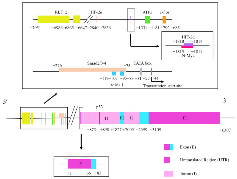
The leukemia inhibitory factor is a secretory glycoprotein with multiple functions [15]. The first study about LIF was published in 1969, when Ichikawa Y identified a protein derived from mice that could inhibit the proliferation of myeloid leukemia M1 cells in vitro on various conditioned media [16]. In 1987, Gearing DP et al. isolated a protein from murine Krebs sarcoma cell cultures that induced the differentiation of mouse myeloid leukemia M1 cells and inhibited their proliferation, thus naming it leukemia inhibitory factor [17]. It has a wide range of biological roles in the neurological, hepatic, endocrine, inflammatory, and immune systems, including the regulation of embryonic stem cell self-renewal, the promotion of embryonic implantation and placental formation, and the stimulation or inhibition of cell proliferation and differentiation, increasing the malignant progression of tumors [15,18,19]. The human LIF gene localizes to a 76 kb segment on chromosome 22q12.1-12.2 [20], which consists of three exons, two introns, and a 3.2 kb untranslated region, and yields a 4.1 kb mRNA product [21]. LIF genes in humans, mice, and other mammals are highly homologous in their coding and non-coding regions [22], indicating that LIF is a highly conserved molecule. Precursor proteins with 202 amino acids are synthesized by LIF mRNA translation, and 22 amino acids are removed from the N-terminal. Finally, the placement of the three disulfide bonds and N-terminal glycosylation steps yield the matured LIF glycoprotein [15,23]. The molecular weight of the non-glycosylated LIF protein is 20-25 kDa, while the glycosylated form ranges from 37 to 63 kDa in weight [24]. In vitro, the biological function of LIF seems to be independent of the degree of glycosylation, but whether glycosylation affects the stability of LIF remains to be determined [23]. Several transcription factors can target LIF promoters or enhancers to regulate LIF expression. Transforming growth factor beta (TGF-β) activates the LIF promoter located at −276/−73 to increase self-renewal in glioma-initiating cells via Smad2/3/4 [25]. Moreover, p53 can regulate maternal reproduction by binding to the intron, located in +873/+898 [26]. The detailed gene structure of LIF is shown in Figure 1 [25,27,28,29,30,31,32,33].
Figure 1.
The structure of the cytokine LIF gene. The rectangular box exhibits the transcriptional regulatory elements of the promoter. The lengths of the exons and introns are shown in base pairs.
LIF belongs to the IL-6 family, which includes oncostatin M, ciliary neurotrophic factor, Charcot–Leyden crystal galactose agglutinin, calcitonin 1, and IL-11 [20,34]. LIF and these cytokines intersect functionally because they share the receptor subunit gp130, which is why they are classified into the same family [22]. In contrast, LIF binds more closely to its specific LIF receptor (LIFR) [7,23]. Existing studies have shown that LIFR can be expressed on many cell surfaces, such as on breast epithelial cells, macrophages, adipocytes, liver cells, and muscle [24,35]. LIF releases via paracrine or autocrine mechanisms, binds to LIFR and the gp130 dimer receptors of the target cells, and then selectively activates signal transduction pathways, including JAK/STAT, MAPK/MEK/ERK, PI3K/AKT, and mTOR, to perform biological functions depending on the cell and tissue conditions [20,36].
3. LIF and Cancer-Associated Cachexia
In some cases, LIF has been thought to be a factor in the origin of cachexia. Nude mice carrying melanoma G361 and SEKI cells expressing large amounts of LIF show a cachectic state, whereas nude mice carrying A375 and MEWO cells without LIF expression are not cachectic [37]. In nude mice that have been inoculated with the neuroepithelioma cell line NAGAI, large amounts of LIF are also detected in association with the induction of cachexia [38]. After the surgical treatment of a nude mouse model of gastric cancer cachexia MKN45c185, the initially elevated LIF in plasma is no longer detectable, and cachexia symptoms are eliminated [39]. LIF is also a key factor that is required for the mouse C26 colon cancer cachexia model [40]. Thena, an anaplastic thyroid cancer cell, induces cachexia in mice by expressing higher levels of IL-6, LIF, and TGF-β [41]. In addition, cachexia models that are mediated by highly expressed LIF include the intracerebral injection of human OVCAR3 ovarian carcinoma, A431 epidermoid carcinoma, and GBLF glioma cells in mice [42]. In the organoids produced by pancreatic cancer patients, LIF, IL-8, and growth differentiation factor 15 (GDF15) are prominently up-regulated in cachectic patients [43]. It has been shown that numerous cancers, including pancreatic, colorectal, esophageal, ovarian, renal, gastric, uterine squamous, and testicular cancers, could aberrantly highly express LIF [44]. In addition, because cachexia is a systemic disease, elevated serum LIF is seen as an indicator of poor prognosis for cancer patients, further demonstrating its potential key role in this pathological process [45,46,47]. Studies have shown that some cancer deaths are attributed to cancer-associated cachexia (CAC), such as pancreatic, esophageal, stomach, lung, liver, and colon cancers [2]. Therefore, we suspect that the cachexia in pancreatic, colon, esophageal and gastric cancers can be treated by targeting the abnormally high expression of LIF.
However, the role of LIF in tumors goes beyond cachexia. Tumor-derived LIF can promote the malignant behavior of tumors in an autocrine manner. KRAS mutation could induce LIF expression in human pancreatic ductal adenocarcinoma (PDAC) cells, and LIF inhibits the intracellular Hippo pathway and promotes tumorigenesis by facilitating YAP/TAZ-TEAD interaction and up-regulating the expression of the YAP1 target genes CNTF and ANKRAD. Neutralizing LIF attenuates pancreatic cancer development and improves the sensitivity of cancer cells to drugs [48]. Colon cancer cell-derived LIF can regulate the expression of other cytokines, such as granulocyte-colony stimulating factor (G-CSF) and IL-6, to promote tumor progression [49]. LIF promotes the stem cell properties of osteosarcoma through the NOTCH1 signaling pathway [50] and enhances the growth and invasion of osteosarcoma by activating STAT3 signaling, while blocking STAT3 signaling can inhibit the development of osteosarcoma [51]. LIF derived from breast and colorectal cancer cells can up-regulate miR-21 expression to promote EMT via STAT3 [52]. LIF over-expression promotes colorectal cancer chemoresistance by reducing the level and function of p53 [53]. Increased serum LIF concentrations can increase tumor cell radiation resistance, inhibit DNA repair, and promote tumor recurrence [54]. LIF mediates STAT3 phosphorylation via the autocrine system during TGF-β regulation and promotes PDAC invasiveness through ECM remodeling, maintaining inflammatory fibroblast phenotype activation [55].
Meanwhile, LIF mediates the crosstalk between cancer-associated stromal cells and tumor cells, which is significantly associated with advanced tumor stage, tumor volume, and a short overall survival time [50,51]. LIF is involved in the interaction between cancer cells and cancer-associated fibroblasts (CAF), forming a feedback loop between cancer cells and CAF [56]. Pancreatic stellate cells (PSC) are widely distributed around pancreatic tumors, and their interaction could transform PSC into CAF [55]. CAF-secreted LIF inhibits pancreatic cancer cell differentiation and maintains their stemness, while blocking LIF using neutralizing antibodies and knocking down LIFR both prolong the survival of KPf/fCL multiple mutant mice [57]. TGF-β can induce CAF to produce LIF, leading to fibroblast activation and the promotion of tumor cell invasiveness [58]. TNF and other inflammatory cytokines that are produced by macrophages in the tumor microenvironment stimulate tumor cells to produce IL-6 and LIF, contributing to tumor growth [59]. LIF could partially control cancer cell immune tolerance by affecting monocytes. Recombinant LIF converts monocytes into tumor-associated macrophage (TAM)-like cells to promote ovarian cancer immunosuppression by inhibiting the expression of the monocyte colony-stimulating factor [60]. LIF blockade results in TAM phenotypic changes, in which C-X-C motif chemokine ligand 9 (CXCL9) expression is elevated, and CD8+ T cells are recruited to the tumor, suggesting that LIF is involved in resistance to immune checkpoint blockade. Neutralizing antibodies to LIF combined with a PD1 immune checkpoint inhibitor could strengthen the immune memory of the host and improve overall survival [61]. Bone marrow mesenchymal stem cells (BM-MSCs) perform a crucial role in supporting the hematopoietic process. It has been shown that BM-MSCs can enhance angiogenesis by releasing LIF to activate the ERK1/2 pathway and induce cancer cells to express vascular endothelial growth factors [62]. In the ovarian cancer microenvironment, cancer-associated mesenchymal stem cells activate the JAK/STAT pathway in ovarian cancer cells by secreting LIF, promoting cancer cell growth, and maintaining their stem cell properties [63]. In addition, our previous research has verified that LIF is highly expressed in cancer-associated adipocytes (CAA) and forms a feedback loop with the CXCLs derived from breast cancer to promote the invasion and metastasis of breast cancer [64].
In summary, LIF promotes tumor progression by being aberrantly expressed in the early stages of cancer and increases the degree of malignancy by affecting the tumor microenvironment. Tumor-derived LIF is a vital initiation factor of cachexia, leading to the progression of cachexia and a poor prognosis for patients with late stages of cancer.
4. LIF and Alterations of Adipose Tissue in Cachexia
According to the type of cells present in adipose tissue, it can be classified into white adipose tissue (WAT), brown adipose tissue (BAT), and beige adipose tissue. WAT constitutes the majority of body fat and stores for energy in the form of triglycerides (TG), while BAT generates heat through uncoupled mitochondrial respiration [65]. In recent years, adipose tissue has also been identified as an endocrine organ that secretes a large number of cytokines that are important for the regulation of the body’s systemic metabolism [66]. In cachexia, adipocyte variation has been widely discussed, with studies confirming adipocyte atrophy, suppressed adipocyte differentiation, or the dedifferentiation of mature adipocytes, WAT browning, and extracellular matrix remodeling [67]. Meanwhile, LIF plays a non-negligible role in adipocyte alterations in cachexia, which could bind to LIFR to exert their effects on adipocytes by activating the JAK/STAT and MAPK (ERK1/2) signaling pathways [68].
Morphological changes in adipocyte area size are primarily produced through lipid hydrolysis within the adipocytes [9]. In adipose tissue, TG is mobilized by adipose triglyceride lipase (ATGL), hormone-sensitive lipase, and monoacylglycerol lipase [69]. Among them, ATGL triggers lipolysis, which releases fatty acids from TG [70]. Moreover, lipoprotein lipase (LPL) is the rate-limiting enzyme for triglyceride hydrolysis [71]. Lipolysis, the catabolism of TG, could lead to fat loss, contributing to cachexia [2,72]. Noncoding RNAs such as circPTK2 [73], infection [74], and cytokines such as LIF contribute to lipolysis in cachexia [26]. Available experiments have demonstrated that elevated plasma LIF concentrations are associated with lipolytic enzymes, such as in murine cachexia models bearing SEK1 and NAGAI cells, and that a reduction in LPL activity caused by LIF can regulate lipolysis [75,76]. LIF activates JAK/STAT signaling, mainly the phosphorylation of STAT1 and STAT3, to promote lipolysis via ATGL [77]. Moreover, JAK inhibitors could effectively alleviate adipose loss and improve overall survival, mainly by inhibiting STAT3 phosphorylation [78].
WAT browning is an energy-releasing process that is accompanied by a rise in UCP1 expression that is commonly seen in CAC to meet tumor energy expenditure, which can be mobilized by cytokines, usually IL-6 [9]. WAT browning facilitates CAC due to energy consumption [79]. Moreover, LIF can also promote thermogenesis by facilitating adipocyte browning during exercise [79,80].
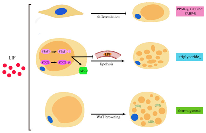
Adipocyte differentiation also has a significant impact on cachexia. The inhibition of adipocyte differentiation has been seen in cachexia mediated by infection [74]; various cytokines [81]; Zinc-α2-glycoprotein [82]; and miRNA, including miR-146b-5p [83], miR-410-3P [84], and miR-155 [85]. LIF may negatively regulate adipocyte differentiation. In preadipocytes, LIFR knockdown results in the reduced expression of the adipocyte differentiation marker genes peroxisome proliferator-activated receptor gamma (PPAR-γ), CCAAT/enhancer-binding protein alpha (C/EBP-α), and fatty acid-binding protein 4 (FABP4) during adipocyte maturation [86]. Moreover, the dedifferentiation of mature adipocytes has also been observed in cachexia and is mediated by tumor-derived proliferin-1 [87] and CircPTK2 [73]. CAA is a type of adipocyte that is formed by the dedifferentiation of the mature adipocytes present in the tumor microenvironment [88]. Some of the alterations that occur in cachectic adipocytes include adipocytes that are small in size or that have decreased TG stores, lower PPAR-γ and C/EBP-α expression, and higher IL-6, TNF-α, IL-1β, and UCP1 expression, which also occur in CAA [89]. We speculate that LIF can promote adipocyte dedifferentiation in the tumor microenvironment and in the case of cachectic adipocytes. The effect of LIF on adipocytes is shown in Figure 2.
Figure 2.
The role of LIF in adipocytes. LIF can activate the phosphorylation of STAT1 and STAT3, increase ATGL to lead to lipolysis, and is associated with decreased LPL activity. Finally, it results in triglyceride loss in adipocytes. LIF also can induce WAT browning, a thermogenic process. Meanwhile, LIF may inhibit preadipocytes’ differentiation to mature adipocytes, as evidenced by the inhibition of PPAR-γ, C/EBP-α, and FABP4 expression (by Figdraw “www.figdraw.com”, accessed on 5 May 2022).
5. LIF and Anorexia Nervosa in Cachexia
Anorexia nervosa can be seen in various diseases, and its pathophysiological processes mainly manifested as weight loss [90]. Anorexia is closely associated with an imbalance between the orexigenic and anorexigenic neuropeptides in the central nervous system [91] and abnormally elevated levels of leptin [92]. Anorexia nervosa has multiple complications that can affect various organs throughout the body [93]. Many inflammatory cytokines are involved in anorexia nervosa, such as IL-1, IL-6, interferon-gamma (IFN-γ), TNF-α, and leptin [90,94]. Anorexia can be caused by elevated LIF expression, which, according to different studies, decreases food intake and leads to weight loss [92]. Animal experiments have shown that LPS can lead to the up-regulation of LIF expression in the central nervous system, resulting in extensive inflammation and anorexia, and with the combination of LPS and corticosterone, LIF up-regulation is more prominent [95].
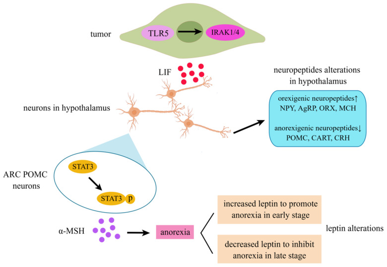
Among the causes of cachexia, tumors account for a considerable proportion. Patients with malignant tumors often suffer from cachexia anorexia syndrome during tumor progression [96], tumor metastasis [97], and radiotherapy and chemotherapy [98,99], with the clinical manifestation including losing appetite. Terawaki K et al. revealed that in gastric cancer, the activation of TLR5 signaling by IRAK-1,4 leads to the up-regulation of LIF expression, which in turn leads to anorexia [100]. Arora G et al. showed that in colon adenocarcinoma, LIF-induced anorexia is associated with the activation of JAK/STAT signaling in the hypothalamus [78].
The hypothalamus is essential for regulating feeding behavior and contributes to the development of anorexia, which occurs in various diseases. Current research has confirmed that LIFR is expressed in proopiomelanocortin (POMC) neurons in the arcuate nucleus (ARC) region in the hypothalamus, which can be activated by LIF and then α-MSH released from ARC POMC neurons to induce anorexia [101]. In addition, LIF derived from tumors interacts extensively with the neuropeptides in anorexia [39]. In gastric cancer cachexia, Terawaki K et al. observed that LIF expression was up-regulated, with the elevated expression of orexigenic neuropeptides including neuropeptide Y (NPY), agouti-related protein (AgRP), orexin (ORX), and melanin-concentrating hormone (MCH), and the inhibited expression of anorexigenic neuropeptides, including POMC, amphetamine-regulated transcript (CART), and corticotropin-releasing hormone (CRH) in the hypothalamus [39]. In the SEKI rat cachexia model, the same phenomenon was observed. Although the expression of anorexigenic peptides was down-regulated in both animal models, the bodyweight of the mice was reduced in a cachectic state. We speculate that the effect of neuropeptides may be the self-regulation of the body in response to the anorexia or cachexia effect produced by LIF.
In addition, the progression of anorexia in cachexia caused by LIF is partly influenced by leptin, which is a crucial adipokine, and changes in the adipose tissue or bodyweight can affect the leptin secretion. The leptin released from adipose tissue acts as a signaling mediator from the periphery to the central nervous system, which is essential in maintaining the balance of adipose tissue weight both in physiology and in the pathologic condition [102]. Leptin and LIF have similar structures and can bind to subunit gp130 to activate STAT3 signaling [103]. Notably, a murine animal model confirmed that adipose loss and anorexia induced by LIF are independent of leptin receptor signaling [77]. It was shown that LIF could promote the expression of leptin at an early stage and generate anorexia [104]. However, in the later stages of LIF-induced anorexia, food intake returns to basal levels as serum and adipose leptin expression decreases and hypothalamic STAT3 phosphorylation activation is reduced [77,78,105]. Furthermore, in mice lacking functional leptin (ob/ob) or leptin receptors (db/db), LIF-induced anorexia persisted without recovery [77]. We speculate that anorexia induced by abnormally high LIF expression may be an acute response. At a very early stage, LIF can increase leptin, leading to anorexia and weight loss, but due to adaptive changes in the body, the effects of LIF are disturbed, leptin expression is suppressed, and anorexic behavior is ameliorated. The decreased circulating leptin is an important “natural” systemic response to combat LIF-induced cachexia and therefore may also be important for the mechanisms of potential cachexia therapies. The effect of LIF on anorexia nervosa is shown in Figure 3.
Figure 3.
LIF-induced anorexia in cancer cachexia. TLR5 can activate IRAK1/4 to promote LIF secretion in tumors. LIF can facilitate α-MSH secretion from ARC POMC neurons in the hypothalamus that are associated with elevated STAT3 phosphorylation. Additionally, up-regulated LIF is associated with increased orexigenic neuropeptides (NPY, AgRP, ORX, and MCH) and decreased anorexigenic neuropeptides (POMC, CART, and CRH) in the hypothalamus. Leptin increases in the early stage to promote anorexia and decreases in the late stage to inhibit anorexia (by Figdraw, “www.figdraw.com”, accessed on 28 May 2022).
6. LIF and Muscle Atrophy in Cachexia
The constant loss and regeneration of skeletal muscle are essential for normal physiological function [106]. In cachexia, the balance between the anabolism and catabolism of proteins is broken, thus promoting lipid oxidation and insulin resistance, inhibiting remodeling, etc. [107]. Patients often suffer from skeletal muscle atrophy [108], especially in CAC. Proinflammatory cytokines such as IL-6, TNF-α, and LIF have been identified to lead to muscle wasting in cachexia [109].
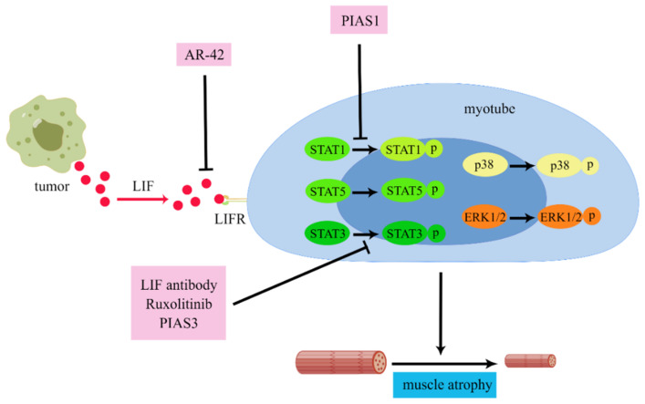
LIF has been observed to be over-expressed in muscle atrophy disease, suggesting that LIF may be related to muscle atrophy [49,110,111]. In colon cancer, LIF can induce muscle atrophy by activating the JAK/STAT [40,112], ERK1/2 [40], and p38 MAPK [112] signaling pathways, resulting in the generation of cachexia. Moreover, the above studies also indicate that inhibiting the JAK/STAT signaling pathways in the myotubes can effectively reduce muscle atrophy, including Ruxolitinib (JAK inhibitor), PIAS3, and the LIF-neutralizing antibody targeting STAT3 and PIAS1 targeting STAT1 [40]. Meanwhile, LIF can also be the target gene of microRNA. MiR-29c can target LIF by combing the area 3′-UTR to down-regulate LIF to inhibit JAK/STAT and MAPK signaling. In the Lewis lung cancer mouse model, the inhibition of lung cancer miR-29c expression resulted in the up-regulation of LIF activity on the myotubes, causing muscle atrophy and cachexia [113].
Under physiological circumstances, both cultured myotubes and muscle can secrete LIF as a response to outside stimulation [114]. However, in pathological conditions, the role of LIF in muscle seems to be paradoxical. LIF can promote the regeneration of muscle atrophy due to denervation [115,116] and injured muscles [117], the proliferation of myoblasts [118], and muscle satellite cells [119]. Clinical trials have demonstrated that exercise inhibits muscle atrophy in cancer patients through the cytokine mediation [120]. LIF expression was significantly up-regulated and accompanied by the down-regulation of myostatin expression, after exercise [121].
Overall, LIF can exert multiple effects on skeletal muscle cells under different conditions [122]. This seems to be related to the crosstalk of other factors in the cachexia, which needs to be further elucidated for better treatment to improve the prognosis of tumor patients. The effect of LIF on skeletal muscle atrophy is shown in Figure 4.
Figure 4.
The mechanisms of LIF in cachexia-induced skeletal muscle atrophy. LIF targeted at myotubes can activate STAT1, STAT3, STAT5, ERK1/2, and p38 signaling. PIAS1 can block STAT1 phosphorylation to inhibit muscle atrophy. The LIF antibody, Ruxolitinib (JAK inhibitor), and PIAS3 can block the phosphorylation of STAT3 to inhibit muscle atrophy. Additionally, AR-42, a histone deacetylation inhibitor, can block LIF to treat CAC-induced muscle atrophy (by Figdraw “www.figdraw.com”, accessed on 5 May 2022).
7. LIF and Other Crucial Factors of Cachexia
Although LIF plays a significant role in cachexia, the pathological process of cachexia is not achieved by LIF alone. Studies at this stage suggest that cancer cachexia is a cross-linked network of inflammatory factors [123]. When a particular inflammatory factor is over-activated, it activates other cachectic factors through this network, causing a cascade reaction that acts on different target organs to form the final pathological state of cachexia. Currently recognized cachexia factors include IL-6, IL-1α, IL-1β, TNF-α, IFN-γ, and GDF15, which are related to the presence of LIF [108]. In rLIF-driven cachexia mouse models, the mRNA levels of IL-6 increased 50-fold, and in both models, this was associated with anorexia and fat loss [108]. In vitro, the lipolysis caused by TNF-α and IL-6 was two-fold and three-fold higher than that caused by LIF, respectively [59]. The separate stimulation of TNF and IL-1β promoted the secretion of LIF and IL-6 in a cachexia study with tumor-bearing mice [124], resulting in a synergistic effect [125]. In addition, LIF can promote the secretion of IL-1α, but the mRNA content does not increase [126]. It is worth noting that there seems to be a consensus that the activation of IL-6 expression follows the up-regulated expression of LIF, thereby promoting the occurrence of anorexia and cachexia syndrome in tumor patients [78,120,121]. However, some research has not detected the elevated expression of IL-6 accompanied by the over-expression of LIF secreted by the tumor in animal models [110,122,127].
Studies have confirmed that TNF-α and IFN-γ are essential cachexia mediators. They can cause a reduction in food intake and bodyweight in mice in sub-lethal amounts [128]. In chordoma, TNF-α has a secretory cycle-promoting effect with LIF that is associated with a synergistic effect and reduced overall patient survival [129]. However, the relationship between LIF and IFN-γ remains unclear in cachexia. IFN-γ can modulate the impact of TNF-α by reducing LIF expression [130], and yet, in encephalitogenic cells, inducing IFN-γ can increase the level of LIF [131].
The most vital role of GDF15 is to regulate energy homeostasis, which can affect normal appetite and cause cachexia when its expression is elevated due to tumors [132]. GDF15 can successively activate the ERK pathway, increase LIF expression through c-Fos binding to the LIF promoter region (−685/−792), and then promote the phosphorylation of STAT3, thus expanding the stemness of glioma cells [33].
Overall, LIF can lead to cachexia through lipolysis, anorexia, and skeletal muscle atrophy in tumors. We summarize the relevant molecular basis of cachexia-associated transcriptional regulation of the LIF gene in Table 1 and genes or enzyme activity regulated by LIF in Table 2.
Table 1.
Transcriptional regulation of the LIF gene.
| Cytokine, Transcription Factor, or microRNA | LIF Expression | Cell Type and Context | References |
|---|---|---|---|
| HIF-2α | up | RKO and HCT116 colorectal cancer cells |
[31] |
| TNF, IL-1β | up | 4JK macrophage derived from tumors | [59] |
| TGF-β | down | mouse PSCs, KPC primary tumor cells |
[55] |
| IL-1 | up | mouse PSCs, KPC primary tumor cells |
[55] |
| TGF-β | up | fibroblasts, SCC12 carcinoma cells | [58] |
| GDF15 | up | glioma stem cells | [33] |
| KRAS | up | BxPC3 and SW1990 pancreatic cancer cells | [48] |
| EBV-encoded protein latent membrane protein 1 | up | nasopharyngeal carcinoma (NPC) serum samples | [54] |
| TLR5, IRAK-1/4 | up | 85As2 gastric cancer cells | [100] |
| CXCLs | up | CAA | [64] |
| miR-29c | down | Lewis lung carcinoma cells | [113] |
Table 2.
Genes, transcription factors, or enzymes regulated by LIF.
| Genes, Transcription Factors, or Enzymes | Regulation by LIF | Function | References |
|---|---|---|---|
| YAP/TAZ-TEAD | up | suppress the Hippo pathway in PDAC cells | [48] |
| IL-6, G-CSF | up | mediate cachexia in colon carcinoma cells | [49] |
| NOTCH1, HEY1, HEYL, HES1, KRT19 | up | keep stem cell-like properties of osteosarcoma | [50] |
| miR-21 | up | promote EMT of breast and colorectal cancer cells |
[52] |
| p53 | down | promote chemoresistance of colorectal cancer | [53] |
| genes of p70S6K signaling, mTOR | up | promote NPC cell growth | [54] |
| CXCL9 | down | inhibit CD8+ T cell tumor infiltration | [61] |
| STAT3 | up | promote ovarian cancer cell growth | [63] |
| CXCLs | up | promote breast cancer cell invasion and metastasis |
[64] |
| IL-6 | up | induce cachexia | [2] |
| STAT1/3, ATGL activity | up | induce lipolysis | [77] |
| LPL activity | down | induce lipolysis | [75,76] |
| PPAR-γ, C/EBP-α, FABP4 | down | inhibit adipocyte differentiation | [86] |
| α-MSH | up | induce anorexia | [101] |
| leptin | up | induce anorexia | [77,104] |
| NPY, AgRP POMC, CART |
up down |
compensate for anorexia | [39,101] |
| p38 | up | induce muscle atrophy | [112] |
| STAT1/3/5, ERK1/2 | up | induce muscle atrophy | [40] |
| mTORC1 | up | induce myotube protein synthesis |
[12] |
8. LIF and Therapeutics in Cachexia
Numerous studies have demonstrated that tumors with abnormally high LIF expression not only cause more rapid tumor proliferation and greater malignancy in migration and invasion in the early stages but that they also induce cachexia in the late stages, resulting in fat loss, anorexia nervosa, and skeletal muscle atrophy. Given this, targeted LIF may reduce tumor progression and the development of late-stage malignancy, allowing patients to have a better quality of life.
In animal experiments, inhibiting JAK, downstream of LIF, can effectively alleviate cachexia [40,78]. At present, histone deacetylation inhibitor AR-42 has been observed to impede muscle atrophy by blocking multiple pro-cachexia drivers, including LIF, to ameliorate cachexia [112]. However, in some cases, LIF can also facilitate the proliferation of muscle cells. Completely suppressing the effect of LIF is not the best option. Therefore, a more thorough study of the dual role of LIF in muscle proliferation and atrophy is warranted. In addition to drug therapy, some attempts have been made to remove LIF from plasma by physical adsorption to treat cachexia, but these studies failed [133].
In addition, blocking LIF to improve anorexia nervosa and fat loss should be an effective treatment. The application of Ghrelin resulted in an increase in bodyweight in LIF-induced mice cachexia and a corresponding increase in serum leptin levels [134]. In addition, Guo et al. demonstrated that LIFR knockdown can decrease LIF-induced lipolysis and contribute to the reduction of browning markers, ultimately leading to an increased fat mass and bodyweight in mice [135]. Notably, lipolysis and anorexia are indirectly related. Decreased protein intake and insufficient energy intake due to anorexia can compensate for energy metabolism through lipolysis. However, Arora GK et al. observed that there was a significant reduction in both fat and bodyweight in mice injected with rLIF in the absence of differences in food intake compared to the control group, suggesting that LIF-induced lipolysis may have mechanisms that are independent of changes in feeding [77]. This means that simply improving the symptoms of anorexia does not stop cachexia from continuing, resulting in the continuation of fat loss. Weight maintenance is the ultimate goal in the treatment of cachexia; therefore, we need to consider the therapeutic potential of LIF in cachexia in a holistic manner. Improving fat loss, anorexia, and skeletal muscle atrophy during the course of cachexia, as well as treating the tumor, the cause of the cachexia, could better improve patient prognosis.
9. Conclusions
Overall, the available studies have confirmed that LIF can lead to weight loss through skeletal muscle atrophy, fat loss, and anorexia nervosa, which contributes to the progression of cachexia. However, there are still some interesting and important questions that deserve further exploration.
First, in recent years, immunotherapy has been applied to cachexia. Immune cells in the tumor microenvironment, including TAM, tumor-infiltrating lymphocytes, and tumor-associated neutrophils, are considered to be capable of secreting large amounts of circulating factors such as IL-6 and IL-1 to promote the malignant progression of cachexia [136]. It has been shown that abnormally high LIF expression in the tumor microenvironment can promote macrophage aggregation, which could secrete IL6 and IL-1α to induce muscle atrophy and cachexia [61,137]. It is well known that most patients with advanced PDAC are in a cachectic state [138], perhaps because LIF is highly expressed in the tumor cells, macrophages, and mast cells in the tumor microenvironment of PDAC patients [139]. In animal experiments, the removal of macrophages prevented and alleviated cachexia [140]. Therefore, perhaps we can achieve a similar effect by targeting LIF inhibition. Unfortunately, there is little research on LIF in this immunotherapy for cachexia, and more in-depth studies can provide more methods for treating cachexia.
Second, the generation and development of cachexia is a highly complex network of inflammatory cross-linkages. Identifying the most crucial malignancy triggers according to disease type and blocking the cascade of inflammatory pathways at the source may be valuable for treating this pathological process. As previously stated, LIF is one of the sources in some diseases. In current clinical studies, good progress has been made in improving cachexia with therapies that counteract key cachexia factors such as IL-6, IL-1β, and IL-1α [14,141,142]. Recently, EC330, a targeted inhibitor of LIF, has been observed to significantly inhibit the malignant behavior of tumors at both the cellular and animal levels and may also be used in cachexia-related studies [143].
Finally, LIF research in this area is still focused on the cellular and animal levels. More needs to be invested in research, especially clinically relevant trials. LIF plays a broad role in cachexia, so we believe that targeted LIF has excellent potential for treating cachexia. Clinical trials may bring hope to patients. Of course, cachexia treatment is multidimensional, and it is not the best option to look at only one aspect of the problem. However, a more thorough study of LIF and therapeutic drugs in cachexia would definitely complement current treatments.
Author Contributions
R.Z. and C.T. contributed equally to this work. R.Z. performed background research, drafted and edited this review, and contributed to the creation of figures; C.T. performed background research, drafted and edited this review, and contributed to the creation of figures; X.X. approved the final draft and reviewed drafts of the paper. All authors have read and agreed to the published version of the manuscript.
Conflicts of Interest
The authors declare no conflict of interest.
Funding Statement
This work was supported by grants from the National Natural Science Foundation of China (81760509) and the Natural Science Foundation of Jiangxi Province of China (20181BAB205043) to Xiangyang Xiong.
Footnotes
Publisher’s Note: MDPI stays neutral with regard to jurisdictional claims in published maps and institutional affiliations.
References
- 1.Ni J., Zhang L. Cancer Cachexia: Definition, Staging, and Emerging Treatments. Cancer Manag. Res. 2020;12:5597–5605. doi: 10.2147/CMAR.S261585. [DOI] [PMC free article] [PubMed] [Google Scholar]
- 2.Baracos V.E., Martin L., Korc M., Guttridge D.C., Fearon K. Cancer-associated cachexia. Nat. Rev. Dis. Primers. 2018;4:17105. doi: 10.1038/nrdp.2017.105. [DOI] [PubMed] [Google Scholar]
- 3.Berardi E., Madaro L., Lozanoska-Ochser B., Adamo S., Thorrez L., Bouche M., Coletti D. A Pound of Flesh: What Cachexia Is and What It Is Not. Diagnostics. 2021;11:116. doi: 10.3390/diagnostics11010116. [DOI] [PMC free article] [PubMed] [Google Scholar]
- 4.Tisdale M.J. Cancer cachexia. Curr. Opin. Gastroenterol. 2010;26:146–151. doi: 10.1097/MOG.0b013e3283347e77. [DOI] [PubMed] [Google Scholar]
- 5.Fearon K.C. Cancer cachexia: Developing multimodal therapy for a multidimensional problem. Eur. J. Cancer. 2008;44:1124–1132. doi: 10.1016/j.ejca.2008.02.033. [DOI] [PubMed] [Google Scholar]
- 6.Fearon K., Strasser F., Anker S.D., Bosaeus I., Bruera E., Fainsinger R.L., Jatoi A., Loprinzi C., Macdonald N., Mantovani G., et al. Definition and classification of cancer cachexia: An international consensus. Lancet Oncol. 2011;12:489–495. doi: 10.1016/S1470-2045(10)70218-7. [DOI] [PubMed] [Google Scholar]
- 7.Kazemi-Bajestani S.M., Mazurak V.C., Baracos V. Computed tomography-defined muscle and fat wasting are associated with cancer clinical outcomes. Semin. Cell Dev. Biol. 2016;54:2–10. doi: 10.1016/j.semcdb.2015.09.001. [DOI] [PubMed] [Google Scholar]
- 8.Tsoli M., Swarbrick M.M., Robertson G.R. Lipolytic and thermogenic depletion of adipose tissue in cancer cachexia. Semin. Cell Dev. Biol. 2016;54:68–81. doi: 10.1016/j.semcdb.2015.10.039. [DOI] [PubMed] [Google Scholar]
- 9.Han J., Meng Q., Shen L., Wu G. Interleukin-6 induces fat loss in cancer cachexia by promoting white adipose tissue lipolysis and browning. Lipids Health Dis. 2018;17:14. doi: 10.1186/s12944-018-0657-0. [DOI] [PMC free article] [PubMed] [Google Scholar]
- 10.Argiles J.M., Stemmler B., Lopez-Soriano F.J., Busquets S. Inter-tissue communication in cancer cachexia. Nat. Rev. Endocrinol. 2018;15:9–20. doi: 10.1038/s41574-018-0123-0. [DOI] [PubMed] [Google Scholar]
- 11.Nishikawa H., Goto M., Fukunishi S., Asai A., Nishiguchi S., Higuchi K. Cancer Cachexia: Its Mechanism and Clinical Significance. Int. J. Mol. Sci. 2021;22:8491. doi: 10.3390/ijms22168491. [DOI] [PMC free article] [PubMed] [Google Scholar]
- 12.Gao S., Durstine J.L., Koh H.J., Carver W.E., Frizzell N., Carson J.A. Acute myotube protein synthesis regulation by IL-6-related cytokines. Am. J. Physiol. Cell Physiol. 2017;313:C487–C500. doi: 10.1152/ajpcell.00112.2017. [DOI] [PMC free article] [PubMed] [Google Scholar]
- 13.Patel H.J., Patel B.M. TNF-alpha and cancer cachexia: Molecular insights and clinical implications. Life Sci. 2017;170:56–63. doi: 10.1016/j.lfs.2016.11.033. [DOI] [PubMed] [Google Scholar]
- 14.Laird B.J., Mcmillan D., Skipworth R., Fallon M.T., Paval D.R., Mcneish I., Gallagher I.J. The Emerging Role of Interleukin 1beta (IL-1beta) in Cancer Cachexia. Inflammation. 2021;44:1223–1228. doi: 10.1007/s10753-021-01429-8. [DOI] [PMC free article] [PubMed] [Google Scholar]
- 15.Nicola N.A., Babon J.J. Leukemia inhibitory factor (LIF) Cytokine Growth Factor Rev. 2015;26:533–544. doi: 10.1016/j.cytogfr.2015.07.001. [DOI] [PMC free article] [PubMed] [Google Scholar]
- 16.Ichikawa Y. Differentiation of a cell line of myeloid leukemia. J. Cell. Physiol. 1969;74:223–234. doi: 10.1002/jcp.1040740303. [DOI] [PubMed] [Google Scholar]
- 17.Gearing D.P., Gough N.M., King J.A., Hilton D.J., Nicola N.A., Simpson R.J., Nice E.C., Kelso A., Metcalf D. Molecular cloning and expression of cDNA encoding a murine myeloid leukaemia inhibitory factor (LIF) Embo J. 1987;6:3995–4002. doi: 10.1002/j.1460-2075.1987.tb02742.x. [DOI] [PMC free article] [PubMed] [Google Scholar]
- 18.Hirai H., Karian P., Kikyo N. Regulation of embryonic stem cell self-renewal and pluripotency by leukaemia inhibitory factor. Biochem. J. 2011;438:11–23. doi: 10.1042/BJ20102152. [DOI] [PMC free article] [PubMed] [Google Scholar]
- 19.Salleh N., Giribabu N. Leukemia inhibitory factor: Roles in embryo implantation and in nonhormonal contraception. Sci. World J. 2014;2014:201514. doi: 10.1155/2014/201514. [DOI] [PMC free article] [PubMed] [Google Scholar]
- 20.Taupin J.L., Pitard V., Dechanet J., Miossec V., Gualde N., Moreau J.F. Leukemia inhibitory factor: Part of a large ingathering family. Int. Rev. Immunol. 1998;16:397–426. doi: 10.3109/08830189809043003. [DOI] [PubMed] [Google Scholar]
- 21.Stahl J., Gearing D.P., Willson T.A., Brown M.A., King J.A., Gough N.M. Structural organization of the genes for murine and human leukemia inhibitory factor. Evolutionary conservation of coding and non-coding regions. J. Biol. Chem. 1990;265:8833–8841. doi: 10.1016/S0021-9258(19)38963-X. [DOI] [PubMed] [Google Scholar]
- 22.Willson T.A., Metcalf D., Gough N.M. Cross-species comparison of the sequence of the leukaemia inhibitory factor gene and its protein. Eur. J. Biochem. 1992;204:21–30. doi: 10.1111/j.1432-1033.1992.tb16601.x. [DOI] [PubMed] [Google Scholar]
- 23.Hinds M.G., Maurer T., Zhang J.G., Nicola N.A., Norton R.S. Solution structure of leukemia inhibitory factor. J. Biol. Chem. 1998;273:13738–13745. doi: 10.1074/jbc.273.22.13738. [DOI] [PubMed] [Google Scholar]
- 24.Yue X., Wu L., Hu W. The regulation of leukemia inhibitory factor. Cancer Cell Microenviron. 2015;2 doi: 10.14800/ccm.877. [DOI] [PMC free article] [PubMed] [Google Scholar]
- 25.Penuelas S., Anido J., Prieto-Sanchez R.M., Folch G., Barba I., Cuartas I., Garcia-Dorado D., Poca M.A., Sahuquillo J., Baselga J., et al. TGF-beta increases glioma-initiating cell self-renewal through the induction of LIF in human glioblastoma. Cancer Cell. 2009;15:315–327. doi: 10.1016/j.ccr.2009.02.011. [DOI] [PubMed] [Google Scholar]
- 26.Saini A., Al-Shanti N., Stewart C.E. Waste management—cytokines, growth factors and cachexia. Cytokine Growth Factor Rev. 2006;17:475–486. doi: 10.1016/j.cytogfr.2006.09.006. [DOI] [PubMed] [Google Scholar]
- 27.Cheng X., Liu J., Shan H., Sun L., Huang C., Yan Q., Jiang R., Ding L., Jiang Y., Zhou J., et al. Activating transcription factor 3 promotes embryo attachment via up-regulation of leukemia inhibitory factor in vitro. Reprod. Biol. Endocrinol. 2017;15:42. doi: 10.1186/s12958-017-0260-7. [DOI] [PMC free article] [PubMed] [Google Scholar]
- 28.Huang C., Sun H., Wang Z., Liu Y., Cheng X., Liu J., Jiang R., Zhang X., Zhen X., Zhou J., et al. Increased Kruppel-like factor 12 impairs embryo attachment via downregulation of leukemia inhibitory factor in women with recurrent implantation failure. Cell Death Discov. 2018;4:23. doi: 10.1038/s41420-018-0088-8. [DOI] [PMC free article] [PubMed] [Google Scholar]
- 29.Bamberger A.M., Jenatschke S., Schulte H.M., Ellebrecht I., Beil F.U., Bamberger C.M. Regulation of the human leukemia inhibitory factor gene by ETS transcription factors. Neuroimmunomodulation. 2004;11:10–19. doi: 10.1159/000072964. [DOI] [PubMed] [Google Scholar]
- 30.Cotterman R., Knoepfler P.S. N-Myc regulates expression of pluripotency genes in neuroblastoma including lif, klf2, klf4, and lin28b. PLoS ONE. 2009;4:e5799. doi: 10.1371/journal.pone.0005799. [DOI] [PMC free article] [PubMed] [Google Scholar]
- 31.Wu L., Yu H., Zhao Y., Zhang C., Wang J., Yue X., Yang Q., Hu W. HIF-2alpha mediates hypoxia-induced LIF expression in human colorectal cancer cells. Oncotarget. 2015;6:4406–4417. doi: 10.18632/oncotarget.3017. [DOI] [PMC free article] [PubMed] [Google Scholar]
- 32.Hu W., Feng Z., Teresky A.K., Levine A.J. p53 regulates maternal reproduction through LIF. Nature. 2007;450:721–724. doi: 10.1038/nature05993. [DOI] [PubMed] [Google Scholar]
- 33.Zhu S., Yang N., Guan Y., Wang X., Zang G., Lv X., Deng S., Wang W., Li T., Chen J. GDF15 promotes glioma stem cell-like phenotype via regulation of ERK1/2-c-Fos-LIF signaling. Cell Death Discov. 2021;7:3. doi: 10.1038/s41420-020-00395-8. [DOI] [PMC free article] [PubMed] [Google Scholar]
- 34.Jones S.A., Jenkins B.J. Recent insights into targeting the IL-6 cytokine family in inflammatory diseases and cancer. Nat. Rev. Immunol. 2018;18:773–789. doi: 10.1038/s41577-018-0066-7. [DOI] [PubMed] [Google Scholar]
- 35.Heinrich P.C., Behrmann I., Haan S., Hermanns H.M., Muller-Newen G., Schaper F. Principles of interleukin (IL)-6-type cytokine signalling and its regulation. Biochem. J. 2003;374:1–20. doi: 10.1042/bj20030407. [DOI] [PMC free article] [PubMed] [Google Scholar]
- 36.Gough N.M., Gearing D.P., King J.A., Willson T.A., Hilton D.J., Nicola N.A., Metcalf D. Molecular cloning and expression of the human homologue of the murine gene encoding myeloid leukemia-inhibitory factor. Proc. Natl. Acad. Sci. USA. 1988;85:2623–2627. doi: 10.1073/pnas.85.8.2623. [DOI] [PMC free article] [PubMed] [Google Scholar]
- 37.Mori M., Yamaguchi K., Honda S., Nagasaki K., Ueda M., Abe O., Abe K. Cancer cachexia syndrome developed in nude mice bearing melanoma cells producing leukemia-inhibitory factor. Cancer Res. 1991;51:6656–6659. [PubMed] [Google Scholar]
- 38.Kajimura N., Iseki H., Tanaka R., Ohue C., Otsubo K., Gyoutoku M., Sasaki K., Akiyama Y., Yamaguchi K. Toxohormones responsible for cancer cachexia syndrome in nude mice bearing human cancer cell lines. Cancer Chemother. Pharmacol. 1996;38:S48–S52. doi: 10.1007/s002800051038. [DOI] [PubMed] [Google Scholar]
- 39.Terawaki K., Sawada Y., Kashiwase Y., Hashimoto H., Yoshimura M., Suzuki M., Miyano K., Sudo Y., Shiraishi S., Higami Y., et al. New cancer cachexia rat model generated by implantation of a peritoneal dissemination-derived human stomach cancer cell line. Am. J. Physiol. Endocrinol. Metab. 2014;306:E373–E387. doi: 10.1152/ajpendo.00116.2013. [DOI] [PubMed] [Google Scholar]
- 40.Seto D.N., Kandarian S.C., Jackman R.W. A Key Role for Leukemia Inhibitory Factor in C26 Cancer Cachexia. J. Biol. Chem. 2015;290:19976–19986. doi: 10.1074/jbc.M115.638411. [DOI] [PMC free article] [PubMed] [Google Scholar]
- 41.Chang J.W., Yeh K.Y., Shen Y.C., Hsieh J.J., Chuang C.K., Liao S.K., Tsai L.H., Wang C.H. Production of multiple cytokines and induction of cachexia in athymic nude mice by a new anaplastic thyroid carcinoma cell line. J. Endocrinol. 2003;179:387–394. doi: 10.1677/joe.0.1790387. [DOI] [PubMed] [Google Scholar]
- 42.Negri D.R., Mezzanzanica D., Sacco S., Gadina M., Benigni F., Cajola L., Finocchiaro G., Ghezzi P., Canevari S. Role of cytokines in cancer cachexia in a murine model of intracerebral injection of human tumours. Cytokine. 2001;15:27–38. doi: 10.1006/cyto.2001.0899. [DOI] [PubMed] [Google Scholar]
- 43.Vaes R., van Dijk D., Welbers T., Blok M.J., Aberle M.R., Heij L., Boj S.F., Olde D.S., Rensen S.S. Generation and initial characterization of novel tumour organoid models to study human pancreatic cancer-induced cachexia. J. Cachexia Sarcopenia Muscle. 2020;11:1509–1524. doi: 10.1002/jcsm.12627. [DOI] [PMC free article] [PubMed] [Google Scholar]
- 44.Jorgensen M.M., de la Puente P. Leukemia Inhibitory Factor: An Important Cytokine in Pathologies and Cancer. Biomolecules. 2022;12:217. doi: 10.3390/biom12020217. [DOI] [PMC free article] [PubMed] [Google Scholar]
- 45.Hu C., Xia R., Zhang X., Li T., Ye Y., Li G., He R., Li Z., Lin Q., Zheng S., et al. circFARP1 enables cancer-associated fibroblasts to promote gemcitabine resistance in pancreatic cancer via the LIF/STAT3 axis. Mol. Cancer. 2022;21:24. doi: 10.1186/s12943-022-01501-3. [DOI] [PMC free article] [PubMed] [Google Scholar]
- 46.Jiang W., Bai W., Li J., Liu J., Zhao K., Ren L. Leukemia inhibitory factor is a novel biomarker to predict lymph node and distant metastasis in pancreatic cancer. Int. J. Cancer. 2021;148:1006–1013. doi: 10.1002/ijc.33291. [DOI] [PubMed] [Google Scholar]
- 47.Loriot Y., Marabelle A., Guegan J.P., Danlos F.X., Besse B., Chaput N., Massard C., Planchard D., Robert C., Even C., et al. Plasma proteomics identifies leukemia inhibitory factor (LIF) as a novel predictive biomarker of immune-checkpoint blockade resistance. Ann. Oncol. 2021;32:1381–1390. doi: 10.1016/j.annonc.2021.08.1748. [DOI] [PubMed] [Google Scholar]
- 48.Wang M.T., Fer N., Galeas J., Collisson E.A., Kim S.E., Sharib J., Mccormick F. Blockade of leukemia inhibitory factor as a therapeutic approach to KRAS driven pancreatic cancer. Nat. Commun. 2019;10:3055. doi: 10.1038/s41467-019-11044-9. [DOI] [PMC free article] [PubMed] [Google Scholar]
- 49.Kandarian S.C., Nosacka R.L., Delitto A.E., Judge A.R., Judge S.M., Ganey J.D., Moreira J.D., Jackman R.W. Tumour-derived leukaemia inhibitory factor is a major driver of cancer cachexia and morbidity in C26 tumour-bearing mice. J. Cachexia Sarcopenia Muscle. 2018;9:1109–1120. doi: 10.1002/jcsm.12346. [DOI] [PMC free article] [PubMed] [Google Scholar]
- 50.Lu B., He Y., He J., Wang L., Liu Z., Yang J., Gao Z., Lu G., Zou C., Zhao W. Epigenetic Profiling Identifies LIF as a Super-enhancer-Controlled Regulator of Stem Cell-like Properties in Osteosarcoma. Mol. Cancer Res. 2020;18:57–67. doi: 10.1158/1541-7786.MCR-19-0470. [DOI] [PubMed] [Google Scholar]
- 51.Liu B., Lu Y., Li J., Liu Y., Liu J., Wang W. Leukemia inhibitory factor promotes tumor growth and metastasis in human osteosarcoma via activating STAT3. Apmis. 2015;123:837–846. doi: 10.1111/apm.12427. [DOI] [PubMed] [Google Scholar]
- 52.Yue X., Zhao Y., Zhang C., Li J., Liu Z., Liu J., Hu W. Leukemia inhibitory factor promotes EMT through STAT3-dependent miR-21 induction. Oncotarget. 2016;7:3777–3790. doi: 10.18632/oncotarget.6756. [DOI] [PMC free article] [PubMed] [Google Scholar]
- 53.Yu H., Yue X., Zhao Y., Li X., Wu L., Zhang C., Liu Z., Lin K., Xu-Monette Z.Y., Young K.H., et al. LIF negatively regulates tumour-suppressor p53 through Stat3/ID1/MDM2 in colorectal cancers. Nat. Commun. 2014;5:5218. doi: 10.1038/ncomms6218. [DOI] [PMC free article] [PubMed] [Google Scholar]
- 54.Liu S.C., Tsang N.M., Chiang W.C., Chang K.P., Hsueh C., Liang Y., Juang J.L., Chow K.P., Chang Y.S. Leukemia inhibitory factor promotes nasopharyngeal carcinoma progression and radioresistance. J. Clin. Investig. 2013;123:5269–5283. doi: 10.1172/JCI63428. [DOI] [PMC free article] [PubMed] [Google Scholar]
- 55.Biffi G., Oni T.E., Spielman B., Hao Y., Elyada E., Park Y., Preall J., Tuveson D.A. IL1-Induced JAK/STAT Signaling Is Antagonized by TGFbeta to Shape CAF Heterogeneity in Pancreatic Ductal Adenocarcinoma. Cancer Discov. 2019;9:282–301. doi: 10.1158/2159-8290.CD-18-0710. [DOI] [PMC free article] [PubMed] [Google Scholar]
- 56.Yoshida G.J. Regulation of heterogeneous cancer-associated fibroblasts: The molecular pathology of activated signaling pathways. J. Exp. Clin. Cancer Res. 2020;39:112. doi: 10.1186/s13046-020-01611-0. [DOI] [PMC free article] [PubMed] [Google Scholar]
- 57.Shi Y., Gao W., Lytle N.K., Huang P., Yuan X., Dann A.M., Ridinger-Saison M., Delgiorno K.E., Antal C.E., Liang G., et al. Targeting LIF-mediated paracrine interaction for pancreatic cancer therapy and monitoring. Nature. 2019;569:131–135. doi: 10.1038/s41586-019-1130-6. [DOI] [PMC free article] [PubMed] [Google Scholar]
- 58.Albrengues J., Bourget I., Pons C., Butet V., Hofman P., Tartare-Deckert S., Feral C.C., Meneguzzi G., Gaggioli C. LIF mediates proinvasive activation of stromal fibroblasts in cancer. Cell Rep. 2014;7:1664–1678. doi: 10.1016/j.celrep.2014.04.036. [DOI] [PubMed] [Google Scholar]
- 59.Billingsley K.G., Fraker D.L., Strassmann G., Loeser C., Fliot H.M., Alexander H.R. Macrophage-derived tumor necrosis factor and tumor-derived of leukemia inhibitory factor and interleukin-6: Possible cellular mechanisms of cancer cachexia. Ann. Surg. Oncol. 1996;3:29–35. doi: 10.1007/BF02409048. [DOI] [PubMed] [Google Scholar]
- 60.Duluc D., Delneste Y., Tan F., Moles M.P., Grimaud L., Lenoir J., Preisser L., Anegon I., Catala L., Ifrah N., et al. Tumor-associated leukemia inhibitory factor and IL-6 skew monocyte differentiation into tumor-associated macrophage-like cells. Blood. 2007;110:4319–4330. doi: 10.1182/blood-2007-02-072587. [DOI] [PubMed] [Google Scholar]
- 61.Pascual-Garcia M., Bonfill-Teixidor E., Planas-Rigol E., Rubio-Perez C., Iurlaro R., Arias A., Cuartas I., Sala-Hojman A., Escudero L., Martinez-Ricarte F., et al. LIF regulates CXCL9 in tumor-associated macrophages and prevents CD8(+) T cell tumor-infiltration impairing anti-PD1 therapy. Nat. Commun. 2019;10:2416. doi: 10.1038/s41467-019-10369-9. [DOI] [PMC free article] [PubMed] [Google Scholar]
- 62.Zhu W., Huang L., Li Y., Zhang X., Gu J., Yan Y., Xu X., Wang M., Qian H., Xu W. Exosomes derived from human bone marrow mesenchymal stem cells promote tumor growth in vivo. Cancer Lett. 2012;315:28–37. doi: 10.1016/j.canlet.2011.10.002. [DOI] [PubMed] [Google Scholar]
- 63.Mclean K., Tan L., Bolland D.E., Coffman L.G., Peterson L.F., Talpaz M., Neamati N., Buckanovich R.J. Leukemia inhibitory factor functions in parallel with interleukin-6 to promote ovarian cancer growth. Oncogene. 2019;38:1576–1584. doi: 10.1038/s41388-018-0523-6. [DOI] [PMC free article] [PubMed] [Google Scholar]
- 64.Zhou C., He X., Tong C., Li H., Xie C., Wu Y., Wang L., Yan X., Luo D., Tang Y., et al. Cancer-associated adipocytes promote the invasion and metastasis in breast cancer through LIF/CXCLs positive feedback loop. Int. J. Biol. Sci. 2022;18:1363–1380. doi: 10.7150/ijbs.65227. [DOI] [PMC free article] [PubMed] [Google Scholar]
- 65.Kahn C.R., Wang G., Lee K.Y. Altered adipose tissue and adipocyte function in the pathogenesis of metabolic syndrome. J. Clin. Investig. 2019;129:3990–4000. doi: 10.1172/JCI129187. [DOI] [PMC free article] [PubMed] [Google Scholar]
- 66.Giralt M., Cereijo R., Villarroya F. Adipokines and the Endocrine Role of Adipose Tissues. Handb. Exp. Pharmacol. 2016;233:265–282. doi: 10.1007/164_2015_6. [DOI] [PubMed] [Google Scholar]
- 67.Mannelli M., Gamberi T., Magherini F., Fiaschi T. The Adipokines in Cancer Cachexia. Int. J. Mol. Sci. 2020;21:4860. doi: 10.3390/ijms21144860. [DOI] [PMC free article] [PubMed] [Google Scholar]
- 68.Zvonic S., Baugh J.J., Arbour-Reily P., Mynatt R.L., Stephens J.M. Cross-talk among gp130 cytokines in adipocytes. J. Biol. Chem. 2005;280:33856–33863. doi: 10.1074/jbc.M508020200. [DOI] [PubMed] [Google Scholar]
- 69.Li X., Sun K. Regulation of Lipolysis in Adipose Tissue and Clinical Significance. Adv. Exp. Med. Biol. 2018;1090:199–210. doi: 10.1007/978-981-13-1286-1_11. [DOI] [PubMed] [Google Scholar]
- 70.Cerk I.K., Wechselberger L., Oberer M. Adipose Triglyceride Lipase Regulation: An Overview. Curr. Protein Pept. Sci. 2018;19:221–233. doi: 10.2174/1389203718666170918160110. [DOI] [PMC free article] [PubMed] [Google Scholar]
- 71.Basu D., Goldberg I.J. Regulation of lipoprotein lipase-mediated lipolysis of triglycerides. Curr. Opin. Lipidol. 2020;31:154–160. doi: 10.1097/MOL.0000000000000676. [DOI] [PMC free article] [PubMed] [Google Scholar]
- 72.Zechner R., Zimmermann R., Eichmann T.O., Kohlwein S.D., Haemmerle G., Lass A., Madeo F. FAT SIGNALS-lipases and lipolysis in lipid metabolism and signaling. Cell Metab. 2012;15:279–291. doi: 10.1016/j.cmet.2011.12.018. [DOI] [PMC free article] [PubMed] [Google Scholar]
- 73.Ding Z., Sun D., Han J., Shen L., Yang F., Sah S., Sui X., Wu G. Novel noncoding RNA CircPTK2 regulates lipolysis and adipogenesis in cachexia. Mol. Metab. 2021;53:101310. doi: 10.1016/j.molmet.2021.101310. [DOI] [PMC free article] [PubMed] [Google Scholar]
- 74.Larian N., Ensor M., Thatcher S.E., English V., Morris A.J., Stromberg A., Cassis L.A. Pseudomonas aeruginosa-derived pyocyanin reduces adipocyte differentiation, body weight, and fat mass as mechanisms contributing to septic cachexia. Food Chem. Toxicol. 2019;130:219–230. doi: 10.1016/j.fct.2019.05.012. [DOI] [PMC free article] [PubMed] [Google Scholar]
- 75.Iseki H., Kajimura N., Ohue C., Tanaka R., Akiyama Y., Yamaguchi K. Cytokine production in five tumor cell lines with activity to induce cancer cachexia syndrome in nude mice. Jpn. J. Cancer Res. 1995;86:562–567. doi: 10.1111/j.1349-7006.1995.tb02435.x. [DOI] [PMC free article] [PubMed] [Google Scholar]
- 76.Mori M., Yamaguchi K., Abe K. Purification of a lipoprotein lipase-inhibiting protein produced by a melanoma cell line associated with cancer cachexia. Biochem. Biophys. Res. Commun. 1989;160:1085–1092. doi: 10.1016/S0006-291X(89)80114-7. [DOI] [PubMed] [Google Scholar]
- 77.Arora G.K., Gupta A., Narayanan S., Guo T., Iyengar P., Infante R.E. Cachexia-associated adipose loss induced by tumor-secreted leukemia inhibitory factor is counterbalanced by decreased leptin. JCI Insight. 2018;3 doi: 10.1172/jci.insight.121221. [DOI] [PMC free article] [PubMed] [Google Scholar]
- 78.Arora G., Gupta A., Guo T., Gandhi A., Laine A., Williams D., Ahn C., Iyengar P., Infante R. JAK Inhibitors Suppress Cancer Cachexia-Associated Anorexia and Adipose Wasting in Mice. JCSM Rapid Commun. 2020;3:115–128. doi: 10.1002/rco2.24. [DOI] [PMC free article] [PubMed] [Google Scholar]
- 79.Xie H., Heier C., Meng X., Bakiri L., Pototschnig I., Tang Z., Schauer S., Baumgartner V.J., Grabner G.F., Schabbauer G., et al. An immune-sympathetic neuron communication axis guides adipose tissue browning in cancer-associated cachexia. Proc. Natl. Acad. Sci. USA. 2022;119:e2112840119. doi: 10.1073/pnas.2112840119. [DOI] [PMC free article] [PubMed] [Google Scholar]
- 80.Kirk B., Feehan J., Lombardi G., Duque G. Muscle, Bone, and Fat Crosstalk: The Biological Role of Myokines, Osteokines, and Adipokines. Curr. Osteoporos. Rep. 2020;18:388–400. doi: 10.1007/s11914-020-00599-y. [DOI] [PubMed] [Google Scholar]
- 81.Batista M.J., Peres S.B., Mcdonald M.E., Alcantara P.S., Olivan M., Otoch J.P., Farmer S.R., Seelaender M. Adipose tissue inflammation and cancer cachexia: Possible role of nuclear transcription factors. Cytokine. 2012;57:9–16. doi: 10.1016/j.cyto.2011.10.008. [DOI] [PubMed] [Google Scholar]
- 82.Zhu H.J., Ding H.H., Deng J.Y., Pan H., Wang L.J., Li N.S., Wang X.Q., Shi Y.F., Gong F.Y. Inhibition of preadipocyte differentiation and adipogenesis by zinc-alpha2-glycoprotein treatment in 3T3-L1 cells. J. Diabetes Investig. 2013;4:252–260. doi: 10.1111/jdi.12046. [DOI] [PMC free article] [PubMed] [Google Scholar]
- 83.Di W., Zhang W., Zhu B., Li X., Tang Q., Zhou Y. Colorectal cancer prompted adipose tissue browning and cancer cachexia through transferring exosomal miR-146b-5p. J. Cell. Physiol. 2021;236:5399–5410. doi: 10.1002/jcp.30245. [DOI] [PubMed] [Google Scholar]
- 84.Sun D., Ding Z., Shen L., Yang F., Han J., Wu G. miR-410-3P inhibits adipocyte differentiation by targeting IRS-1 in cancer-associated cachexia patients. Lipids Health Dis. 2021;20:115. doi: 10.1186/s12944-021-01530-9. [DOI] [PMC free article] [PubMed] [Google Scholar]
- 85.Wu Q., Sun S., Li Z., Yang Q., Li B., Zhu S., Wang L., Wu J., Yuan J., Yang C., et al. Tumour-originated exosomal miR-155 triggers cancer-associated cachexia to promote tumour progression. Mol. Cancer. 2018;17:155. doi: 10.1186/s12943-018-0899-5. [DOI] [PMC free article] [PubMed] [Google Scholar] [Retracted]
- 86.Wang T., Yan R., Xu X., Yu H., Wu J., Yang Y., Li W. Effects of leukemia inhibitory factor receptor on the adipogenic differentiation of human bone marrow mesenchymal stem cells. Mol. Med. Rep. 2019;19:4719–4726. doi: 10.3892/mmr.2019.10140. [DOI] [PMC free article] [PubMed] [Google Scholar]
- 87.Nguyen T.D., Miyatake Y., Yoshida T., Kawahara H., Hanayama R. Tumor-secreted proliferin-1 regulates adipogenesis and lipolysis in cachexia. Int. J. Cancer. 2021;148:1982–1992. doi: 10.1002/ijc.33418. [DOI] [PubMed] [Google Scholar]
- 88.Tang Y., Zhang W., Sheng T., He X., Xiong X. Overview of the molecular mechanisms contributing to the formation of cancerassociated adipocytes (Review) Mol. Med. Rep. 2021;24:768. doi: 10.3892/mmr.2021.12408. [DOI] [PMC free article] [PubMed] [Google Scholar]
- 89.Rybinska I., Agresti R., Trapani A., Tagliabue E., Triulzi T. Adipocytes in Breast Cancer, the Thick and the Thin. Cells. 2020;9:560. doi: 10.3390/cells9030560. [DOI] [PMC free article] [PubMed] [Google Scholar]
- 90.Schorr M., Miller K.K. The endocrine manifestations of anorexia nervosa: Mechanisms and management. Nat. Rev. Endocrinol. 2017;13:174–186. doi: 10.1038/nrendo.2016.175. [DOI] [PMC free article] [PubMed] [Google Scholar]
- 91.Perboni S., Inui A. Anorexia in cancer: Role of feeding-regulatory peptides. Philos. Trans. R. Soc. B Biol. Sci. 2006;361:1281–1289. doi: 10.1098/rstb.2006.1863. [DOI] [PMC free article] [PubMed] [Google Scholar]
- 92.Prima V., Tennant M., Gorbatyuk O.S., Muzyczka N., Scarpace P.J., Zolotukhin S. Differential modulation of energy balance by leptin, ciliary neurotrophic factor, and leukemia inhibitory factor gene delivery: Microarray deoxyribonucleic acid-chip analysis of gene expression. Endocrinology. 2004;145:2035–2045. doi: 10.1210/en.2003-1376. [DOI] [PubMed] [Google Scholar]
- 93.Hobbs W.L., Johnson C.A. Anorexia nervosa: An overview. Am. Fam. Physician. 1996;54:1273–1279, 1284–1286. [PubMed] [Google Scholar]
- 94.Corcos M., Guilbaud O., Chaouat G., Cayol V., Speranza M., Chambry J., Paterniti S., Moussa M., Flament M., Jeammet P. Cytokines and anorexia nervosa. Psychosom. Med. 2001;63:502–504. doi: 10.1097/00006842-200105000-00021. [DOI] [PubMed] [Google Scholar]
- 95.Kelly K.A., Michalovicz L.T., Miller J.V., Castranova V., Miller D.B., O’Callaghan J.P. Prior exposure to corticosterone markedly enhances and prolongs the neuroinflammatory response to systemic challenge with LPS. PLoS ONE. 2018;13:e190546. doi: 10.1371/journal.pone.0190546. [DOI] [PMC free article] [PubMed] [Google Scholar]
- 96.Nicolini A., Ferrari P., Masoni M.C., Fini M., Pagani S., Giampietro O., Carpi A. Malnutrition, anorexia and cachexia in cancer patients: A mini-review on pathogenesis and treatment. Biomed. Pharmacother. 2013;67:807–817. doi: 10.1016/j.biopha.2013.08.005. [DOI] [PubMed] [Google Scholar]
- 97.Archid R., Solass W., Tempfer C., Konigsrainer A., Adolph M., Reymond M.A., Wilson R.B. Cachexia Anorexia Syndrome and Associated Metabolic Dysfunction in Peritoneal Metastasis. Int. J. Mol. Sci. 2019;20:5444. doi: 10.3390/ijms20215444. [DOI] [PMC free article] [PubMed] [Google Scholar]
- 98.Zhang F., Shen A., Jin Y., Qiang W. The management strategies of cancer-associated anorexia: A critical appraisal of systematic reviews. BMC Complement. Altern. Med. 2018;18:236. doi: 10.1186/s12906-018-2304-8. [DOI] [PMC free article] [PubMed] [Google Scholar]
- 99.Irune E., Dwivedi R.C., Nutting C.M., Harrington K.J. Treatment-related dysgeusia in head and neck cancer patients. Cancer Treat. Rev. 2014;40:1106–1117. doi: 10.1016/j.ctrv.2014.06.011. [DOI] [PubMed] [Google Scholar]
- 100.Terawaki K., Kashiwase Y., Uzu M., Nonaka M., Sawada Y., Miyano K., Higami Y., Yanagihara K., Yamamoto M., Uezono Y. Leukemia inhibitory factor via the Toll-like receptor 5 signaling pathway involves aggravation of cachexia induced by human gastric cancer-derived 85As2 cells in rats. Oncotarget. 2018;9:34748–34764. doi: 10.18632/oncotarget.26190. [DOI] [PMC free article] [PubMed] [Google Scholar]
- 101.Grossberg A.J., Scarlett J.M., Zhu X., Bowe D.D., Batra A.K., Braun T.P., Marks D.L. Arcuate nucleus proopiomelanocortin neurons mediate the acute anorectic actions of leukemia inhibitory factor via gp130. Endocrinology. 2010;151:606–616. doi: 10.1210/en.2009-1135. [DOI] [PMC free article] [PubMed] [Google Scholar]
- 102.Suzuki H., Asakawa A., Amitani H., Nakamura N., Inui A. Cancer cachexia—Pathophysiology and management. J. Gastroenterol. 2013;48:574–594. doi: 10.1007/s00535-013-0787-0. [DOI] [PMC free article] [PubMed] [Google Scholar]
- 103.Dozio E., Ruscica M., Galliera E., Corsi M.M., Magni P. Leptin, ciliary neurotrophic factor, leukemia inhibitory factor and interleukin-6: Class-I cytokines involved in the neuroendocrine regulation of the reproductive function. Curr. Protein Pept. Sci. 2009;10:577–584. doi: 10.2174/138920309789630561. [DOI] [PubMed] [Google Scholar]
- 104.Sarraf P., Frederich R.C., Turner E.M., Ma G., Jaskowiak N.T., Rivet D.R., Flier J.S., Lowell B.B., Fraker D.L., Alexander H.R. Multiple cytokines and acute inflammation raise mouse leptin levels: Potential role in inflammatory anorexia. J. Exp. Med. 1997;185:171–175. doi: 10.1084/jem.185.1.171. [DOI] [PMC free article] [PubMed] [Google Scholar]
- 105.Beretta E., Dhillon H., Kalra P.S., Kalra S.P. Central LIF gene therapy suppresses food intake, body weight, serum leptin and insulin for extended periods. Peptides. 2002;23:975–984. doi: 10.1016/S0196-9781(02)00021-9. [DOI] [PubMed] [Google Scholar]
- 106.Tipton K.D., Wolfe R.R. Exercise, protein metabolism, and muscle growth. Int. J. Sport Nutr. Exerc. Metab. 2001;11:109–132. doi: 10.1123/ijsnem.11.1.109. [DOI] [PubMed] [Google Scholar]
- 107.Rohm M., Zeigerer A., Machado J., Herzig S. Energy metabolism in cachexia. EMBO Rep. 2019;20 doi: 10.15252/embr.201847258. [DOI] [PMC free article] [PubMed] [Google Scholar]
- 108.Argiles J.M., Busquets S., Stemmler B., Lopez-Soriano F.J. Cancer cachexia: Understanding the molecular basis. Nat. Rev. Cancer. 2014;14:754–762. doi: 10.1038/nrc3829. [DOI] [PubMed] [Google Scholar]
- 109.Webster J.M., Kempen L., Hardy R.S., Langen R. Inflammation and Skeletal Muscle Wasting During Cachexia. Front. Physiol. 2020;11:597675. doi: 10.3389/fphys.2020.597675. [DOI] [PMC free article] [PubMed] [Google Scholar]
- 110.Dolly A., Dumas J.F., Servais S. Cancer cachexia and skeletal muscle atrophy in clinical studies: What do we really know? J. Cachexia Sarcopenia Muscle. 2020;11:1413–1428. doi: 10.1002/jcsm.12633. [DOI] [PMC free article] [PubMed] [Google Scholar]
- 111.Reardon K.A., Davis J., Kapsa R.M., Choong P., Byrne E. Myostatin, insulin-like growth factor-1, and leukemia inhibitory factor mRNAs are upregulated in chronic human disuse muscle atrophy. Muscle Nerve. 2001;24:893–899. doi: 10.1002/mus.1086. [DOI] [PubMed] [Google Scholar]
- 112.Tseng Y.C., Kulp S.K., Lai I.L., Hsu E.C., He W.A., Frankhouser D.E., Yan P.S., Mo X., Bloomston M., Lesinski G.B., et al. Preclinical Investigation of the Novel Histone Deacetylase Inhibitor AR-42 in the Treatment of Cancer-Induced Cachexia. J. Natl. Cancer Inst. 2015;107:v274. doi: 10.1093/jnci/djv274. [DOI] [PMC free article] [PubMed] [Google Scholar]
- 113.Xie K., Xiong H., Xiao W., Xiong Z., Hu W., Ye J., Xu N., Shi J., Yuan C., Chen Z., et al. Downregulation of miR-29c promotes muscle wasting by modulating the activity of leukemia inhibitory factor in lung cancer cachexia. Cancer Cell Int. 2021;21:627. doi: 10.1186/s12935-021-02332-w. [DOI] [PMC free article] [PubMed] [Google Scholar]
- 114.Hunt L.C., White J. The Role of Leukemia Inhibitory Factor Receptor Signaling in Skeletal Muscle Growth, Injury and Disease. Adv. Exp. Med. Biol. 2016;900:45–59. doi: 10.1007/978-3-319-27511-6_3. [DOI] [PubMed] [Google Scholar]
- 115.Kami K., Morikawa Y., Kawai Y., Senba E. Leukemia inhibitory factor, glial cell line-derived neurotrophic factor, and their receptor expressions following muscle crush injury. Muscle Nerve. 1999;22:1576–1586. doi: 10.1002/(SICI)1097-4598(199911)22:11<1576::AID-MUS14>3.0.CO;2-T. [DOI] [PubMed] [Google Scholar]
- 116.Kami K., Senba E. Localization of leukemia inhibitory factor and interleukin-6 messenger ribonucleic acids in regenerating rat skeletal muscle. Muscle Nerve. 1998;21:819–822. doi: 10.1002/(SICI)1097-4598(199806)21:6<819::AID-MUS20>3.0.CO;2-M. [DOI] [PubMed] [Google Scholar]
- 117.White J.D., Bower J.J., Kurek J.B., Austin L. Leukemia inhibitory factor enhances regeneration in skeletal muscles after myoblast transplantation. Muscle Nerve. 2001;24:695–697. doi: 10.1002/mus.1057. [DOI] [PubMed] [Google Scholar]
- 118.White J.D., Davies M., Grounds M.D. Leukaemia inhibitory factor increases myoblast replication and survival and affects extracellular matrix production: Combined in vivo and In vitro studies in post-natal skeletal muscle. Cell Tissue Res. 2001;306:129–141. doi: 10.1007/s004410100432. [DOI] [PubMed] [Google Scholar]
- 119.Spangenburg E.E., Booth F.W. Multiple signaling pathways mediate LIF-induced skeletal muscle satellite cell proliferation. Am. J. Physiol. Cell Physiol. 2002;283:C204–C211. doi: 10.1152/ajpcell.00574.2001. [DOI] [PubMed] [Google Scholar]
- 120.Broholm C., Pedersen B.K. Leukaemia inhibitory factor—An exercise-induced myokine. Exerc. Immunol. Rev. 2010;16:77–85. [PubMed] [Google Scholar]
- 121.Pedersen B.K., Febbraio M.A. Muscles, exercise and obesity: Skeletal muscle as a secretory organ. Nat. Rev. Endocrinol. 2012;8:457–465. doi: 10.1038/nrendo.2012.49. [DOI] [PubMed] [Google Scholar]
- 122.Barbalho S.M., Prado N.E., De Alvares G.R., Bechara M.D., Baisi C.E., Audi M., Guissoni C.L., Landgraf G.E., Buchaim R.L., Buchaim D.V., et al. Myokines: A descriptive review. J. Sports Med. Phys. Fit. 2020;60:1583–1590. doi: 10.23736/S0022-4707.20.10884-3. [DOI] [PubMed] [Google Scholar]
- 123.Tsoli M., Robertson G. Cancer cachexia: Malignant inflammation, tumorkines, and metabolic mayhem. Trends Endocrinol. Metab. 2013;24:174–183. doi: 10.1016/j.tem.2012.10.006. [DOI] [PubMed] [Google Scholar]
- 124.Feingold K.R., Doerrler W., Dinarello C.A., Fiers W., Grunfeld C. Stimulation of lipolysis in cultured fat cells by tumor necrosis factor, interleukin-1, and the interferons is blocked by inhibition of prostaglandin synthesis. Endocrinology. 1992;130:10–16. doi: 10.1210/endo.130.1.1370149. [DOI] [PubMed] [Google Scholar]
- 125.Auernhammer C.J., Chesnokova V., Melmed S. Leukemia inhibitory factor modulates interleukin-1beta-induced activation of the hypothalamo-pituitary-adrenal axis. Endocrinology. 1998;139:2201–2208. doi: 10.1210/endo.139.5.6017. [DOI] [PubMed] [Google Scholar]
- 126.Fouladi-Nashta A.A., Mohamet L., Heath J.K., Kimber S.J. Interleukin 1 signaling is regulated by leukemia inhibitory factor (LIF) and is aberrant in Lif-/- mouse uterus. Biol. Reprod. 2008;79:142–153. doi: 10.1095/biolreprod.107.065219. [DOI] [PubMed] [Google Scholar]
- 127.Jackman R.W., Floro J., Yoshimine R., Zitin B., Eiampikul M., El-Jack K., Seto D.N., Kandarian S.C. Continuous Release of Tumor-Derived Factors Improves the Modeling of Cachexia in Muscle Cell Culture. Front. Physiol. 2017;8:738. doi: 10.3389/fphys.2017.00738. [DOI] [PMC free article] [PubMed] [Google Scholar]
- 128.Langstein H.N., Doherty G.M., Fraker D.L., Buresh C.M., Norton J.A. The roles of gamma-interferon and tumor necrosis factor alpha in an experimental rat model of cancer cachexia. Cancer Res. 1991;51:2302–2306. [PubMed] [Google Scholar]
- 129.Gulluoglu S., Tuysuz E.C., Sahin M., Yaltirik C.K., Kuskucu A., Ozkan F., Dalan A.B., Sahin F., Ture U., Bayrak O.F. The role of TNF-alpha in chordoma progression and inflammatory pathways. Cell. Oncol. 2019;42:663–677. doi: 10.1007/s13402-019-00454-y. [DOI] [PubMed] [Google Scholar]
- 130.Spratte J., Oemus A., Zygmunt M., Fluhr H. Interferon-gamma differentially modulates the impact of tumor necrosis factor-alpha on human endometrial stromal cells. Reprod. Biol. 2015;15:146–153. doi: 10.1016/j.repbio.2015.07.003. [DOI] [PubMed] [Google Scholar]
- 131.Stark J.L., Lyons J.A., Cross A.H. Interferon-gamma produced by encephalitogenic cells induces suppressors of cytokine signaling in primary murine astrocytes. J. Neuroimmunol. 2004;151:195–200. doi: 10.1016/j.jneuroim.2004.03.005. [DOI] [PubMed] [Google Scholar]
- 132.Tsai V., Husaini Y., Sainsbury A., Brown D.A., Breit S.N. The MIC-1/GDF15-GFRAL Pathway in Energy Homeostasis: Implications for Obesity, Cachexia, and Other Associated Diseases. Cell Metab. 2018;28:353–368. doi: 10.1016/j.cmet.2018.07.018. [DOI] [PubMed] [Google Scholar]
- 133.Ishiko O., Sumi T., Yoshida H., Hirai K., Honda K., Matsumoto Y., Ogita S. Anemia-inducing substance is related to elimination of lipolytic hyperactivity by cyclic plasma perfusion in human cancer cachexia. Nutr. Cancer. 2000;37:169–172. doi: 10.1207/S15327914NC372_8. [DOI] [PubMed] [Google Scholar]
- 134.Hanada T., Toshinai K., Kajimura N., Nara-Ashizawa N., Tsukada T., Hayashi Y., Osuye K., Kangawa K., Matsukura S., Nakazato M. Anti-cachectic effect of ghrelin in nude mice bearing human melanoma cells. Biochem. Biophys. Res. Commun. 2003;301:275–279. doi: 10.1016/S0006-291X(02)03028-0. [DOI] [PubMed] [Google Scholar]
- 135.Guo T., Gupta A., Yu J., Granados J.Z., Gandhi A.Y., Evers B.M., Iyengar P., Infante R.E. LIFR-alpha-dependent adipocyte signaling in obesity limits adipose expansion contributing to fatty liver disease. iscience. 2021;24:102227. doi: 10.1016/j.isci.2021.102227. [DOI] [PMC free article] [PubMed] [Google Scholar]
- 136.Kasprzak A. The Role of Tumor Microenvironment Cells in Colorectal Cancer (CRC) Cachexia. Int. J. Mol. Sci. 2021;22:1565. doi: 10.3390/ijms22041565. [DOI] [PMC free article] [PubMed] [Google Scholar]
- 137.Shukla S.K., Markov S.D., Attri K.S., Vernucci E., King R.J., Dasgupta A., Grandgenett P.M., Hollingsworth M.A., Singh P.K., Yu F., et al. Macrophages potentiate STAT3 signaling in skeletal muscles and regulate pancreatic cancer cachexia. Cancer Lett. 2020;484:29–39. doi: 10.1016/j.canlet.2020.04.017. [DOI] [PMC free article] [PubMed] [Google Scholar]
- 138.Gumpper-Fedus K., Hart P.A., Belury M.A., Crowe O., Cole R.M., Pita G.V., Badi N., Liva S., Hinton A., Coss C., et al. Altered Plasma Fatty Acid Abundance Is Associated with Cachexia in Treatment-Naive Pancreatic Cancer. Cells. 2022;11:910. doi: 10.3390/cells11050910. [DOI] [PMC free article] [PubMed] [Google Scholar]
- 139.Wrona E., Potemski P., Sclafani F., Borowiec M. Leukemia Inhibitory Factor: A Potential Biomarker and Therapeutic Target in Pancreatic Cancer. Arch. Immunol. Ther. Exp. 2021;69:2. doi: 10.1007/s00005-021-00605-w. [DOI] [PMC free article] [PubMed] [Google Scholar]
- 140.Duong L., Radley-Crabb H.G., Gardner J.K., Tomay F., Dye D.E., Grounds M.D., Pixley F.J., Nelson D.J., Jackaman C. Macrophage Depletion in Elderly Mice Improves Response to Tumor Immunotherapy, Increases Anti-tumor T Cell Activity and Reduces Treatment-Induced Cachexia. Front. Genet. 2018;9:526. doi: 10.3389/fgene.2018.00526. [DOI] [PMC free article] [PubMed] [Google Scholar]
- 141.Narsale A.A., Carson J.A. Role of interleukin-6 in cachexia: Therapeutic implications. Curr. Opin. Supportive Palliat. Care. 2014;8:321–327. doi: 10.1097/SPC.0000000000000091. [DOI] [PMC free article] [PubMed] [Google Scholar]
- 142.Mcdonald J.J., Mcmillan D.C., Laird B. Targeting IL-1alpha in cancer cachexia: A narrative review. Curr. Opin. Supportive Palliat. Care. 2018;12:453–459. doi: 10.1097/SPC.0000000000000398. [DOI] [PubMed] [Google Scholar]
- 143.Yue X., Wu F., Wang J., Kim K., Santhamma B., Dileep K.V., Zhang K., Viswanadhapalli S., Vadlamudi R.K., Ahmed G., et al. EC330, a small-molecule compound, is a potential novel inhibitor of LIF signaling. J. Mol. Cell Biol. 2020;12:477–480. doi: 10.1093/jmcb/mjaa008. [DOI] [PMC free article] [PubMed] [Google Scholar]


