Figure 3.
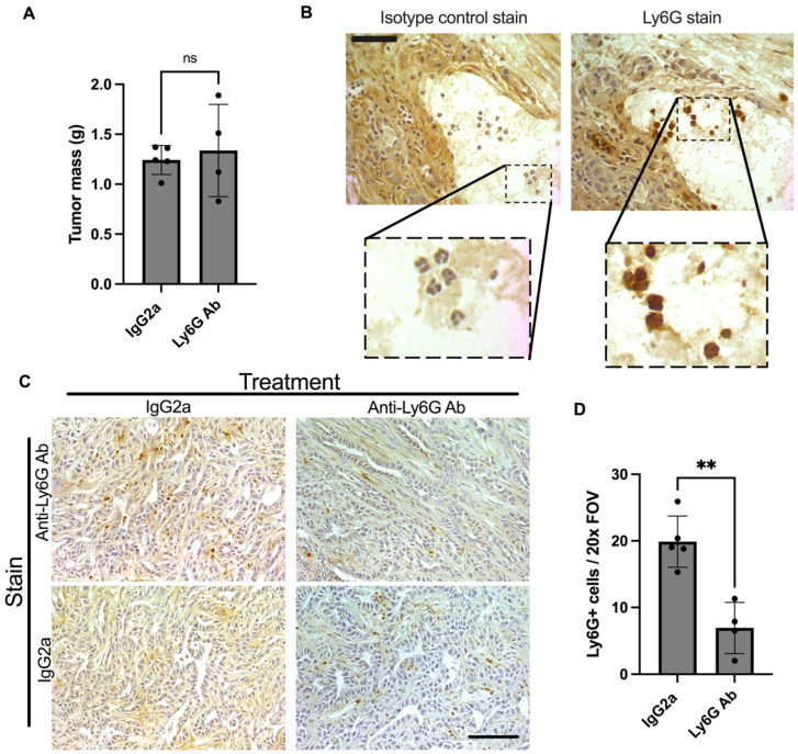
Effect of Ly6G antibody treatment on tumor. Orthotopic KPC tumor-bearing mice were treated with an anti-Ly6G antibody (1A8) or isotype control (2A3) every 3 days starting at 5 days post inoculation. (A) Endpoint tumor mass was not different between groups (ns = not significant). (B) Serial sections were also stained using the isotype control antibody (2A3) in parallel. This shows a high magnification (40×) view of serial sections. Comparing the stains shows that cells with characteristic multilobed nuclei pick up the Ly6G stain. Scale bar = 50 μm. (C) Representative tumor sections stained with anti-Ly6G antibody (1A8) to detect Ly6G+ cells in the tumor. While KPC tumors from isotype control-treated mice did not show positive immunoreactivity in the absence of the Ly6G antibody, KPC tumors from mice treated with anti-Ly6G treatment showed some cells with positive immunoreactivity. These cells likely reflect Ly6G+ cells labeled, but not yet depleted, by the 1A8 antibody in vivo (during treatment). Images were taken at 20× magnification. Scale bar = 100 µm. (D) KPC tumors from isotype control-treated mice (n = 5) had significantly more Ly6G positive cells per field of view (FOV) than those from anti-Ly6G antibody-treated mice (** p < 0.001; n = 4).

