Figure 4.
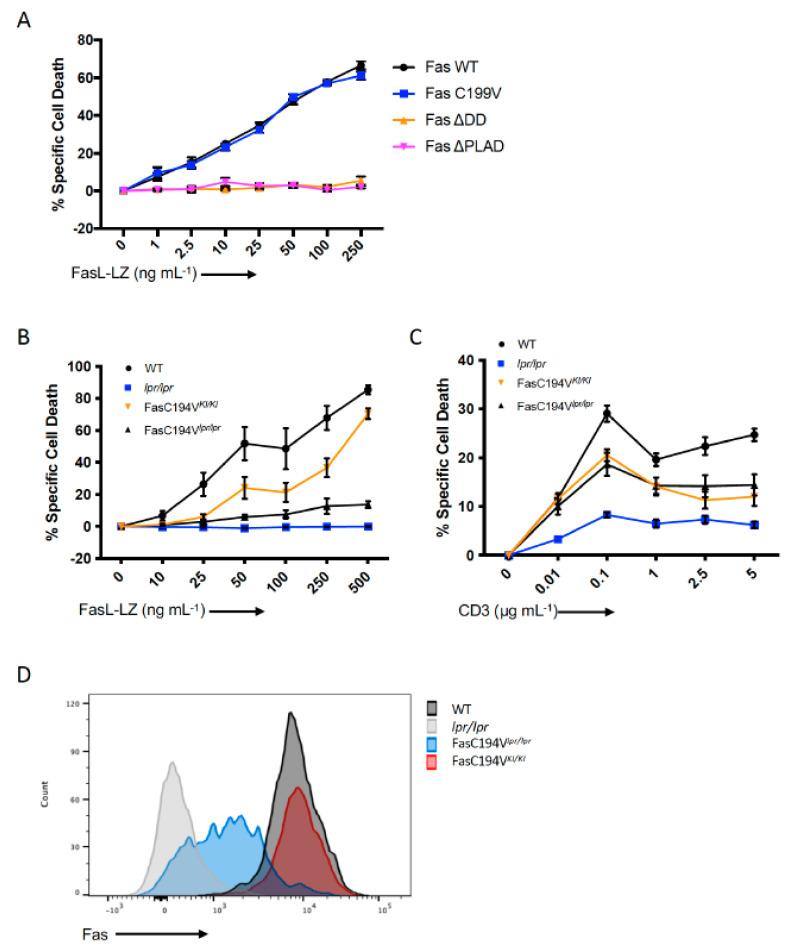
Relationship between Fas clustering and apoptosis induction in T cells. (A) Fas-deficient Jurkat cells (RapoC2) were transfected with mVenus-tagged WT or mutant Fas. 24 h post-transfection, cells were treated with increasing amounts of FasL-LZ for 6 h prior to staining with Annexin V, DiIC(5), and Live/Dead, and being analyzed by flow cytometry. (B) Activated CD4+ T cells from mice with the indicated genotype were stained for Fas surface expression and assessed by flow cytometry. Data are representative of n = 3 independent experiments. (C) CD4 T cells were isolated from mice of the indicated genotypes and activated for 72 h with anti-CD3/28. Cells were cultured with supplemental IL-2 prior to treatment with FasL-LZ at the indicated concentrations for 6 h, then stained for assessment of cell death via flow cytometry as in (A). (D) CD3/28 activated CD4 T cells from mice of the indicated genotypes were rested in IL-2 and subsequently restimulated with plate-bound anti-CD3 antibody at the indicated concentrations for 16 h prior to assessment for death via flow cytometry as in (A). Panels (A–C) are cumulative data from a minimum of three independent experiments (n = 3).

