Abstract
Trophoblast cell surface antigen 2 (Trop-2, encoded by TACSTD2) is the target protein of sacituzumab govitecan, a novel antibody-drug conjugate for locally advanced or metastatic urothelial carcinoma. However, the expression status of Trop-2 in upper tract urothelial carcinoma (UTUC) remains unclear. We performed immunohistochemical analysis of 99 UTUC samples to evaluate the expression status of Trop-2 in patients with UTUC and analyze its association with clinical outcomes. Trop-2 was positive in 94 of the 99 UTUC samples, and high Trop-2 expression was associated with favorable progression-free survival (PFS) and cancer-specific survival (p = 0.0011, 0.0046). Multivariate analysis identified high Trop-2 expression as an independent predictor of favorable PFS (all cases, p = 0.045; high-risk group (pT3≤ or presence of lymphovascular invasion or lymph node metastasis), p = 0.014). Gene expression analysis using RNA sequencing data from 72 UTUC samples demonstrated the association between high TACSTD2 expression and favorable PFS (all cases, p = 0.069; high-risk group, p = 0.029). In conclusion, we demonstrated that Trop-2 is widely expressed in UTUC. Although high Trop-2 expression was a favorable prognostic factor in UTUC, its widespread expression suggests that sacituzumab govitecan may be effective for a wide range of UTUC.
Keywords: trophoblast cell surface antigen 2, TACSTD2, upper tract urothelial carcinoma, immunohistochemical analysis, RNA sequencing, sacituzumab govitecan
1. Introduction
Upper tract urothelial carcinoma (UTUC) is a relatively rare disease, accounting for 5–10% of all urothelial carcinomas (UC) [1,2]. Patients with UTUC often present with invasive or metastatic disease at diagnosis and, therefore, have a poorer prognosis than those with urothelial bladder cancer (UBC) [1]. Phenotypic and genetic differences between both malignancies have also been found, and increasing evidence suggests that UTUC could be a distinct disease from UBC [2]. Thus, it is important to develop optimal management strategies for UTUC.
Locally advanced or metastatic UC, including UTUC, is an incurable disease with poor survival. Until recently, patients with metastatic UC have had limited treatment options and exhibit tumor progression after platinum chemotherapy, immune checkpoint inhibitor treatment, or both [3,4]. However, the therapeutic potential for metastatic UC was expanded by the accelerated approval of enfortumab vedotin, an antibody–drug conjugate (ADC) targeting Nectin-4, by the US Food and Drug Administration (FDA) in 2019 [5]. In April 2021, sacituzumab govitecan, an ADC targeting trophoblast cell surface antigen 2 (Trop-2), became a treatment option for patients with locally advanced or metastatic UC who previously received a platinum-containing chemotherapy and a programmed cell death 1 (PD-1) or PD-1 ligand 1 (PD-L1) inhibitor [6]. Sacituzumab govitecan, which is an anti-Trop-2 monoclonal antibody conjugated to SN-38, is an active metabolite of irinotecan that creates breaks in double-stranded DNA and leads to apoptosis [7]. Previous studies have indicated that cells overexpressing Trop-2 are highly sensitive to sacituzumab govitecan [8,9,10,11]. Therefore, it is clinically important to evaluate the expression of Trop-2 in solid tumors to predict the efficacy of sacituzumab govitecan.
Trop-2 is a 40-kDa transmembrane glycoprotein encoded by the single-exon gene TACSTD2 and is highly expressed on the surface of various epithelial cancer cells, including UBC [10,11,12,13,14,15,16,17,18,19]. However, the expression status of Trop-2 and its prognostic significance in UTUC have not been fully investigated. Therefore, in this study, we investigated the expression pattern of Trop-2 using tissue microarray (TMA) specimens of 99 patients with UTUCs. Furthermore, we analyzed the association of Trop-2 expression with clinical outcomes in UTUC. We also performed gene expression analysis using the RNA-sequencing data from 72 UTUC samples and compared clinical outcomes between the high and low TACSTD2 expression groups.
2. Materials and Methods
2.1. Patients and Tissue Samples
For the immunohistochemical analysis, the UTUC-TMA was constructed using spotted triplicate UTUC samples from dominant tumors/invasive components, if present, of 99 patients with non-metastatic UTUC. These patients underwent radical nephroureterectomies performed at Osaka General Medical Center between 1997 and 2011, as described previously [20,21,22,23,24,25].
For the gene expression analysis, 72 patients with non-metastatic UTUC who underwent radical nephroureterectomy at Osaka University Hospital between 2016 and 2020 were enrolled. Surgically resected UTUC samples were immersed in RNAlater tissue storage reagent (Thermo Fisher Scientific, Waltham, MA, USA) and stored at −20 °C.
Ethical approval for the study was obtained from each local institutional review board (IRB), including: Osaka General Medical Center Institutional Review Board (IRB), Protocol Number 25–2014 and Osaka University Hospital IRB, Protocol Number #13397-14. Written informed consent was obtained from all patients before recruitment. Tumor progression was defined as the development of non-lower urinary tract lesions, including recurrence at the site of nephroureterectomy and lymph node or visceral metastasis. The high-risk group consisted of patients who had a pathologic stage of ≥pT3 or positive lymphatic invasion or lymph node metastasis [26].
2.2. Immunohistochemical Analysis
Immunohistochemical staining for Trop-2 was performed using 4 μm-thick paraffin-embedded tissue sections from the UTUC-TMA, which were deparaffinized using xylene and a graded series of ethanol concentrations. For Trop-2 antigen retrieval, the sections were treated with Tris-ethylenediaminetetraacetic acid (EDTA) buffer (pH 7.0) and steamed by placing them above boiling water for 20 min. Endogenous peroxidase activity was blocked by incubating the sections with 0.3% hydrogen peroxide for 5 min, followed by overnight incubation with primary antibodies against Trop-2 (1:200; SC-376181, Santa Cruz Biotechnology, Dallas, TX, USA) at 4 °C.
Then, we used the EnVision + system-horseradish peroxidase (HRP)-labeled polymer anti-mouse (DAKO) according to the manufacturer’s instructions. Sections were counterstained with hematoxylin, dehydrated using a graded series of ethanol concentrations, cleared in xylene, and mounted for viewing under a microscope. The intensity and extent of Trop-2 expression were determined using the histochemical scoring system (H-score), which was based on the product of the staining intensity (score, 0–3+) and percentage of stained cells (0–100%) at a given intensity. Specimens were subsequently classified as negative (0+, H-score 0–14), low (1+, H-score 15–99), moderate (2+, H-score 100–199), and high (3+; H-score, 200–300).
2.3. TACSTD2 Gene Expression Analysis in UTUC
RNA sequencing analysis was performed as previously reported [27]. Briefly, total RNA was isolated from 73 UTUC tumor samples using the RNeasy mini kit (QIAGEN, Venlo, Netherlands) according to the manufacturer’s protocol. The RNA integrity was verified using an Agilent 2100 bioanalyzer with RNA nano reagents (Agilent Technologies, La Jolla, CA, USA), and RNA was subjected to polyA+ selection and chemical fragmentation. The 100-bp RNA fraction was used to construct cDNA libraries using the TruSeq Stranded mRNA Prep kit (Illumina, San Diego, CA, USA) and the obtained paired-end libraries were sequenced using the Illumina NovaSeq6000 platform with a standard 100-bp paired-end read protocol at Macrogen Japan. TACSTD2 gene expression values were estimated from the RNA sequencing data using the Genomon pipeline (https://github.com/Genomon-Project/genomon-docs/tree/v2.0 (accessed on 4 January 2022). The alignment was performed with the STAR aligner (v.2.5.2a) against the hg19 human genome. BAM files named Aligned.sortedByCoord.out.bam, generated using the STAR software, were used to quantify the expression data using GenomonExpression. Patients were divided into high and low TACSTD2 expression groups (n = 36 each) based on the median value to evaluate the association between TACSTD2 expression and prognosis.
2.4. Statistical Analyses
Statistical analyses were performed using JMP Pro 16.0.0 (SAS Institute Inc., Cary, NC, USA) and data were visualized using GraphPad Prism version 7.05 (GraphPad Software, San Diego, CA, USA). Fisher’s exact test was used to evaluate the association between categorized variables. The survival rates were determined using the Kaplan–Meier method, whereas the log-rank test was used for comparison between groups. The Cox proportional hazards model was used to determine the statistical significance of prognostic indicators in univariate and multivariate settings. p-values < 0.05 were considered statistically significant and p-values < 0.1 were considered statistically trending.
3. Results
3.1. Patient Characteristics
The clinicopathological characteristics and outcomes of the 99 and 72 patients (using immunohistochemical and RNA sequencing analyses, respectively) are summarized in Table 1. Sixty of 99 patients (60.6%) and 33 of 72 patients (45.8%) were classified in the high-risk group. None of the patients received neoadjuvant therapy before tissue collection; however, 26 of 99 patients (26.3%) and 12 of 72 patients (16.7%) underwent adjuvant chemotherapy. Tumor extension was observed in 38 of 99 patients (38.4%) and 25 of 72 patients (34.7%).
Table 1.
Clinicopathologic characteristics and outcome of patients with upper tract urothelial carcinoma.
| Variable | Immunohistochemical Analysis | RNA Sequencing Analysis |
|---|---|---|
| n = 99 | n = 72 | |
| Age (year), median (range) | 71 (48–87) | 73 (45–89) |
| Sex, n (%) | ||
| Male | 60 (60.6) | 55 (76.4) |
| Female | 39 (39.4) | 17 (23.6) |
| Laterality, n (%) | ||
| Right | 43 (43.4) | 38 (52.8) |
| Left | 56 (56.6) | 34 (47.2) |
| Tumor location, n (%) | ||
| Renal pelvis | 45 (45.5) | 27 (37.5) |
| Ureter | 50 (50.5) | 43 (59.7) |
| Both | 4 (4.0) | 2 (2.8) |
| Tumor grade, n (%) | ||
| Low-grade | 15 (15.2) | 8 (11.1) |
| High-grade | 84 (85.9) | 64 (88.9) |
| Pathological T stage, n (%) | ||
| pTa | 19 (19.2) | 14 (19.4) |
| pT1 | 18 (18.2) | 22 (30.6) |
| pT2 | 8 (8.1) | 10 (13.9) |
| pT3 | 48 (48.5) | 23 (31.9) |
| pT4 | 6 (6.1) | 3 (4.2) |
| Lymphovascular invasion, n (%) | ||
| Yes | 40 (40.4) | 50 (69.4) |
| No | 59 (59.6) | 22 (30.6) |
| Lymph node metastasis, n (%) | ||
| pN0 | 84 (84.8) | 63 (87.5) |
| pN+ | 12 (12.1) | 9 (12.5) |
| pNx | 3 (3.0) | 0 (0.0) |
| High-risk group †, n (%) | ||
| Yes | 60 (60.6) | 33 (45.8) |
| No | 36 (36.4) | 39 (54.2) |
| Unknown | 3 (3.0) | 0 (0.0) |
| Adjuvant chemotherapy, n (%) | ||
| Yes | 26 (26.3) | 12 (16.7) |
| No | 63 (63.6) | 60 (83.3) |
| Progression, n (%) | ||
| Yes | 38 (38.4) | 25 (34.7) |
| No | 61 (61.6) | 47 (65.2) |
| Follow-up (month), median (range) | 37 (1–173) | 28 (2–88) |
† High-risk group is defined as patients with a pathologic stage of ≥pT3 or positive lymphatic invasion or lymph node metastasis.
3.2. Trop-2 Expression in UTUC and Association with Clinicopathological Characteristics
Typical patterns of Trop-2 immunohistochemical expression in UTUC-TMA specimens are portrayed in Figure 1. Trop-2 positivity was detected in 94 (94.9%; 25 [25.3%] low, 42 [42.4%] moderate, 27 [27.3%] high) of the 99 UTUC samples. Next, our analysis of the association of Trop-2 expression with the clinicopathological profiles of 99 patients with UTUC (Table 2) indicated no association with patient sex, tumor location, tumor grade, lymphovascular invasion, or lymph node metastasis (0 vs. 1+/2+/3+; 0/1+ vs. 2+/3+; 0/1+/2+ vs. 3+). Moreover, high Trop-2 expression was detected at a significantly higher level in non-muscle-invasive (40.5%) than in muscle-invasive (19.4%) tumors (p = 0.020).
Figure 1.
Immunohistochemical expression of trophoblast cell surface antigen 2 (Trop-2) in upper tract urothelial carcinoma (UTUC) tissue microarray specimens. UTUC tissue with (A) high (3+), (B) moderate (2+), (C) low (1+), and (D) negative (0+) expression.
Table 2.
Association of Trop-2 expression with clinicopathological characteristics of upper tract urothelial carcinoma.
| Variable | n | Trop-2 expression | p | |||||
|---|---|---|---|---|---|---|---|---|
| 0 + (%) | 1 + (%) | 2 + (%) | 3 + (%) | 0 vs. 1 +/2 +/3 + | 0/1 + vs. 2 +/3 + | 0/1 +/2 + vs. 3 + | ||
| Sex | 0.077 | 0.37 | 0.82 | |||||
| Male | 60 | 1 (1.7) | 15 (25.0) | 26 (43.3) | 18 (30.0) | |||
| Female | 39 | 4 (10.3) | 10 (25.6) | 16 (41.0) | 9 (23.1) | |||
| Tumor site | 0.37 | 0.66 a | 0.82 a | |||||
| Renal pelvis | 45 | 1 (2.2) | 12 (26.7) | 20 (44.4) | 12 (26.7) | |||
| Ureter | 50 | 4 (8.0) | 13 (26.0) | 20 (40.0) | 13 (26.0) | |||
| Both | 4 | 0 (0.0) | 0 (0.0) | 2 (50.0) | 2 (50.0) | |||
| Tumor grade | 0.57 | 0.54 | 1.00 | |||||
| Low-grade | 15 | 1 (6.7) | 2 (13.3) | 8 (53.3) | 4 (26.7) | |||
| High-grade | 84 | 4 (4.8) | 23 (27.4) | 34 (40.5) | 23 (27.4) | |||
| Pathological stage | 0.65 b | 1.00 b | 0.020 b ** | |||||
| pTa | 19 | 1 (5.3) | 5 (26.3) | 4 (21.1) | 9 (47.4) | |||
| pT1 | 18 | 0 (0.0) | 5 (27.8) | 7 (38.9) | 6 (33.3) | |||
| pT2 | 8 | 0 (0.0) | 2 (25.0) | 4 (50.0) | 2 (25.0) | |||
| pT3 | 48 | 3 (6.3) | 12 (25.0) | 24 (50.0) | 9 (18.8) | |||
| pT4 | 6 | 1 (16.7) | 1 (16.7) | 3 (50.0) | 1 (16.7) | |||
| Lymphovascular invasion | 0.39 | 1.00 | 0.069 | |||||
| No | 59 | 2 (3.4) | 16 (27.1) | 21 (35.6) | 20 (33.9) | |||
| Yes | 40 | 3 (7.5) | 9 (22.5) | 21 (52.5) | 7 (17.5) | |||
| Lymph node involvement | 0.075 c | 0.74 | 0.50 c | |||||
| pN0 | 84 | 2 (2.4) | 22 (26.2) | 35 (41.7) | 25 (29.8) | |||
| pN+ | 12 | 2 (16.7) | 2 (16.7) | 6 (50.0) | 2 (16.7) | |||
| pNx | 3 | 1 (33.3) | 1 (33.3) | 1 (33.3) | 0 (0.0) | |||
a Renal pelvis vs. ureter; b pTa + pT1 vs. pT2 + pT3 + pT4; c pN0 vs. pN+; ** p < 0.05.
3.3. Immunohistochemical Analysis of Associations of Trop-2 Expression with Patient Outcomes
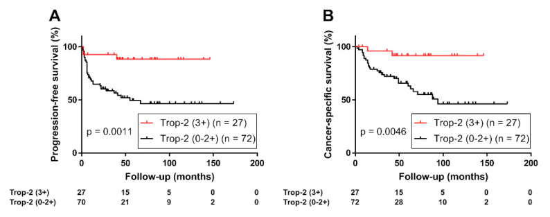
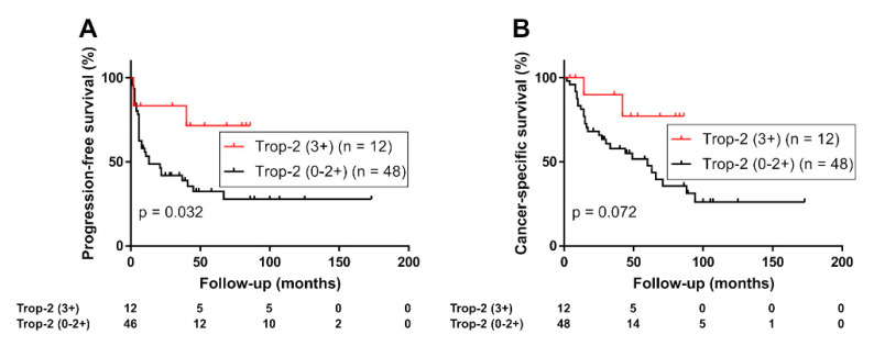
The Kaplan–Meier analysis of the prognostic value of Trop-2 expression status in UTUC revealed that patients with high Trop-2 expression (n = 27) had a significantly lower risk of tumor progression (Log-rank test, p = 0.0011; Figure 2A) or cancer-specific mortality (Log-rank test, p = 0.0046; Figure 2B) than those with moderate, low, or negative Trop-2 tumor expression (n = 72). In a sub-group of patients with muscle-invasive tumors (n = 62), high Trop-2 expression was also associated with favorable PFS (p = 0.032, Supplementary Figure S1A) and cancer-specific survival (p = 0.061, Supplementary Figure S1B). In addition, although limited to the high-risk group (n = 60), highTrop-2 expression was associated with favorable PFS (p = 0.032, Figure 3A) and cancer-specific survival (p = 0.072, Figure 3B).
Figure 2.
(A) Progression-free survival and (B) cancer-specific survival of 99 patients with upper tract urothelial carcinoma stratified using expression of trophoblast cell surface antigen 2 (Trop-2, 0/1+/2+ vs. 3+).
Figure 3.
(A) Progression-free survival and (B) cancer-specific survival of 60 patients with high-risk upper tract urothelial carcinoma (with a pathologic stage of ≥pT3 or positive lymphatic invasion or lymph node metastasis) stratified using expression of Trop-2 (0/1+/2+ vs. 3+).
Next, the multivariate analysis with the Cox proportional hazard model was performed to determine whether highTrop-2 expression was an independent prognostic factor in patients with UTUC. The results demonstrated that highTrop-2 expression was an independent predictor of favorable PFS in UTUC (all cases: hazard ratio [HR], 0.29; 95% confidence interval [CI], 0.087–0.97; p = 0.045]; high-risk group: HR, 0.27 [95% CI, 0.081–0.88; p = 0.031, Table 3 and Table 4).
Table 3.
Univariate and multivariate analyses of progression-free survival and cancer-specific survival in patients with upper tract urothelial carcinoma.
| Progression-Free Survival | Cancer-Specific Survival | |||||||||||
|---|---|---|---|---|---|---|---|---|---|---|---|---|
| Variable | Univariate | Multivariate | Univariate | Multivariate | ||||||||
| HR | 95 %CI | p | HR | 95 %CI | p | HR | 95 %CI | p | HR | 95 %CI | p | |
| All cases (n = 99) | ||||||||||||
| Sex (male/female) | 1.33 | 0.68–2.61 | 0.40 | 1.33 | 0.62–2.85 | 0.46 | ||||||
| Age (70</≤70) | 1.38 | 0.72–2.64 | 0.33 | 1.65 | 0.78–3.48 | 0.19 | ||||||
| Tumor site (pelvis/ureter) | 0.80 | 0.41–1.55 | 0.50 | 0.83 | 0.40–1.72 | 0.61 | ||||||
| Tumor grade (high/low) | 4.44 | 1.07–18.51 | 0.041 ** | 5.52 | 1.29–23.72 | 0.022 ** | 7.77 | 1.06–57.18 | 0.044 ** | 6.95 | 0.94–51.23 | 0.057 * |
| pT stage (MI/NMI) | 17.30 | 4.15–72.14 | <0.001 ** | 8.5 | 1.94–37.31 | 0.0046 ** | 1.09 × 10⁹ | 13.52–Inf | <0.001 ** | 5.76 × 10⁹ | (6.39–7.60) × 1038 | <0.001 ** |
| Lymphovascular invasion (Yes/No) | 5.84 | 2.88–11.83 | <0.001 ** | 2.54 | 1.10–5.84 | 0.028 ** | 5.62 | 2.50–12.66 | <0.001 ** | 1.86 | 0.77–4.48 | 0.16 |
| Lymph node metastasis (Yes/No) | 4.40 | 2.07–9.36 | <0.001 ** | 2.06 | 0.95–4.47 | 0.068 * | 2.74 | 1.17–6.40 | 0.020 ** | 1.07 | 0.43–2.63 | 0.89 |
| Adjuvant chemotherapy (Yes/No) | 1.13 | 0.56–2.28 | 0.73 | 1.08 | 0.49–2.37 | 0.85 | ||||||
| Trop-2 strong expression | 0.18 | 0.055–0.58 | 0.0043 ** | 0.29 | 0.087–0.97 | 0.045 ** | 0.16 | 0.039–0.69 | 0.014 ** | 0.31 | 0.072–1.36 | 0.12 |
Abbreviations: CI, confidence interval; HR, hazard ratio; Inf, Infinity; MI, muscle invasive; NMI, non–muscle invasive; ** p < 0.05 and * p < 0.1.
Table 4.
Univariate and multivariate analyses of progression-free survival and cancer-specific survival in patients with high-risk upper tract urothelial carcinoma.
| Progression-Free Survival | Cancer-Specific Survival | |||||||||||
|---|---|---|---|---|---|---|---|---|---|---|---|---|
| Variable | Univariate | Multivariate | Univariate | Multivariate | ||||||||
| HR | 95 %CI | p | HR | 95 %CI | p | HR | 95 %CI | p | HR | 95 %CI | p | |
| High-risk group † (n = 60) | ||||||||||||
| Sex (male/female) | 1.35 | 0.67–2.72 | 0.40 | 1.23 | 0.58–2.65 | 0.6 | ||||||
| Age (70</≤70) years | 1.11 | 0.56–2.18 | 0.77 | 1.57 | 0.73–3.37 | 0.24 | ||||||
| Tumor site (pelvis/ureter) | 0.95 | 0.48–1.91 | 0.89 | 0.97 | 0.46–2.04 | 0.93 | ||||||
| Tumor grade (high/low) | 6.96 | 0.95–51.06 | 0.056 * | 7.91 | 1.07–58.25 | 0.042 ** | 6.41 | 0.87–47.47 | 0.069 * | 7.09 | 0.96–52.49 | 0.055 * |
| Adjuvant chemotherapy (Yes/No) | 0.71 | 0.35–1.46 | 0.36 | 0.63 | 0.28–1.41 | 0.26 | ||||||
| Trop-2 strong expression | 0.30 | 0.092–0.99 | 0.048 ** | 0.268 | 0.081–0.88 | 0.031 ** | 0.29 | 0.069–1.23 | 0.093 * | 0.26 | 0.061–1.10 | 0.067 * |
† High-risk group is defined as patients with a pathologic stage of ≥pT3 or positive lymphatic invasion or lymph node metastasis; ** p < 0.05 and * p < 0.1. Abbreviations: CI, confidence interval; HR, hazard ratio.
3.4. Associations of TACSTD2 Gene Expression with Patient Outcomes
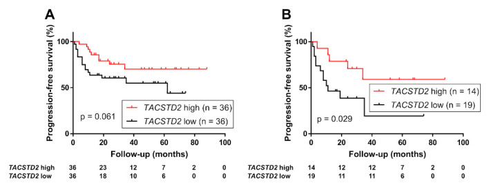
Finally, we evaluated the association between TACSTD2 expression and prognosis using the RNA sequencing data in a different patient cohort (n = 72). The Kaplan–Meier analysis demonstrated that the group with high TACSTD2 expression had a more favorable PFS than the low expression group (Log-rank test, all cases, p = 0.061; high-risk group, p = 0.029, Figure 4). In the multivariate analysis with Cox proportional hazard model, high TACSTD2 expression was identified as an independent predictor of favorable PFS in high-risk UTUC (HR, 0.21; 95% CI, 0.057–0.75; p = 0.017, Supplementary Table S1).
Figure 4.
Progression-free survival in (A) 72 patients with upper tract urothelial carcinoma (UTUC) and (B) 33 patients with high-risk UTUC (with a pathologic stage of ≥pT3 or positive lymphatic invasion or lymph node metastasis) stratified using expression of TACSTD2 gene (high vs. low).
4. Discussion
In this study, we demonstrated that Trop-2, the target protein of sacituzumab govitecan, was widely expressed in UTUC and that its high expression was associated with a better prognosis. To date, it has been reported that Trop-2 is highly expressed in various types of solid tumors, such as ovarian [10], cervical [11,12], colorectal [13,14], gastric [15], pancreatic [16], and breast cancers [17]. Sacituzumab govitecan was approved by FDA for patients with metastatic triple negative breast cancer who received at least two prior therapies in April 2021 [28]. Additionally, elevated expression of Trop-2 has also been reported in UC [18,19]. The TROPHY-U-01 phase II trial evaluated the efficacy of sacituzumab govitecan in locally advanced or metastatic UC and found an objective response rate (ORR) of 27% (31 of 113 patients) and a decrease in detectable disease in 77% of the patients [29]. Thus, sacituzumab govitecan has become a novel therapeutic option for metastatic UC after prior platinum-based therapies and immune checkpoint inhibitors [6]. However, previous studies have focused only on UBC; therefore, the expression of Trop-2 in UTUC has not been fully investigated. UTUC has been suggested to be a distinct disease from UBC [2]; as such, assessing the expression of proteins in UTUC is important even if their expression in UBC is well understood. Indeed, we previously indicated that Nectin-4, the target protein of enfortumab vedotin, is expressed at lower levels in UTUC than in UBC [25]. This finding supports the results of a subgroup analysis in a phase 3 trial, which indicated that enfortumab vedotin was less effective in UTUC than in UBC [30]. Our present study indicated that Trop-2 was highly expressed in UTUC and UBC, where it was reported to be 83% [31]. This finding suggests that sacituzumab govitecan may be a treatment option for advanced UTUC in addition to UBC.
Additionally, we revealed that the impact of Trop-2 on cancer prognosis may differ between UBC and UTUC. Overexpression of Trop-2 has been associated with increased tumor aggressiveness and poor prognosis in several types of cancers, including UBC [12,13,14,15,16,17,18,19,32]. Furthermore, Avellini et al. [18] reported that Trop-2 expression increases with increasing severity of UBC, and Trop-2 enhances proliferation and migration of UBC in vitro. Zhang et al. [19] found high expression of Trop-2 was associated with shorter recurrence-free survival in non-muscle-invasive UBC patients. However, in the present study, we revealed that high Trop-2 expression was associated with a good prognosis in UTUC. This finding is inconsistent with that reported in UBC; therefore, in addition to the immunohistochemical analysis, we also evaluated the gene expression of another cohort. The results of the analysis indicated that the high TASCSTD2 expression group still indicated a favorable prognosis. The loss of Trop-2 has been reported to promote carcinogenesis and features of epithelial to mesenchymal transition in specific cancers such as head and neck squamous cell carcinoma [33]. Consequently, the association between high Trop-2 expression and favorable prognosis in UTUC might be a unique feature of UTUC that differs from what occurs with UBC. Further studies are expected to improve knowledge of the differences between UTUC and UBC [34].
The present study has some limitations that are worth mentioning. First, the UTUC specimens analyzed were obtained using radical nephroureterectomy and included samples from low-risk UTUC patients as well. Metastatic UTUC possesses a more aggressive phenotype than the localized form and may exhibit differential marker expression. Second, immunohistochemical analysis is a semi-quantitative evaluation method, thus, the staining pattern may not represent the actual expression level of the protein. Third, considering that the response rate of patients with advanced bladder cancer to sacituzumab govitecan was limited to 27% in the TROPHY-U-01 trial [29], the expression of Trop-2 does not ensure the efficacy of sacituzumab govitecan. An initial small pilot study (IMMU-132) suggested that high Trop-2 expression in UC was positively correlated with treatment response [31]. However, the relationship between Trop-2 expression pattern and the actual patient response rate to sacituzumab govitecan must be confirmed in future clinical trials.
In conclusion, our study indicated that Trop-2 is widely expressed in UTUC. Although Trop-2 expression was identified as a favorable prognostic factor, its broad expression suggests that sacituzumab govitec may be a potential therapeutic option in a wide spectrum of patients with UTUC. Our future task is to evaluate the actual response rate of sacituzumab govitecan for UTUC in clinical practice and to assess its relationship with Trop-2 expression patterns.
Acknowledgments
We thank Satoshi Nojima for providing support regarding the assessments of the tissue microarray.
Supplementary Materials
The following supporting information can be downloaded at: https://www.mdpi.com/article/10.3390/curroncol29060312/s1. Table S1: Univariate and multivariate analyses of progression-free survival in patients with upper tract urothelial carcinoma in RNA sequencing analysis, Figure S1: Progression-free survival (A) and cancer-specific survival (B) in 62 patients with muscle-invasive upper tract urothelial carcinoma stratified by the expression of Trop-2 (0/1+/2+ vs. 3+).
Author Contributions
Conceptualization, K.F. and E.T.; methodology, K.F. and E.T.; software, K.K. (Ken Kuwahara) and S.I.; validation, K.K. (Kotoe Katayama), T.M., T.K., K.H., A.K., M.U., K.Y. and R.I.; investigation, E.T. and K.N.; resources, M.U, T.T. and H.F.; data curation, E.T. and K.N.; writing—original draft preparation, E.T.; writing—review and editing, K.F.; visualization, E.T.; supervision, H.U. and N.N.; project administration, K.F. All authors have read and agreed to the published version of the manuscript.
Institutional Review Board Statement
The study was conducted in accordance with the Declaration of Helsinki and approved by the Institutional Review Board of OSAKA GENERAL MEDICAL CENTER (Protocol Number 25–2014) and OSAKA UNIVERSITY HOSPITAL (Protocol Number #13397-14).
Informed Consent Statement
Informed consent was obtained from all subjects involved in the study.
Data Availability Statement
The data presented in this study are available on request from the corresponding author.
Conflicts of Interest
The authors declare no conflict of interest.
Funding Statement
This research received no external funding.
Footnotes
Publisher’s Note: MDPI stays neutral with regard to jurisdictional claims in published maps and institutional affiliations.
References
- 1.Rouprêt M., Babjuk M., Compérat E., Zigeuner R., Sylvester R.J., Burger M., Cowan N.C., Gontero P., Van Rhijn B.W., Mostafid A.H., et al. European Association of Urology Guidelines on Upper Urinary Tract Urothelial Carcinoma: 2017 Update. Eur. Urol. 2018;73:111–122. doi: 10.1016/j.eururo.2017.07.036. [DOI] [PubMed] [Google Scholar]
- 2.Leow J.J., Chong K.T., Chang S.L., Bellmunt J. Upper tract urothelial carcinoma: A different disease entity in terms of management. ESMO Open. 2016;1:e000126. doi: 10.1136/esmoopen-2016-000126. [DOI] [PMC free article] [PubMed] [Google Scholar]
- 3.Siefker-Radtke A., Curti B. Immunotherapy in metastatic urothelial carcinoma: Focus on immune checkpoint inhibition. Nat. Rev. Urol. 2018;15:112–124. doi: 10.1038/nrurol.2017.190. [DOI] [PubMed] [Google Scholar]
- 4.Özen H. Bladder cancer. Curr. Opin. Oncol. 1999;11:207–212. doi: 10.1097/00001622-199905000-00013. [DOI] [PubMed] [Google Scholar]
- 5.US Food & Drug Administration FDA Grants Accelerated Approval to Enfortumab Vedotin-Ejfv for Metastatic Urothelial Cancer. [(accessed on 25 May 2022)]; Available online: https://www.fda.gov/drugs/resources-information-approved-drugs/fda-grants-accelerated-approval-enfortumab-vedotinejfv-metastatic-urothelial-cancer.
- 6.US Food & Drug Administration FDA Grants Accelerated Approval to Sacituzumab Govitecan for Advanced Urothelial Cancer. [(accessed on 25 May 2022)]; Available online: https://www.fda.gov/drugs/resources-information-approved-drugs/fda-grants-accelerated-approval-sacituzumab-govitecan-advanced-urothelial-cancer.
- 7.Goldenberg D.M., Sharkey R.M. Antibody-drug conjugates targeting TROP-2 and incorporating SN-38: A case study of anti-TROP-2 sacituzumab govitecan. MAbs. 2019;11:987–995. doi: 10.1080/19420862.2019.1632115. [DOI] [PMC free article] [PubMed] [Google Scholar]
- 8.Cardillo T.M., Sharkey R.M., Rossi D.L., Arrojo R., Mostafa A.A., Goldenberg D.M. Synthetic Lethality Exploitation by an Anti–Trop-2-SN-38 Antibody–Drug Conjugate, IMMU-132, Plus PARP Inhibitors in BRCA1/2 –wild-type Triple-Negative Breast Cancer. Clin. Cancer Res. 2017;23:3405–3415. doi: 10.1158/1078-0432.CCR-16-2401. [DOI] [PubMed] [Google Scholar]
- 9.Perrone E., Manara P., Lopez S., Bellone S., Bonazzoli E., Manzano A., Zammataro L., Bianchi A., Zeybek B., Buza N., et al. Sacituzumab govitecan, an antibody-drug conjugate targeting trophoblast cell-surface antigen 2, shows cytotoxic activity against poorly differentiated endometrial adenocarcinomas in vitro and in vivo. Mol. Oncol. 2020;14:645–656. doi: 10.1002/1878-0261.12627. [DOI] [PMC free article] [PubMed] [Google Scholar]
- 10.Perrone E., Lopez S., Zeybek B., Bellone S., Bonazzoli E., Pelligra S., Zammataro L., Manzano A., Manara P., Bianchi A., et al. Preclinical Activity of Sacituzumab Govitecan, an Antibody-Drug Conjugate Targeting Trophoblast Cell-Surface Antigen 2 (Trop-2) Linked to the Active Metabolite of Irinotecan (SN-38), in Ovarian Cancer. Front. Oncol. 2020;10:118. doi: 10.3389/fonc.2020.00118. [DOI] [PMC free article] [PubMed] [Google Scholar]
- 11.Zeybek B., Manzano A., Bianchi A., Bonazzoli E., Bellone S., Buza N., Hui P., Lopez S., Perrone E., Manara P., et al. Cervical carcinomas that overexpress human trophoblast cell-surface marker (Trop-2) are highly sensitive to the antibody-drug conjugate sacituzumab govitecan. Sci. Rep. 2020;10:973. doi: 10.1038/s41598-020-58009-3. [DOI] [PMC free article] [PubMed] [Google Scholar]
- 12.Liu T., Liu Y., Bao X., Tian J., Liu Y., Yang X. Overexpression of TROP2 Predicts Poor Prognosis of Patients with Cervical Cancer and Promotes the Proliferation and Invasion of Cervical Cancer Cells by Regulating ERK Signaling Pathway. PLoS ONE. 2013;8:e75864. doi: 10.1371/journal.pone.0075864. [DOI] [PMC free article] [PubMed] [Google Scholar]
- 13.Ohmachi T., Tanaka F., Mimori K., Inoue H., Yanaga K., Mori M. Clinical significance of TROP2 expression in colorectal cancer. Clin. Cancer Res. 2006;12:3057–3063. doi: 10.1158/1078-0432.CCR-05-1961. [DOI] [PubMed] [Google Scholar]
- 14.Zhao P., Yu H.Z., Cai J.H. Clinical investigation of TROP-2 as an independent biomarker and potential therapeutic target in colon cancer. Mol. Med. Rep. 2015;12:4364–4369. doi: 10.3892/mmr.2015.3900. [DOI] [PubMed] [Google Scholar]
- 15.Liu F., He Y., Cao Q., Liu N., Zhang W. Trop2 is overexpressed in gastric cancer and predicts poor prognosis. Oncotarget. 2016;2016:1–7. doi: 10.1155/2016/2436518. [DOI] [PMC free article] [PubMed] [Google Scholar]
- 16.Fong D., Moser P., Krammel C., Gostner J., Margreiter R., Mitterer M., Gastl G., Spizzo G. High expression of TROP2 correlates with poor prognosis in pancreatic cancer. Br. J. Cancer. 2008;99:1290–1295. doi: 10.1038/sj.bjc.6604677. [DOI] [PMC free article] [PubMed] [Google Scholar]
- 17.Aslan M., Hsu E.-C., Garcia-Marques F.J., Bermudez A., Liu S., Shen M., West M., Zhang C.A., Rice M.A., Brooks J.D., et al. Oncogene-mediated metabolic gene signature predicts breast cancer outcome. npj Breast Cancer. 2021;7:141. doi: 10.1038/s41523-021-00341-6. [DOI] [PMC free article] [PubMed] [Google Scholar]
- 18.Avellini C., Licini C., Lazzarini R., Gesuita R., Guerra E., Tossetta G., Castellucci C., Giannubilo S.R., Procopio A., Alberti S., et al. The trophoblast cell surface antigen 2 and miR-125b axis in urothelial bladder cancer. Oncotarget. 2017;8:58642–58653. doi: 10.18632/oncotarget.17407. [DOI] [PMC free article] [PubMed] [Google Scholar]
- 19.Zhang L., Yang G., Jiang H., Liu M. TROP2 is associated with the recurrence of patients with non-muscle invasive bladder cancer. Int. J. Clin. Exp. Med. 2017;10:1643–1650. [Google Scholar]
- 20.Munari E., Fujita K., Faraj S., Chaux A., Gonzalez-Roibon N., Hicks J., Meeker A., Nonomura N., Netto G.J. Dysregulation of mammalian target of rapamycin pathway in upper tract urothelial carcinoma. Hum. Pathol. 2013;44:2668–2676. doi: 10.1016/j.humpath.2013.07.008. [DOI] [PubMed] [Google Scholar]
- 21.Inoue S., Mizushima T., Fujita K., Meliti A., Ide H., Yamaguchi S., Fushimi H., Netto G.J., Nonomura N., Miyamoto H. GATA3 immunohistochemistry in urothelial carcinoma of the upper urinary tract as a urothelial marker and a prognosticator. Hum. Pathol. 2017;64:83–90. doi: 10.1016/j.humpath.2017.04.003. [DOI] [PubMed] [Google Scholar]
- 22.Kashiwagi E., Fujita K., Yamaguchi S., Fushimi H., Ide H., Inoue S., Mizushima T., Reis L.O., Sharma R., Netto G.J., et al. Expression of steroid hormone receptors and its prognostic significance in urothelial carcinoma of the upper urinary tract. Cancer Biol. Ther. 2016;17:1188–1196. doi: 10.1080/15384047.2016.1235667. [DOI] [PMC free article] [PubMed] [Google Scholar]
- 23.Fujita K., Ujike T., Nagahara A., Uemura M., Tanigawa G., Shimazu K., Fushimi H., Yamaguchi S., Nonomura N. Endoglin expression in upper urinary tract urothelial carcinoma is associated with intravesical recurrence after radical nephroureterectomy. Int. J. Urol. 2015;22:463–467. doi: 10.1111/iju.12719. [DOI] [PubMed] [Google Scholar]
- 24.Matsuzaki K., Fujita K., Hayashi Y., Matsushita M., Nojima S., Jingushi K., Kato T., Kawashima A., Ujike T., Nagahara A., et al. STAT3 expression is a prognostic marker in upper urinary tract urothelial carcinoma. PLoS ONE. 2018;13:e0201256. doi: 10.1371/journal.pone.0201256. [DOI] [PMC free article] [PubMed] [Google Scholar]
- 25.Tomiyama E., Fujita K., Pena M.R., Taheri D., Banno E., Kato T., Hatano K., Kawashima A., Ujike T., Uemura M., et al. Expression of Nectin-4 and PD-L1 in Upper Tract Urothelial Carcinoma. Int. J. Mol. Sci. 2020;21:5390. doi: 10.3390/ijms21155390. [DOI] [PMC free article] [PubMed] [Google Scholar]
- 26.Fujita K., Taneishi K., Inamoto T., Ishizuya Y., Takada S., Tsujihata M., Tanigawa G., Minato N., Nakazawa S., Takada T., et al. Adjuvant chemotherapy improves survival of patients with high-risk upper urinary tract urothelial carcinoma: A propensity score-matched analysis. BMC Urol. 2017;17:4–9. doi: 10.1186/s12894-017-0305-4. [DOI] [PMC free article] [PubMed] [Google Scholar]
- 27.Nakano K., Koh Y., Yamamichi G., Yumiba S., Tomiyama E., Matsushita M., Hayashi Y., Wang C., Ishizuya Y., Yamamoto Y., et al. Perioperative Circulating Tumor DNA Enables Identification of Patients with Poor Prognosis in Upper Tract Urothelial Carcinoma. Cancer Sci. 2022;113:1830–1842. doi: 10.1111/cas.15334. [DOI] [PMC free article] [PubMed] [Google Scholar]
- 28.US Food & Drug Administration FDA Grants Regular Approval to Sacituzumab Govitecan for Triple-Negative Breast Cancer. [(accessed on 25 May 2022)]; Available online: https://www.fda.gov/drugs/resources-information-approved-drugs/fda-grants-regular-approval-sacituzumab-govitecan-triple-negative-breast-cancer.
- 29.Tagawa S.T., Balar A.V., Petrylak D.P., Kalebasty A.R., Loriot Y., Fléchon A., Jain R.K., Agarwal N., Bupathi M., Barthelemy P., et al. TROPHY-U-01: A Phase II Open-Label Study of Sacituzumab Govitecan in Patients with Metastatic Urothelial Carcinoma Progressing After Platinum-Based Chemotherapy and Checkpoint Inhibitors. J. Clin. Oncol. 2021;39:2474–2485. doi: 10.1200/JCO.20.03489. [DOI] [PMC free article] [PubMed] [Google Scholar]
- 30.Powles T., Rosenberg J.E., Sonpavde G.P., Loriot Y., Durán I., Lee J.-L., Matsubara N., Vulsteke C., Castellano D., Wu C., et al. Enfortumab Vedotin in Previously Treated Advanced Urothelial Carcinoma. N. Engl. J. Med. 2021;384:1125–1135. doi: 10.1056/NEJMoa2035807. [DOI] [PMC free article] [PubMed] [Google Scholar]
- 31.Faltas B., Goldenberg D.M., Ocean A.J., Govindan S.V., Wilhelm F., Sharkey R.M., Hajdenberg J., Hodes G., Nanus D.M., Tagawa S.T. Sacituzumab Govitecan, a Novel Antibody–Drug Conjugate, in Patients with Metastatic Platinum-Resistant Urothelial Carcinoma. Clin. Genitourin. Cancer. 2016;14:e75–e79. doi: 10.1016/j.clgc.2015.10.002. [DOI] [PubMed] [Google Scholar]
- 32.Hsu E.-C., Rice M.A., Bermudez A., Marques F.J.G., Aslan M., Liu S., Ghoochani A., Zhang C.A., Chen Y.-S., Zlitni A., et al. Trop2 is a driver of metastatic prostate cancer with neuroendocrine phenotype via PARP1. Proc. Natl. Acad. Sci. USA. 2020;117:2032–2042. doi: 10.1073/pnas.1905384117. [DOI] [PMC free article] [PubMed] [Google Scholar]
- 33.Wang J., Zhang K., Grabowska D., Li A., Dong Y., Day R., Humphrey P., Lewis J., Kladney R.D., Arbeit J.M., et al. Loss of Trop2 Promotes Carcinogenesis and Features of Epithelial to Mesenchymal Transition in Squamous Cell Carcinoma. Mol. Cancer Res. 2011;9:1686–1695. doi: 10.1158/1541-7786.MCR-11-0241. [DOI] [PMC free article] [PubMed] [Google Scholar]
- 34.Audenet F., Isharwal S., Cha E.K., Donoghue M.T., Drill E.N., Ostrovnaya I., Pietzak E.J., Sfakianos J.P., Bagrodia A., Murugan P., et al. Clonal Relatedness and Mutational Differences between Upper Tract and Bladder Urothelial Carcinoma. Clin. Cancer Res. 2018;25:967–976. doi: 10.1158/1078-0432.CCR-18-2039. [DOI] [PMC free article] [PubMed] [Google Scholar]
Associated Data
This section collects any data citations, data availability statements, or supplementary materials included in this article.
Supplementary Materials
Data Availability Statement
The data presented in this study are available on request from the corresponding author.


