Abstract
Transcranial direct current stimulation (tDCS) has emerged as an appealing rehabilitative approach to improve brain function, with promising data on gait and balance in people with multiple sclerosis (MS). However, single variable weights have not yet been adequately assessed. Hence, the aim of this pilot randomized controlled trial was to evaluate the tDCS effects on balance and gait in patients with MS through a machine learning approach. In this pilot randomized controlled trial (RCT), we included people with relapsing–remitting MS and an Expanded Disability Status Scale >1 and <5 that were randomly allocated to two groups—a study group, undergoing a 10-session anodal motor cortex tDCS, and a control group, undergoing a sham treatment. Both groups underwent a specific balance and gait rehabilitative program. We assessed as outcome measures the Berg Balance Scale (BBS), Fall Risk Index and timed up-and-go and 6-min-walking tests at baseline (T0), the end of intervention (T1) and 4 (T2) and 6 weeks after the intervention (T3) with an inertial motion unit. At each time point, we performed a multiple factor analysis through a machine learning approach to allow the analysis of the influence of the balance and gait variables, grouping the participants based on the results. Seventeen MS patients (aged 40.6 ± 14.4 years), 9 in the study group and 8 in the sham group, were included. We reported a significant repeated measures difference between groups for distances covered (6MWT (meters), p < 0.03). At T1, we showed a significant increase in distance (m) with a mean difference (MD) of 37.0 [−59.0, 17.0] (p = 0.003), and in BBS with a MD of 2.0 [−4.0, 3.0] (p = 0.03). At T2, these improvements did not seem to be significantly maintained; however, considering the machine learning analysis, the Silhouette Index of 0.34, with a low cluster overlap trend, confirmed the possible short-term effects (T2), even at 6 weeks. Therefore, this pilot RCT showed that tDCS may provide non-sustained improvements in gait and balance in MS patients. In this scenario, machine learning could suggest evidence of prolonged beneficial effects.
Keywords: multiple sclerosis, tDCS, neurorehabilitation, rehabilitation, gait analysis, mobility, machine learning, multiple factor analysis
1. Introduction
Multiple sclerosis (MS) is a chronic neurodegenerative disease characterized by the presence of demyelinating lesions affecting the central nervous system [1]. With a predominantly young age of onset, MS is currently the leading cause of disability in young–middle-aged adults in the developed world [2]. MS is heterogeneous in both its symptomatic presentation and clinical progression, with motor, sensory and cognitive impairments frequently observed in varying magnitudes [3]. It is estimated that 40% of MS patients show gait impairment, worsening over time and interfering with daily activities [4,5,6]. Additionally, 60–70% of people with MS report problems with balance and an increased risk of falls, with a consequent disability [7,8,9,10].
Walking changes in MS include reduced gait speed, step length, cadence and joint motion and impaired walking endurance, as well as impaired coordination and increased metabolic cost of walking [11,12,13]. Gait impairment in MS might be due to cerebellar or sensory ataxia, spasticity, weakness and apraxia, including cognitive impairments, and vision disorders from optic atrophy could also contribute [14]. These impairments are most commonly assessed with performance measures such as the 6-min walk test (6MWT) and the timed up-and-go (TUG) tests, reflecting walking endurance and speed [15]. MS can also result in impairments in vestibular function, proprioception, vision and coordination, as well as in the integration of these functions, thereby leading to balance disorders [7,8,9,10]. A reduced ability to maintain position, slowed regulation of movement within the limits of stability, reduced adaptive responses to changes in body position and external perturbations represent the elements that best characterize balance disturbances in MS [16].
The most effective non-pharmacological approach to manage walking impairment and improve balance is the practice of physical exercise [17,18]. The rehabilitation of balance and gait deficits usually relies on the principles of neuroplasticity and motor learning strategies [19]. It is widely demonstrated that the rehabilitation could exploit the individual patient’s potential to restore nervous functions beyond the spontaneous mechanisms of recovery from MS-related damage, regardless of the disability level and tissue damage [20,21,22,23,24]. The goal of rehabilitation interventions is to minimize motor impairments while facilitating the activation of neural pathways in order to achieve the long-term recovery of walking [20,25]. While traditional rehabilitation techniques remain the benchmark, new technologies are emerging, allowing improved management of disabling symptoms [26,27,28,29].
Recently, the role of transcranial direct current stimulation (tDCS) in movement disorders has been taking on intriguing perspectives in dystonia, tremor and Parkinson’s disease, as well as degenerative cerebellar ataxia [30]. Among the non-invasive neurostimulation techniques, tDCS represents the predominant approach to motor and non-motor symptoms in MS [31]. It is a neurorehabilitation technique able to modulate cortical excitability and cortico-spinal circuits, inducing a subthreshold modification of neuronal resting membrane potentials [32]. Classically, anodal tDCS increases cortical excitability, whereas cathodal tDCS reduces it, both leading to long-lasting changes in neuronal activities of specific brain networks [33].
Anodal tDCS determines improvements in gait parameters both during and after stimulation in people with MS [34,35]. Applied to the primary motor cortex (M1), it ameliorates motor performance, leading to synergistic effects when combined with a conventional rehabilitation treatment [36,37]. Nevertheless, the exact influence of tDCS on gait in MS patients is mixed, being influenced by the degree of disability, the number of stimulation sessions and the type of parameters measured to define its effects [35,38]. Patients with MS with a low EDSS appear to benefit most from a combined treatment of neurostimulation and aerobic exercise and of a number of stimulation sessions that should be no less than 10 sessions [34,36,39]. Workman, et al. [40] demonstrated that stimulation during the 6MWT, which is a clinical measure of gait impairment for MS patients, significantly increased walking speed. Pilloni, et al. [34] showed that even a single session of anodal tDCS to the M1 during aerobic exercise was unable to improve gait speed and distance walked in 2 min at a 4-week follow-up [36].
The difficulty in analyzing literature data increases when comparing the effects on gait parameters. These are often based on subjective parameters, i.e., based on self-assessment questionnaires of gait quality, or on parameters that investigate only few aspects of the complex system that governs walking (short distances, long distances, posture maintenance) [41].
In the management of motor symptoms using tDCS, the main debate concerns the duration of the effects induced by the tDCS. Several studies conducted on stroke, cerebral palsy and Parkinson’s disease have found benefits on motor outcomes that are continuous and durable after repeated tDCS treatments [42,43,44]. On the other hand, the literature on MS is inconsistent, with significant data on short-term effects obtained via stimulation during motor task performance and prolonged effects obtained only by assessing motor task performance after stimulation [40].
In recent years, machine learning studies have been successfully improved its diagnostic capability in a wide range of medical applications, also being enhanced by support vector machines (SVM) [45,46,47,48]. Such relatively simple representatives of machine learning algorithms perform quite well when extracting the most relevant information from complex data, providing a more sophisticated approach to classification problems by often mimicking neural networks [20,49]. Machine learning methods, such as k-nearest neighbors, SVM and neural networks, were developed to identify, collect and evaluate pathological gait patterns [50]. Piryonesi, et al. demonstrated that machine processing can predict falls and injuries in MS patients using decision trees and gradient boosted trees [51].
In this scenario, the multiple factor analysis (MFA) is a multivariant technique in statistics that allows the analysis of several groups of continuous variables of different natures, allowing the clustering of individuals via a machine learning model [52,53]. The MFA adopts a the geometric approach based on a set of variables, vectorizing the inertia of each factor on the Cartesian axes [54].
To date, although the data concerning the use of tDCS in the treatment of motor symptoms are complex, tDCS appears to be one of the most applied technologies with an interesting implication in terms of balance alterations. Indeed, walking is a complex task involving the cooperation of several bodily functional systems, including postural control, on which the reference literature on neurostimulation seems not to provide exhaustive information.
To the best of our knowledge, this is the first study evaluating the impact of tDCS in MS patients with a combination of balance and locomotion parameters using a cluster analysis. Our hypothesis is to consider improvements in the functional parameters of balance and gait by stimulating the motor area, and confirming this with a cluster analysis.
Therefore, in such a complex scenario, the objective of the present study is to evaluate the tDCS effects over time on balance and gait in patients with relapsing–remitting MS by measuring single variable weights through a machine learning approach.
2. Materials and Methods
2.1. Study Design
This is a pilot randomized control trial (RCT) with a double-blind, sham-controlled design of tDCS therapy paired with therapeutic exercise for gait and balance performance. Ethical approval was obtained from the Institutional Review Board Committee (number 303/2019). The study was performed in accordance with the Ethical Principles for Medical Research Involving Human Subjects outlined in the Declaration of Helsinki. All participants were fully informed about all experimental procedures and signed a written informed consent form prior to participation.
2.2. Participants
Relapsing–remitting MS patients were recruited from the Outpatient Physical and Rehabilitative Unit of the University Hospital “Mater Domini” of Catanzaro, Italy. Inclusion criteria were: (1) diagnosis of relapsing–remitting MS according to the McDonald Criteria [55]; (2) 18–70 years of age; (3) an Expanded Disability Status Scale (EDSS) >1 and <5. We excluded patients with the following characteristics: (1) severe cognitive impairment (Mini-Mental Status Examination <24); (2) changes in disease-modifying medications within the last 21 days; (3) documented history of seizures and brain aneurysms; (4) metal prostheses in the skull or hearing aids; (5) consumption of medications that might influence neuroplasticity (i.e., benzodiazepines, anti-cholinergic medication, selective serotonin reuptake inhibitors, GABAergic medications).
2.3. Intervention
After the enrollment, all study participants were divided through a randomization scheme with 1:1 allocation into 2 groups: the experimental group, undergoing tDCS and physical therapy, and the control group, undergoing a sham tDCS and physical therapy.
In more detail, the interventional protocol was structured in a daily tDCS session with active or simulated stimulation lasting 20 min for 5 days a week for two weeks (10 sessions in total). The tDCS was delivered with a battery-powered electrical stimulator (Brainstim, EMS s.r.l., Bologna, Italy) connected to a pair of saline-soaked 25-cm2 synthetic surface sponge electrodes placed on the scalp. The sponges were soaked in saline solution until saturation. An M1-SO electrode montage was applied with the positive electrode (i.e., anode) over C3 (M1 cortex) and the negative electrode (i.e., cathode) over Fp2 (supraorbital margin) according to the 10–20 EEG system [32]. The left or right position of the anode electrode was determined according to the side of the body that was most affected. In the experimental group, the active tDCS condition consisted of 20 min of continuous stimulation at a maximum intensity of 2.0 mA. At the beginning of the stimulation, the current was increased over a 30 s period. Participants were instructed to notify the investigator if they felt any uncomfortable sensations arising from the stimulation. At the end of the session, the current was automatically ramped down to 0.0 mA over a 30 s period. On the other hand, in the control group, the device was programmed without the current being delivered during the session.
Both groups underwent conventional physical therapy at the end of each tDCS session, performing 30 min of balance training followed by 30 min of resistance training of the lower extremities. Specific balance programs were performed and consisted of proprioceptive, static and dynamic balance exercises with a computerized board (Balance Board, Biodex Medical Systems, Shirley, NY, USA) [56] under the supervision of an experienced physical therapist. Later, patients performed resistance training of the lower extremities that included combined aerobic and resistance exercises, consisting of 5 to 10 repetitions at 60% of predicted 1-RM for lower limb exercises, including knee flexion and extension, plantarflexion, trunk flexion and trunk extension in that order every time. Exercises were performed at a self-selected, comfortable pace with at least 1 min of rest between exercises [57]. Lastly, after a 5-min pause the program was concluded with a 3-min training with treadmill system (RTM 600 Biodex Medical System, Shirley, NY, USA) [58].
2.4. Outcome Measures
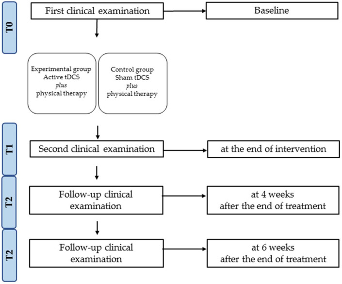
The enrolled study participants were assessed at the baseline (T0), at the end of intervention (T1), at 4 weeks (T2) and 6 weeks after the end of treatment (T3), in terms of balance and gait, through TUG [59], 6MWT [60] (distance, velocity and cadence), Berg Balance Scale (BBS) and Fall Risk Index (FRI), obtained using the Biodex Balance System® (as depicted by Figure 1).
Figure 1.
Study design and timeline.
In more detail, the 6MWT and TUG tests were measured using a single wearable inertial sensor (G-Sensor® BTS Bioengineering S.p.A., Milan, Italy) to instrumentally confirm the time taken (seconds) in carrying out the TUG and the meters traveled during the 6MWT. This inertial sensor was positioned on the participant’s waist using a semi-elastic belt covering on the L1–L2 intervertebral disc for the TUG test and the L4–L5 region for walking assessment, providing acceleration values along three orthogonal axes. For the TUG assessment, participants were instructed to sit on a chair with back support and without armrests, then they stood up, walked for 3 m at self-selected speed, performed a 180° turn around at an obstacle, walked back to the chair and performed a second 180° turn to sit down [44]. The patient was also asked to perform a self-paced submaximal walk along a hard surface, trying to cover as much distance as possible and measuring the distance traveled for 6 min [61]. Each participant received no specific warm-up activity and a researcher walked to one side and behind the participant, close enough for safety purposes; the distance was recorded in meters [62]. Post-processing of the data using dedicated software (BTS G-Studio; BTS Bioengineering S.p.A.) allowed the following parameters to be computed: gait velocity and gait cycle obtained performing the 6MWT and TUG tests [63].
The BBS is a 14-item scale widely used to assess both dynamic and static balance disorders in MS, providing information about a patient’s balance-related abilities [64]. The BBS is scored on a 5-point ordinal scale (0 cannot perform–4 normal performance), with a higher score indicating better performance and with a maximum total score of 56. Lastly, to assess balance, this study used a commercially available balance device, the Biodex Balance System (Biodex Medical Systems, Shirley, NY, USA), to evaluate an individual’s ability to maintain dynamic postural stability [65]. The Biodex Balance System is a circular platform that moves freely and simultaneously about the anteroposterior and mediolateral axes. Patients were instructed to maintain the vertical projection with their center of gravity in the midpoint of the platform by observing a vertical screen located 30 cm in front of their face. Each assessment took 20 s, with 10 s rest periods in between. Patients stood barefoot on the platform with eyes open and the board was set to constant instability. The average of the results from 3 assessments was obtained as the normalized Fall Risk Index (FRI), a score obtained as the ratio between the FRI and the maximum predictive FRI for the relevant age, indicating a greater risk of fall than expected for that age (normalized FRI > 1) and those with an equal or even lesser risk of fall than expected for that age (Normalized FRI ≤ 1)[66].
2.5. Statistical Analysis
The collected data were analyzed using the statistical package R 3.5.2 (R foundation, Vienna, Austria). Descriptive analyses were generated for the demographic and clinical variables of the two arms. The normal distribution of the continuous variables was assessed via Shapiro–Wilk test. Because of the non-parametric distribution, a Mann–Whitney U test was performed to examine the effect of the intergroup treatment (active, sham) and the Wilcoxon test in an intra-group evaluation (pre-evaluation and postevaluation). The type I error (α) was set to 0.05, and the effect sizes were assessed using the rank biserial correlation (RBC). In addition, we conducted the Friedman test, a repeated measures statistical test of the ANOVA type, for non-parametric data. A post hoc evaluation was conducted to assess treatment or time point differences with an MFA and cluster evaluation.
To explore the dataset in more depth, revealing the existence of a difference at a certain time point, we used a factorial analysis and hierarchical clustering. An MFA is a multivariate data analysis method for summarizing and visualizing complex data, whereby individuals are described by several sets of variables [52,53,67]. An MFA takes into account the contributions of all variables to define the distances between individuals on a Cartesian chart through the proportion of variance (eigenvalues) of each variable attained by the different dimensions (axes) [68]. The variables with larger variance values contribute the most to the definition of the dimensions (axes) [69]. Thus, variables that contribute the most to dimension 1 (abscissa) and dimension 2 (ordinate) are the most important in explaining the variability and the position of individuals in the chart [70]. Given a graph of variables (correlation circle), it is possible to show the relationship between the variables and the dimensions [54,69,71] by computing the classical Euclidean distance from the principal coordinates. Lastly, we performed the MFA, a machine learning cluster method that weights the distances of participants influenced by variables, grouping individuals into clusters and computing the classical Euclidean distances from the principal coordinates [68,72]. We computed and visualized the multiple factor analysis in R software using FactoMineR (for the analysis) and fact extra (for data visualization).
To validate the MFA clustering, we performed K-means clustering as a machine learning algorithm, a model capable of dividing data in such a way that the degree of similarity between two data observations is maximal if they belong to the same group and minimal if they do not [73]. With the use of JASP v0.16 (JASP Team, Amsterdam, Netherlands), we obtained the R2, a score that indicates the amount of variance explained by the model; the Akaike Information Criterion (AIC) score, where lower values represent better clustering outputs; and the silhouette score, a value ranging from −1 to 1, where 1 represents dense and well-separated clusters [74].
3. Results
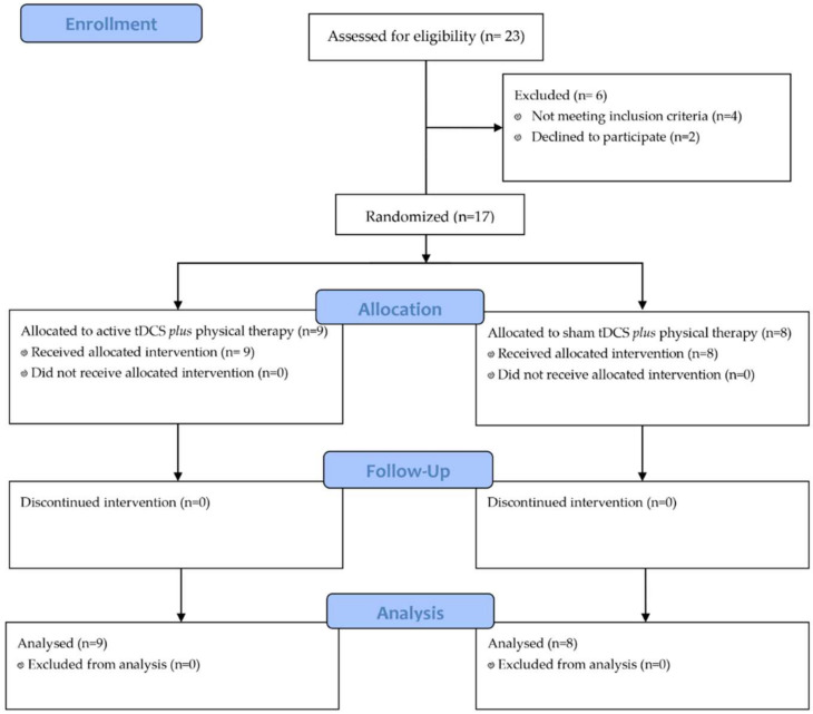
Out of the 23 patients assessed for eligibility, 6 were excluded (4 did not meet the inclusion criteria for the diagnosis of secondary progressive MS, while 2 patients declined to participate). Seventeen patients were enrolled after providing informed consent and were randomized into two groups: tDCS plus physical therapy (n = 9), or the control group, undergoing sham tDCS and physical therapy (n = 8), as depicted in Figure 2.
Figure 2.
Study flow-chart.
There were no statistically significant differences between groups in terms of the demographic and clinical features at baseline (see Table 1 for further details).
Table 1.
Baseline characteristics of the experimental group (tDCS plus physical therapy) and control group (sham tDCS plus physical therapy).
| Experimental Group (n = 9) | Control Group (n = 8) | p Value | |
|---|---|---|---|
| Male/Female | 3/6 | 2/6 | p = 0.213 |
| Age (years) | 43.22 ± 10.46 | 39.75 ± 8.39 | p = 0.081 |
| EDSS | 3 [1] | 3 [0] | p = 0.154 |
| 6MWT (meters) | 300.1 ± 18.8 | 294.5 ± 13.39 | p = 0.134 |
| Gait velocity (m/s) | 0.87 ± 0.26 | 0.92 ± 0.24 | p = 0.070 |
| Gait cycle (meters) | 1.01 ± 0.17 | 0.96 ± 0.46 | p = 0.076 |
| TUG (seconds) | 14.11 ± 3.5 | 12.8 ± 1.1 | p = 0.072 |
| BBS | 48.33 ± 7.4 | 50.4 ± 1.4 | p = 0.356 |
| FRI | 1.31 ± 0.3 | 1.25 ± 0.3 | p = 0.069 |
Continuous variables and parametric data are expressed as means ± standard deviations; categorical variables are expressed as counts (percentages) or medians (interquartile ranges) for non-parametric data; ratios are expressed as x/y. Respectively, we performed the X2 and Mann–Whitney U tests. Abbreviations: 6MWT: 6 min walking test; BBS: Berg Balance Scale; EDSS: Expanded Disability Status Scale; FRI: Fall Risk Index; tDCS: transcranial direct current stimulation; TUG: timed up-and-go.
Stimulation was well-tolerated across participants, with no relevant side effects for any participant. We demonstrated significant intra-group improvements for 6MWT in ΔT0-T1 (experimental group: p = 0.006; control group: p = 0.009), ∆T1-T2 (experimental group: p = 0.007; control group: p = 0.006) and ∆T2-T3 (experimental group: p = 0.006); for TUG in ∆T0-T1 (experimental group: p = 0.031; control group: p = 0.043) and ∆T2-T3 (experimental group: p = 0.042; control group: p = 0.045); and for BBS in ∆T0-T1 (experimental group: p = 0.023) (see Table 2).
Table 2.
Intra-group differences in the outcome measures for experimental (tDCS plus physical therapy) and control (sham tDCS plus physical therapy) groups.
| T0 | T1 | ∆T0-T1 p-Value |
T2 | ∆T1-T2 p-Value |
T3 | ∆T2-T3 p-Value |
||
|---|---|---|---|---|---|---|---|---|
| 6MWT (meters) | experimental | 300.1 ± 18.8 | 322.0 ± 17.0 | 0.006 | 317.3 ± 19.1 | 0.007 | 303.7 ± 16.9 | 0.006 |
| control | 294.5 ± 13.4 | 299.5 ± 14.5 | 0.009 | 284.2 ± 14.9 | 0.006 | 281.5 ± 13.7 | 0.084 | |
| Gait velocity (m/s) | experimental | 0.9 ± 0.3 | 0.8 ± 0.3 | 0.331 | 0.8 ± 0.5 | 0.105 | 0.8 ± 0.5 | 0.312 |
| control | 0.9 ± 0.2 | 0.9 ± 0.4 | 0.227 | 0.8 ± 1.2 | 0.109 | 0.7 ± 0.4 | 0.059 | |
| Gait cycle (meters) | experimental | 1.0 ± 0.2 | 0.8 ± 0.4 | 0.451 | 0.8 ± 0.41 | 0.245 | 0.8 ± 0.2 | 0.677 |
| control | 1.0 ± 0.5 | 1.1 ± 0.4 | 0.423 | 1.0 ± 0.52 | 0.311 | 0.9 ± 0.5 | 0.546 | |
| TUG (seconds) | experimental | 14.1 ± 3.5 | 12.8 ± 5.0 | 0.031 | 13.0 ± 3.7 | 0.154 | 12.0 ± 3.9 | 0.042 |
| control | 12.8 ± 1.1 | 11.3 ± 3.9 | 0.043 | 10.3 ± 2.6 | 0.06 | 11.3 ± 2.1 | 0.044 | |
| BBS | experimental | 48.3 ± 7.4 | 53.3 ± 3.9 | 0.023 | 52.2 ± 5.1 | 0.095 | 50.3 ± 6.8 | 0.083 |
| control | 50.4 ± 1.4 | 51.4 ± 4.4 | 0.162 | 52.4 ± 3.3 | 0.094 | 49.8 ± 2.0 | 0.079 | |
| FRI | experimental | 1.3 ± 0.3 | 1.0 ± 0.8 | 0.099 | 1.0 ± 0.6 | 0.876 | 1.0 ± 0.5 | 0.765 |
| control | 1.3 ± 0.3 | 1.2 ± 0.5 | 0.203 | 1.2 ± 0.6 | 0.785 | 0.1 ± 0.6 | 0.672 |
Continuous variables are expressed as means ± standard deviations. Wilcoxon’s paired t-Test was used for intra-group differences. Note: p-values are significant if <0.05 (expressed in bold type). Abbreviations: 6MWT: 6 min walking test; BBS: Berg Balance Scale; FRI: Fall Risk Index; tDCS: transcranial direct current stimulation; TUG: timed up-and-go.
However, the between-group analysis showed statistically significant differences in terms of gait (6MWT: RBC = 0.8; p = 0.003) and balance (BBS: RBC = 0.4; (p = 0.031) at the end of treatment (T1). Nevertheless, there were significant between-group differences in terms of the 6MWT scores, even at T2 (RBC = 0.93; p = 0.001) and T3 (RBC = 0.84; p = 0.007), with a positive but not significant difference between BBS and TUG at T2 and T3.
Subsequently, we performed a non-parametric repeated measures Friedman test and Kendall’s W test. The experimental group showed a significant difference in repeated measures compared to the control (experimental group: p-value = 0.03, W = 0.34 versus p-value = 0.15, W = 0.20). Moreover, we reported a significant decrease in gait velocity (m/s) in the control group (p = 0.01, W = 0.77), and a significant increase in gait cycle (meters) in the experimental group (p = 0.04, W = 0.31). Lastly, we demonstrated similar significant reductions in TUG (seconds) in both groups, as depicted in Table 3.
Table 3.
Repeated measure differences (Friedman test) in the outcome measures for the experimental (tDCS plus physical therapy) and control (sham tDCS plus physical therapy) groups.
| Chi-Squared | p-Value | Kendall’s W | ||
|---|---|---|---|---|
| 6MWT | experimental | 9.14 | 0.03 | 0.34 |
| control | 5.35 | 0.15 | 0.20 | |
| between-group | 9.24 | 0.03 | 0.23 | |
| Gait velocity | experimental | 2.00 | 0.57 | 0.07 |
| control | 20.92 | 0.01 | 0.77 | |
| between-group | 2.43 | 0.49 | 0.34 | |
| Gait cycle | experimental | 8.43 | 0.04 | 0.31 |
| control | 1.25 | 0.74 | 0.05 | |
| between-group | 0.60 | 0.90 | 0.05 | |
| TUG | experimental | 9.93 | 0.02 | 0.37 |
| control | 11.4 | 0.01 | 0.42 | |
| between-group | 5.64 | 0.13 | 0.35 | |
| BBS | experimental | 4.48 | 0.21 | 0.17 |
| control | 3.32 | 0.34 | 0.12 | |
| between-group | 3.74 | 0.29 | 010 | |
| FRI | experimental | 4.74 | 0.19 | 0.18 |
| control | 4.75 | 0.19 | 0.18 | |
| between-group | 3.24 | 0.36 | 0.12 |
Note: p-values are significant if <0.05 (expressed in bold type). Abbreviations: 6MWT: 6 min walking test; BBS: Berg Balance Scale; FRI: Fall Risk Index; tDCS: transcranial direct current stimulation; TUG: timed up-and-go.
Given these positive results at the end of the treatment (T1), we assessed the benefits one month after the end of the tDCS application. Regardless of the intervention (active or sham), BBS and FRI did not change over time. Despite the Mann–Whitney U and rank biserial correlation calculations, we examined the weights of the variables at the various timepoints to understand how they could have changed over time with respect to the two groups.
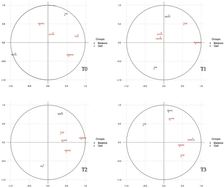
As shown in Figure 3, we divided the parameters of balance (green) and gait (orange), then we evaluated the weights of the single variables through linear regression, representing them two-dimensionally on a Cartesian plane. Then, the variables were arranged as vectors based on their weight and proximity to the axes influencing the spatial positions of individuals at the various timepoints analyzed.
Figure 3.
Correlations between quantitative variables and dimensions. The plot depicts the topographical influence in the arrangement of the variables on the graph along the abscissa (Dim1) and the ordinate (Dim2). Abbreviations: BERG: Berg Balance Score; Dim1: dimension 1 (abscissa axis); Dim2, dimension 2 (ordinate axis); g-cycle: gait cycle; TUG: timed up-and-go; FRI: Fall Risk Index.
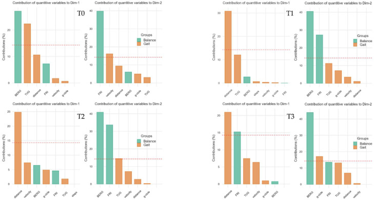
With Figure 4, it is possible to analyze the weight that each single variable gives to the dimension (Cartesian axes). In this scenario, it can be determined how a variable influences the participants in the study groups at each timepoint, conditioning the respective bidimensional position.
Figure 4.
Contributions to the dimension graphs (Cartesian axes). The single weight of each variable in the construction of the Dim1 (abscissa axes of previous figure) and Dim2 (ordinate axes of previous figure) is graphed by bar plots. Abbreviations: BERG: Berg Balance Score; Dim1: dimension 1 (abscissa axis); Dim2, dimension 2 (ordinate axis); g-cycle: gait cycle; TUG: timed up-and-go; FRI: Fall Risk Index.
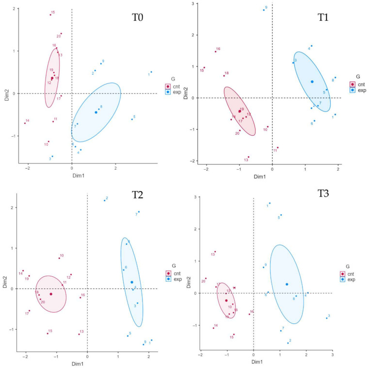
Finally, we analyzed the arrangement of individuals based on the weights and positions of the variables along the Cartesian axes (dimensions), as shown in Figure 4. The cluster arrangement of the two groups can help us to understand the weight and influence of the variables analyzed in the previous figures.
In this scenario, the individuals of the experimental group with active tDCS are placed in the upper right corner, clearly detaching the participants of the control group at the end of the treatment (T1). This characteristic is due to the positive influence of the “distance” variable on the abscissa axis (dimension 1, Dim1) and the positive correlation of the BBS on the ordinates (dimension 2, Dim2). The cluster representation is confirmed with a similar arrangement in T2, and partially due to the influence of the gait outcome and the correlations of balance variables, keeping the experimental cluster in the right upper quadrant detached from the control cluster, as in T1 (for further details see Figure 5).
Figure 5.
Clustered individual factors map. Each individual is positioned according to the Cartesian axes and thickens in specific clusters that reflect the influences of the dimension and each variable. Abbreviations: cnt: control group (sham tDCS + physical therapy); Dim1: dimension 1 (abscissa axis); exp: experimental group (real tDCS + physical therapy); G: groups.
We assessed the quality of clustering by adapting the data to a K-means machine learning approach, reporting an observed R2 value of 0.54, demonstrating that the model shows good reliability; an AIC of 976.45, which demonstrate a moderate quality of fit of the model with several clusters; and a Silhouette Index of 0.34, with a low overlap trend for a small sample.
4. Discussion
The present pilot RCT suggests the potential enhancement of tDCS when supplemented with physical exercise in improving gait and balance in MS patients in the short term.
Our findings reported for the experimental group significant improvements in 6MWT scores for all time-points (∆T0-T1: p = 0.006; ∆T1-T2: p = 0.007; ∆T2-T3: p = 0.006), for TUG in ∆T0-T1 (p = 0.031) and ∆T2-T3 (p = 0.042) and for BBS in ∆T0-T1. The main finding is positive results of the 6MWT in the active intervention, considering that TUG scores significantly changed regardless of the intervention. There were statistically significant differences in 6MWT (p = 0.003) and BBS (p = 0.031) between groups at the end of treatment (T1). Furthermore, there were also significant between-group differences in terms of 6MWT scores at T2 (p = 0.001) and T3 (p = 0.007).
These data were further considered after performing a cluster analysis; considering the weights of each parameter measured, the variables related to walking and balance were similarly disposed both at the end and at 4 weeks after the end of treatment. This disposition might indicate a prolonged beneficial effect induced by the combination of these rehabilitative therapies.
We shed light on the presence and persistence of the effects induced by the proposed treatment, considering the therapeutic exercise as a reference and estimating the modulatory effects over time driven by the addition of tDCS. The statistical analysis showed that exercise interventions moderately improved the walking distances immediately after the end of treatment, losing effect 4 and 6 weeks later. The addition of tDCS appeared to provide an ameliorative effect, with significant increases in the distances walked by patients in the experimental group at T1, as in a recent review [35], and significant increases in BBS for people with MS [75]. At 4 weeks after the end of the treatment, we found a loss of gain compared to T1, considering that it was significantly lower in the experimental group than in the control group. Indeed, in this latter group, the drop in the number of meters covered increased as time progressed, with a discontinuation of the beneficial effect that was previously appreciated. The scientific literature defines therapeutic exercise as the treatment of choice for gait disorders, allowing improvements in autonomy, mobility and consequently quality of life in patients with MS [17,76]. Systematic reviews and meta-analyses have shown small but clinically meaningful effects for exercise training in terms of walking distance, speed, endurance and stride length, being homogeneous across modes of exercise training (i.e., aerobic vs. resistance) [77,78,79]. The effects of exercise appear to require prolonged treatment over time, from 3 weeks to 24 weeks, resulting in significant changes in gait parameters measurable from 12 weeks of treatment onward [80,81]. It is not known how long the advantages acquired by a conventional treatment will last, in light of which a correct therapeutic prescription could be defined. In the present study, a daily treatment shortened in time was able to bring a small gain in walking distance that was not maintained over time, thereby indicating the need to repeat the treatment protocol or to identify a combination treatment that can prolong the effects over time. This is what seemed to have happened in the experimental group, in which the initial benefit was only partially reduced over time, with a return to the baseline condition only 6 weeks after the end of treatment.
The stimulation effect appears to be based on the neuromodulating effect that a direct current of low intensity is able to determine on cortical activity [32]. Stimulating the hemisphere contralateral to the most affected limb, we targeted a cortical region in which neuroplasticity appears to be more difficult to recover, as demonstrated by the work by Chaves, et al. [82]. Exercise alone appears to have less neuroplastic potential on the hemisphere contralateral to the weaker limb [83]. Considering that walking is a complex motor task that requires properly balanced activation of the lower limbs, the addition of a neuromodulating treatment improved the motor performance of the most affected limb by improving the motor task overall.
Furthermore, a protocol of 10 sessions of stimulation associated with gait training represents the current reference, since it is clear that a small number of sessions has proven to be ineffective [34,35,36]. The immediate impact of tDCS is now well documented [40], although what is not well investigated is the persistence of this effect after the end of the stimulation itself. A recent study by Pilloni, et al. [36] shows that multiple sessions of tDCS coupled with aerobic exercise lead to persistent improvements in distance covered by performing the 2MWT, indicating a lack of effect determined by exercise alone. In our pilot RCT, the effect of exercise alone on 6MWT distance is present at the end of treatment and returns to baseline conditions as early as 4 weeks after the end of treatment. The difference between the studies could be due to the different MS patient populations (RR vs. RR + SP) and the degree of EDSS. In a previous study, Savci, et al. [84] included MS patients with a median EDSS score of 4.0 (range 1.5 to 6.0), with longer distances performed in the 6MWT compared to the results reported in our study. This can be explained by the use of median and IQR data in a frequency distribution with a number of extreme values, which among other things pushes the curve towards a lower EDSS score.
In patients with MS, the TUG execution provides additional details on functional mobility and independence, measuring the ability to sit and stand up from a chair, walk in a straight line and change direction in an easy and comfortable way [85,86]. Both the control and experimental groups improved their ambulatory functional performances, with reductions in the time required to perform the TUG test, keeping the results obtained at the end of the treatments stable even at 6 weeks. These results have a positive impact on the patient’s quality of life, being closely associated with the demands and activities of daily living.
The synergy of tDCS and exercise can not only improve motor performance, but also can positively affect postural control, balance and fall risk [87]. Considering the close relationship between walking and balance control, patients underwent a specific rehabilitation protocol. A clinical balance assessment using the BBS revealed an important improvement of the total score in the experimental group, which was not recorded in the control group. Indeed, the control group subjected to sham stimulation in addition to balance training recorded a non-significant improvement of only one point, maintained one month after the end of treatment. This short-lived effect was lost 6 weeks after the completion of treatment. In contrast, the active stimulation resulted in a 5-point gain at the end of treatment and a modest gain at one month, with a loss of effect at 6 weeks. These are encouraging results, considering that a 3-point change in the BBS is the cut-off score to correctly classify a clinically important change in balance performance during activities of daily living [64].
In this pilot study, we showed that tDCS might add positive effects when combined with physical therapy in terms of balance and gait, albeit these findings could not be seen after 4 weeks. However, the machine learning approach, through the analysis of the clusters between the beginning and end of treatment, underlined an influence on BBS in the experimental group. In more detail, the combination of tDCS and physical therapy appeared to maintain the direction of arrangement on the Cartesian axes at T2 in the MS patients in the study group. These findings were not properly in line with the classic statistical analysis, which did not show a significant difference between groups at 4 weeks after the end of treatment (T2). In this scenario, the multiple factor analysis could legitimize the improvements in balance with a common vectorization of the study clusters, in the sense of improvements in BBS, Fall Risk Index and walking performance scores in the active tDCS group compared to the sham tDCS group.
At T3, the improvements appeared to decrease, although the parameters of balance and gait that did not seem to carry over to T2 with an intergroup analysis, as could be found through a machine learning analysis. In fact, through an evaluation of the grouping of the clusters based on the outcomes, the results obtained at the end of the treatment seemed to be similar even 6 weeks after the end of the intervention.
We are aware that our study has some limitations. First, the sample size could be considered relatively small for a machine learning model, although in addition to strict eligibility criteria, it should also be considered that we chose a cluster approach that provides an analysis of the arrangement of groups based on the variables entered, excluding predictive analyses. Second, any drop in measurement quality can prevent machine learning algorithms from accurately modeling the non-linear associations between features. However, it should be noted that we have estimated almost two-thirds of the variance for the two dimensions. Third, in the context of the results obtained, we considered the addition of an intervention that is not always accessible, and which requires skills in execution. Lastly, several tDCS studies in MS have focused on fatigue, although this variable was been included in the present work due to the already in-depth potential of prefrontal area stimulation [88,89,90,91,92,93].
On the other hand, to the best of our knowledge, this is first study that analyzed the effects of tDCS combined to physical therapy in terms of balance and gait outcomes in patients with MS, not only through a standard statistical analysis but also through a machine learning approach.
5. Conclusions
Taken together, the present pilot RCT showed that tDCS coupled with physical therapy might have a significant effect on 6MWT in MS patients. This treatment seemed to not maintain the improvements on balance at one month after the end of treatment, although a similar distribution through the weight of gait and balance outcomes was shown by the cluster-type machine learning analysis. These promising findings might indicate a prolonged beneficial effect induced by the combination of these rehabilitative therapies, although perspectives will be required to consider task-related stimulations, perhaps using coupling a virtual reality approach. Further research is necessary to investigate the efficacy of novel rehabilitative interventions such as tDCS in patients affected by MS, also considering the use of a machine learning approach.
Author Contributions
Conceptualization, N.M., A.d.S. and A.A.; methodology, N.M. and A.d.S.; investigation, C.M., L.M. and A.T.; software, N.M. and A.d.S.; formal analysis, N.M. and A.d.S.; data curation, M.T.I., I.R. and T.P.; writing—original draft preparation, N.M. and C.M.; writing—review and editing, A.d.S. and A.A.; visualization, L.M., M.T.I., I.R., A.T., T.P. and P.V.; supervision, A.d.S., P.V. and A.A. All authors have read and agreed to the published version of the manuscript.
Institutional Review Board Statement
The study was conducted in accordance with the Declaration of Helsinki and Ethical approval was obtained from the Institutional Review Board Committee (number 303/2019).
Informed Consent Statement
Informed consent was obtained from all subjects involved in the study.
Data Availability Statement
Not applicable.
Conflicts of Interest
The authors declare no conflict of interest.
Funding Statement
This research received no external funding.
Footnotes
Publisher’s Note: MDPI stays neutral with regard to jurisdictional claims in published maps and institutional affiliations.
References
- 1.Lassmann H. Multiple Sclerosis Pathology. Cold Spring Harb. Perspect. Med. 2018;8:a028936. doi: 10.1101/cshperspect.a028936. [DOI] [PMC free article] [PubMed] [Google Scholar]
- 2.Filippi M., Bar-Or A., Piehl F., Preziosa P., Solari A., Vukusic S., Rocca M.A. Multiple sclerosis. Nat. Rev. Dis. Prim. 2018;4:43. doi: 10.1038/s41572-018-0041-4. [DOI] [PubMed] [Google Scholar]
- 3.Solaro C., de Sire A., Uccelli M.M., Mueller M., Bergamaschi R., Gasperini C., Restivo D.A., Stabile M.R., Patti F. Efficacy of levetiracetam on upper limb movement in multiple sclerosis patients with cerebellar signs: A multicenter double-blind, placebo-controlled, crossover study. Eur. J. Neurol. 2020;27:2209–2216. doi: 10.1111/ene.14403. [DOI] [PubMed] [Google Scholar]
- 4.Galea M.P., Lizama L.E.C., Butzkueven H., Kilpatrick T.J. Gait and balance deterioration over a 12-month period in multiple sclerosis patients with EDSS scores ≤ 3.0. Neurorehabilitation. 2017;40:277–284. doi: 10.3233/NRE-161413. [DOI] [PubMed] [Google Scholar]
- 5.LaRocca N.G. Impact of walking impairment in multiple sclerosis: Perspectives of patients and care partners. Patient. 2011;4:189–201. doi: 10.2165/11591150-000000000-00000. [DOI] [PubMed] [Google Scholar]
- 6.Frechette M.L., Meyer B.M., Tulipani L.J., Gurchiek R.D., McGinnis R.S., Sosnoff J.J. Next Steps in Wearable Technology and Community Ambulation in Multiple Sclerosis. Curr. Neurol. Neurosci. Rep. 2019;19:80. doi: 10.1007/s11910-019-0997-9. [DOI] [PubMed] [Google Scholar]
- 7.Martin C.L., Phillips B.A., Kilpatrick T.J., Butzkueven H., Tubridy N., McDonald E., Galea M.P. Gait and balance impairment in early multiple sclerosis in the absence of clinical disability. Mult. Scler. 2006;12:620–628. doi: 10.1177/1352458506070658. [DOI] [PubMed] [Google Scholar]
- 8.Gunn H.J., Newell P., Haas B., Marsden J.F., Freeman J.A. Identification of risk factors for falls in multiple sclerosis: A systematic review and meta-analysis. Phys. Ther. 2013;93:504–513. doi: 10.2522/ptj.20120231. [DOI] [PubMed] [Google Scholar]
- 9.Mazumder R., Murchison C., Bourdette D., Cameron M. Falls in people with multiple sclerosis compared with falls in healthy controls. PLoS ONE. 2014;9:e107620. doi: 10.1371/journal.pone.0107620. [DOI] [PMC free article] [PubMed] [Google Scholar]
- 10.Sattelmayer K.M., Chevalley O., Kool J., Wiskerke E., Denkinger L.N., Giacomino K., Opsommer E., Hilfiker R. Development of an exercise programme for balance abilities in people with multiple sclerosis: A development of concept study using Rasch analysis. Arch. Physiother. 2021;11:29. doi: 10.1186/s40945-021-00120-3. [DOI] [PMC free article] [PubMed] [Google Scholar]
- 11.Comber L., Galvin R., Coote S. Gait deficits in people with multiple sclerosis: A systematic review and meta-analysis. Gait Posture. 2017;51:25–35. doi: 10.1016/j.gaitpost.2016.09.026. [DOI] [PubMed] [Google Scholar]
- 12.Cameron M.H., Wagner J.M. Gait abnormalities in multiple sclerosis: Pathogenesis, evaluation, and advances in treatment. Curr. Neurol. Neurosci. Rep. 2011;11:507–515. doi: 10.1007/s11910-011-0214-y. [DOI] [PubMed] [Google Scholar]
- 13.Richmond S.B., Swanson C.W., Peterson D.S., Fling B.W. A temporal analysis of bilateral gait coordination in people with multiple sclerosis. Mult. Scler. Relat. Disord. 2020;45:102445. doi: 10.1016/j.msard.2020.102445. [DOI] [PubMed] [Google Scholar]
- 14.Bethoux F. Gait disorders in multiple sclerosis. Contin. Lifelong Learn. Neurol. 2013;19:1007–1022. doi: 10.1212/01.CON.0000433286.92596.d5. [DOI] [PubMed] [Google Scholar]
- 15.Latimer-Cheung A.E., Pilutti L.A., Hicks A.L., Martin Ginis K.A., Fenuta A.M., MacKibbon K.A., Motl R.W. Effects of exercise training on fitness, mobility, fatigue, and health-related quality of life among adults with multiple sclerosis: A systematic review to inform guideline development. Arch. Phys. Med. Rehabil. 2013;94:1800–1828. doi: 10.1016/j.apmr.2013.04.020. [DOI] [PubMed] [Google Scholar]
- 16.Cameron M.H., Nilsagard Y. Handbook of Clinical Neurology. Volume 159. Elsevier B.V.; Amsterdam, The Netherlands: 2018. Balance, gait, and falls in multiple sclerosis; pp. 237–250. [DOI] [PubMed] [Google Scholar]
- 17.Motl R.W., Sandroff B.M., Kwakkel G., Dalgas U., Feinstein A., Heesen C., Feys P., Thompson A.J. Exercise in patients with multiple sclerosis. Lancet Neurol. 2017;16:848–856. doi: 10.1016/S1474-4422(17)30281-8. [DOI] [PubMed] [Google Scholar]
- 18.Salari N., Hayati A., Kazeminia M., Rahmani A., Mohammadi M., Fatahian R., Shohaimi S. The effect of exercise on balance in patients with stroke, Parkinson, and multiple sclerosis: A systematic review and meta-analysis of clinical trials. Neurol. Sci. 2021;16:848–856. doi: 10.1007/s10072-021-05689-y. [DOI] [PubMed] [Google Scholar]
- 19.Donzé C., Massot C. Rehabilitation in multiple sclerosis in 2021. La Presse Médicale. 2021;50:104066. doi: 10.1016/j.lpm.2021.104066. [DOI] [PubMed] [Google Scholar]
- 20.Prosperini L., di Filippo M. Beyond clinical changes: Rehabilitation-induced neuroplasticity in MS. Mult. Scler. J. 2019;25:1348–1362. doi: 10.1177/1352458519846096. [DOI] [PubMed] [Google Scholar]
- 21.Tomassini V., Matthews P.M., Thompson A.J., Fuglo D., Geurts J.J., Johansen-Berg H., Jones D.K., Rocca M.A., Wise R.G., Barkhof F., et al. Neuroplasticity and functional recovery in multiple sclerosis. Nat. Rev. Neurol. 2012;8:635–646. doi: 10.1038/nrneurol.2012.179. [DOI] [PMC free article] [PubMed] [Google Scholar]
- 22.Tomassini V., Johansen-Berg H., Leonardi L., Paixão L., Jbabdi S., Palace J., Pozzilli C., Matthews P.M. Preservation of motor skill learning in patients with multiple sclerosis. Mult. Scler. J. 2011;17:103–115. doi: 10.1177/1352458510381257. [DOI] [PMC free article] [PubMed] [Google Scholar]
- 23.de Sire A., Bigoni M., Priano L., Baudo S., Solaro C., Mauro A. Constraint-Induced Movement Therapy in multiple sclerosis: Safety and three-dimensional kinematic analysis of upper limb activity. A randomized single-blind pilot study. Neurorehabilitation. 2019;45:247–254. doi: 10.3233/NRE-192762. [DOI] [PubMed] [Google Scholar]
- 24.de Sire A., Mauro A., Priano L., Baudo S., Bigoni M., Solaro C. Effects of constraint-induced movement therapy on upper limb activity according to a bi-dimensional kinematic analysis in progressive multiple sclerosis patients: A randomized single-blind pilot study. Funct. Neurol. 2019;34:151–157. [PubMed] [Google Scholar]
- 25.Prosperini L., Piattella M.C., Giannì C., Pantano P. Functional and Structural Brain Plasticity Enhanced by Motor and Cognitive Rehabilitation in Multiple Sclerosis. Neural Plast. 2015;2015:481574. doi: 10.1155/2015/481574. [DOI] [PMC free article] [PubMed] [Google Scholar]
- 26.Hershey L.A. Comment: Performance improvement with computer training in Parkinson disease. Neurology. 2014;82:1224. doi: 10.1212/WNL.0000000000000299. [DOI] [PubMed] [Google Scholar]
- 27.Marotta N., Demeco A., Indino A., de Scorpio G., Moggio L., Ammendolia A. Nintendo WiiTM versus Xbox KinectTM for functional locomotion in people with Parkinson’s disease: A systematic review and network meta-analysis. Disabil. Rehabil. 2020;44:331–336. doi: 10.1080/09638288.2020.1768301. [DOI] [PubMed] [Google Scholar]
- 28.Marotta N., Demeco A., Moggio L., Ammendolia A. The adjunct of transcranial direct current stimulation to Robot-assisted therapy in upper limb post-stroke treatment. J. Med. Eng. Technol. 2021;45:494–501. doi: 10.1080/03091902.2021.1922527. [DOI] [PubMed] [Google Scholar]
- 29.Calafiore D., Invernizzi M., Ammendolia A., Marotta N., Fortunato F., Paolucci T., Ferraro F., Curci C., Cwirlej-Sozanska A., de Sire A. Efficacy of Virtual Reality and Exergaming in Improving Balance in Patients With Multiple Sclerosis: A Systematic Review and Meta-Analysis. Front. Neurol. 2021;12:773459. doi: 10.3389/fneur.2021.773459. [DOI] [PMC free article] [PubMed] [Google Scholar]
- 30.Ganguly J., Murgai A., Sharma S., Aur D., Jog M. Non-invasive Transcranial Electrical Stimulation in Movement Disorders. Front. Neurosci. 2020;14:522. doi: 10.3389/fnins.2020.00522. [DOI] [PMC free article] [PubMed] [Google Scholar]
- 31.Iodice R., Manganelli F., Dubbioso R. The therapeutic use of non-invasive brain stimulation in multiple sclerosis-a review. Restor. Neurol. Neurosci. 2017;35:497–509. doi: 10.3233/RNN-170735. [DOI] [PubMed] [Google Scholar]
- 32.Sánchez-Kuhn A., Pérez-Fernández C., Cánovas R., Flores P., Sánchez-Santed F. Transcranial direct current stimulation as a motor Neurorehabilitation tool: An empirical review. Biomed. Eng. Online. 2017;16:115–136. doi: 10.1186/s12938-017-0361-8. [DOI] [PMC free article] [PubMed] [Google Scholar]
- 33.Cirillo G., Di Pino G., Capone F., Ranieri F., Florio L., Todisco V., Tedeschi G., Funke K., Di Lazzaro V. Neurobiological after-effects of non-invasive brain stimulation. Brain Stimul. 2017;10:1–18. doi: 10.1016/j.brs.2016.11.009. [DOI] [PubMed] [Google Scholar]
- 34.Pilloni G., Choi C., Coghe G., Cocco E., Krupp L.B., Pau M., Charvet L.E. Gait and Functional Mobility in Multiple Sclerosis: Immediate Effects of Transcranial Direct Current Stimulation (tDCS) Paired With Aerobic Exercise. Front. Neurol. 2020;11:310. doi: 10.3389/fneur.2020.00310. [DOI] [PMC free article] [PubMed] [Google Scholar]
- 35.Hiew S., Nguemeni C., Zeller D. Efficacy of transcranial direct current stimulation in people with multiple sclerosis: A review. Eur. J. Neurol. 2021;29:648–664. doi: 10.1111/ene.15163. [DOI] [PubMed] [Google Scholar]
- 36.Pilloni G., Choi C., Shaw M.T., Coghe G., Krupp L., Moffat M., Cocco E., Pau M., Charvet L. Walking in multiple sclerosis improves with tDCS: A randomized, double-blind, sham-controlled study. Ann. Clin. Transl. Neurol. 2020;7:2310–2319. doi: 10.1002/acn3.51224. [DOI] [PMC free article] [PubMed] [Google Scholar]
- 37.Roche N., Geiger M., Bussel B. Mechanisms underlying transcranial direct current stimulation in rehabilitation. Ann. Phys. Rehabil. Med. 2015;58:214–219. doi: 10.1016/j.rehab.2015.04.009. [DOI] [PubMed] [Google Scholar]
- 38.Swanson C.W., Proessl F., Stephens J.A., Miravalle A.A., Fling B.W. Non-invasive brain stimulation to assess neurophysiologic underpinnings of lower limb motor impairment in multiple sclerosis. J. Neurosci. Methods. 2021;356:109143. doi: 10.1016/j.jneumeth.2021.109143. [DOI] [PubMed] [Google Scholar]
- 39.Halabchi F., Alizadeh Z., Sahraian M.A., Abolhasani M. Exercise prescription for patients with multiple sclerosis; potential benefits and practical recommendations. BMC Neurol. 2017;17:185. doi: 10.1186/s12883-017-0960-9. [DOI] [PMC free article] [PubMed] [Google Scholar]
- 40.Workman C.D., Kamholz J., Rudroff T. Transcranial Direct Current Stimulation (tDCS) to Improve Gait in Multiple Sclerosis: A Timing Window Comparison. Front. Hum. Neurosci. 2019;13:420. doi: 10.3389/fnhum.2019.00420. [DOI] [PMC free article] [PubMed] [Google Scholar]
- 41.Oveisgharan S., Karimi Z., Abdi S., Sikaroodi H. The use of brain stimulation in the rehabilitation of walking disability in patients with multiple sclerosis: A randomized double-blind clinical trial study. Curr. J. Neurol. 2019;18:57. doi: 10.18502/ijnl.v18i2.1289. [DOI] [PMC free article] [PubMed] [Google Scholar]
- 42.Boggio P.S., Nunes A., Rigonatti S.P., Nitsche M.A., Pascual-Leone A., Fregni F. Repeated sessions of noninvasive brain DC stimulation is associated with motor function improvement in stroke patients. Restor. Neurol. Neurosci. 2007;25:123–129. [PubMed] [Google Scholar]
- 43.Grecco L.A.C., de Almeida Carvalho Duarte N., Mendonça M.E., Cimolin V.Ô., Galli M., Fregni F., Oliveira C.S. Transcranial direct current stimulation during treadmill training in children with cerebral palsy: A randomized controlled double-blind clinical trial. Res. Dev. Disabil. 2014;35:2840–2848. doi: 10.1016/j.ridd.2014.07.030. [DOI] [PubMed] [Google Scholar]
- 44.Costa-Ribeiro A., Maux A., Bosford T., Aoki Y., Castro R., Baltar A., Shirahige L., Moura Filho A., Nitsche M.A., Monte-Silva K. Transcranial direct current stimulation associated with gait training in Parkinson’s disease: A pilot randomized clinical trial. Dev. Neurorehabil. 2017;20:121–128. doi: 10.3109/17518423.2015.1131755. [DOI] [PubMed] [Google Scholar]
- 45.Scholz M., Haase R., Trentzsch K., Stölzer--hutsch H., Ziemssen T. Improving digital patient care: Lessons learned from patient-- reported and expert--reported experience measures for the clinical practice of multidimensional walking assessment. Brain Sci. 2021;11:786. doi: 10.3390/brainsci11060786. [DOI] [PMC free article] [PubMed] [Google Scholar]
- 46.Zhao Y., Healy B.C., Rotstein D., Guttmann C.R.G., Bakshi R., Weiner H.L., Brodley C.E., Chitnis T. Exploration of machine learning techniques in predicting multiple sclerosis disease course. PLoS ONE. 2017;12:e0174866. doi: 10.1371/journal.pone.0174866. [DOI] [PMC free article] [PubMed] [Google Scholar]
- 47.Liu H., Guan J., Li H., Bao Z., Wang Q., Luo X., Xue H. Predicting the Disease Genes of Multiple Sclerosis Based on Network Representation Learning. Front. Genet. 2020;11:328. doi: 10.3389/fgene.2020.00328. [DOI] [PMC free article] [PubMed] [Google Scholar]
- 48.Trentzsch K., Schumann P., Śliwiński G., Bartscht P., Haase R., Schriefer D., Zink A., Heinke A., Jochim T., Malberg H., et al. Using machine learning algorithms for identifying gait parameters suitable to evaluate subtle changes in gait in people with multiple sclerosis. Brain Sci. 2021;11:1049. doi: 10.3390/brainsci11081049. [DOI] [PMC free article] [PubMed] [Google Scholar]
- 49.Marzullo A., Kocevar G., Stamile C., Durand-Dubief F., Terracina G., Calimeri F., Sappey-Marinier D. Classification of multiple sclerosis clinical profiles via graph convolutional neural networks. Front. Neurosci. 2019;13:594. doi: 10.3389/fnins.2019.00594. [DOI] [PMC free article] [PubMed] [Google Scholar]
- 50.Martínez-Heras E., Solana E., Prados F., Andorrà M., Solanes A., López-Soley E., Montejo C., Pulido-Valdeolivas I., Alba-Arbalat S., Sola-Valls N., et al. Characterization of multiple sclerosis lesions with distinct clinical correlates through quantitative diffusion MRI. NeuroImage Clin. 2020;28:102411. doi: 10.1016/j.nicl.2020.102411. [DOI] [PMC free article] [PubMed] [Google Scholar]
- 51.Piryonesi S.M., Rostampour S., Piryonesi S.A. Predicting falls and injuries in people with multiple sclerosis using machine learning algorithms. Mult. Scler. Relat. Disord. 2021;49:102740. doi: 10.1016/j.msard.2021.102740. [DOI] [PubMed] [Google Scholar]
- 52.Bécue-Bertaut M., Pagès J. Multiple factor analysis and clustering of a mixture of quantitative, categorical and frequency data. Comput. Stat. Data Anal. 2008;52:3255–3268. doi: 10.1016/j.csda.2007.09.023. [DOI] [Google Scholar]
- 53.Pinto M., Marotta N., Caracò C., Simeone E., Ammendolia A., de Sire A. Quality of Life Predictors in Patients With Melanoma: A Machine Learning Approach. Front. Oncol. 2022;12:843611. doi: 10.3389/fonc.2022.843611. [DOI] [PMC free article] [PubMed] [Google Scholar]
- 54.Husson F., Le S., Pagès J. Exploratory Multivariate Analysis by Example Using R. Chapman and Hall/CRC; New York, NY, USA: 2017. [Google Scholar]
- 55.Thompson A.J., Banwell B.L., Barkhof F., Carroll W.M., Coetzee T., Comi G., Correale J., Fazekas F., Filippi M., Freedman M.S., et al. Diagnosis of multiple sclerosis: 2017 revisions of the McDonald criteria. Lancet Neurol. 2018;17:162–173. doi: 10.1016/S1474-4422(17)30470-2. [DOI] [PubMed] [Google Scholar]
- 56.Ali A.S., Darwish M.H., Shalaby N.M., Abbas R.L., Soubhy H.Z. Efficacy of core stability versus task oriented trainings on balance in ataxic persons with multiple sclerosis. A single blinded randomized controlled trial. Mult. Scler. Relat. Disord. 2021;50:102866. doi: 10.1016/j.msard.2021.102866. [DOI] [PubMed] [Google Scholar]
- 57.White L.J., McCoy S.C., Castellano V., Gutierrez G., Stevens J.E., Walter G.A., Vandenborne K. Resistance training improves strength and functional capacity in persons with multiple sclerosis. Mult. Scler. 2004;10:668–674. doi: 10.1191/1352458504ms1088oa. [DOI] [PubMed] [Google Scholar]
- 58.Cofré Lizama L.E., Bruijn S.M., Galea M.P. Gait stability at early stages of multiple sclerosis using different data sources. Gait Posture. 2020;77:214–217. doi: 10.1016/j.gaitpost.2020.02.006. [DOI] [PubMed] [Google Scholar]
- 59.Sebastião E., Sandroff B.M., Learmonth Y.C., Motl R.W. Validity of the Timed Up and Go Test as a Measure of Functional Mobility in Persons with Multiple Sclerosis. Arch. Phys. Med. Rehabil. 2016;97:1072–1077. doi: 10.1016/j.apmr.2015.12.031. [DOI] [PubMed] [Google Scholar]
- 60.Storm F.A., Cesareo A., Reni G., Biffi E. Wearable inertial sensors to assess gait during the 6-min walk test: A systematic review. Sensors. 2020;20:2660. doi: 10.3390/s20092660. [DOI] [PMC free article] [PubMed] [Google Scholar]
- 61.Goldman M.D., Marrie R.A., Cohen J.A. Evaluation of the six-minute walk in multiple sclerosis subjects and healthy controls. Mult. Scler. 2008;14:383–390. doi: 10.1177/1352458507082607. [DOI] [PubMed] [Google Scholar]
- 62.Wetzel J.L., Fry D.K., Pfalzer L.A. Six-minute walk test for persons with mild or moderate disability from multiple sclerosis: Performance and explanatory factors. Physiother. Can. 2011;63:166–180. doi: 10.3138/ptc.2009-62. [DOI] [PMC free article] [PubMed] [Google Scholar]
- 63.Pau M., Caggiari S., Mura A., Corona F., Leban B., Coghe G., Lorefice L., Marrosu M.G., Cocco E. Clinical assessment of gait in individuals with multiple sclerosis using wearable inertial sensors: Comparison with patient-based measure. Mult. Scler. Relat. Disord. 2016;10:187–191. doi: 10.1016/j.msard.2016.10.007. [DOI] [PubMed] [Google Scholar]
- 64.Ovacık U., Çelik D. Minimal Clinically Important Difference of Berg Balance Scale in People with Multiple Sclerosis. Arch. Phys. Med. Rehabil. 2019;100:1191. doi: 10.1016/j.apmr.2018.12.040. [DOI] [PubMed] [Google Scholar]
- 65.Atteya A., Elwishy A., Kishk N., Ismail R.S., Badawy R. Assessment of postural balance in multiple sclerosis patients. Egypt. J. Neurol. Psychiatry Neurosurg. 2019;55:7. doi: 10.1186/s41983-018-0049-4. [DOI] [Google Scholar]
- 66.Prometti P., Olivares A., Gaia G., Bonometti G., Comini L., Scalvini S. Biodex fall risk assessment in the elderly with ataxia: A new age-dependent derived index in rehabilitation: An observational study. Medicine. 2016;95 doi: 10.1097/MD.0000000000002977. [DOI] [PMC free article] [PubMed] [Google Scholar]
- 67.Abdi H., Williams L.J., Valentin D. Multiple factor analysis: Principal component analysis for multitable and multiblock data sets. Wiley Interdiscip. Rev. Comput. Stat. 2013;5:149–179. doi: 10.1002/wics.1246. [DOI] [Google Scholar]
- 68.Abdullah S.S., Rostamzadeh N., Sedig K., Garg A.X., McArthur E. Visual analytics for dimension reduction and cluster analysis of high dimensional electronic health records. Informatics. 2020;7:17. doi: 10.3390/informatics7020017. [DOI] [Google Scholar]
- 69.Peres-Neto P.R., Jackson D.A., Somers K.M. Giving meaningful interpretation to ordination axes: Assessing loading significance in principal component analysis. Ecology. 2003;84:2347–2363. doi: 10.1890/00-0634. [DOI] [Google Scholar]
- 70.Giacalone D., Ribeiro L.M., Frøst M.B. Consumer-Based Product Profiling: Application of Partial Napping® for Sensory Characterization of Specialty Beers by Novices and Experts. J. Food Prod. Mark. 2013;19:201–218. doi: 10.1080/10454446.2013.797946. [DOI] [Google Scholar]
- 71.Beh E.J. Exploratory multivariate analysis by example using R. J. Appl. Stat. 2012;39:1381–1382. doi: 10.1080/02664763.2012.657409. [DOI] [Google Scholar]
- 72.Pagès-Hélary S., Dujourdy L., Cayot N. Flavor compounds in blackcurrant berries: Multivariate analysis of datasets obtained with natural variability and various experimental parameters. LWT. 2022;153:112425. doi: 10.1016/j.lwt.2021.112425. [DOI] [Google Scholar]
- 73.Tibshirani R., Walther G., Hastie T. Estimating the number of clusters in a data set via the gap statistic. J. R. Stat. Soc. Ser. B Stat. Methodol. 2001;63:411–423. doi: 10.1111/1467-9868.00293. [DOI] [Google Scholar]
- 74.de Sire A., Gallelli L., Marotta N., Lippi L., Fusco N., Calafiore D., Cione E., Muraca L., Maconi A., de Sarro G., et al. Vitamin D Deficiency in Women with Breast Cancer: A Correlation with Osteoporosis? A Machine Learning Approach with Multiple Factor Analysis. Nutrients. 2022;14:1586. doi: 10.3390/nu14081586. [DOI] [PMC free article] [PubMed] [Google Scholar]
- 75.Martino Cinnera A., Bisirri A., Leone E., Morone G., Gaeta A. Effect of dual-task training on balance in patients with multiple sclerosis: A systematic review and meta-analysis. Clin. Rehabil. 2021;35:1399–1412. doi: 10.1177/02692155211010372. [DOI] [PubMed] [Google Scholar]
- 76.Kalb R., Brown T.R., Coote S., Costello K., Dalgas U., Garmon E., Giesser B., Halper J., Karpatkin H., Keller J., et al. Exercise and lifestyle physical activity recommendations for people with multiple sclerosis throughout the disease course. Mult. Scler. J. 2020;26:1459–1469. doi: 10.1177/1352458520915629. [DOI] [PMC free article] [PubMed] [Google Scholar]
- 77.Learmonth Y.C., Ensari I., Motl R.W. Physiotherapy and walking outcomes in adults with multiple sclerosis: Systematic review and meta-analysis. Phys. Ther. Rev. 2016;21:160–172. doi: 10.1080/10833196.2016.1263415. [DOI] [Google Scholar]
- 78.Pearson M., Dieberg G., Smart N. Exercise as a Therapy for Improvement of Walking Ability in Adults with Multiple Sclerosis: A Meta-Analysis. Arch. Phys. Med. Rehabil. 2015;96:1339–1348. doi: 10.1016/j.apmr.2015.02.011. [DOI] [PubMed] [Google Scholar]
- 79.Snook E.M., Motl R.W. Effect of exercise training on walking mobility in multiple sclerosis: A meta-analysis. Neurorehabil. Neural Repair. 2009;23:108–116. doi: 10.1177/1545968308320641. [DOI] [PubMed] [Google Scholar]
- 80.Wonneberger M., Schmidt S. Changes of gait parameters following long-term aerobic endurance exercise in mildly disabled multiple sclerosis patients: An exploratory study. Eur. J. Phys. Rehabil. Med. 2015;51:755–762. [PubMed] [Google Scholar]
- 81.Langeskov--Christensen M., Hvid L.G., Jensen H.B., Nielsen H.H., Petersen T., Stenager E., Dalgas U. Efficacy of high--intensity aerobic exercise on common multiple sclerosis symptoms. Acta Neurol. Scand. 2022;145:229–238. doi: 10.1111/ane.13540. [DOI] [PubMed] [Google Scholar]
- 82.Chaves A.R., Devasahayam A.J., Kelly L.P., Pretty R.W., Ploughman M. Exercise-Induced Brain Excitability Changes in Progressive Multiple Sclerosis: A Pilot Study. J. Neurol. Phys. Ther. 2020;44:132–144. doi: 10.1097/NPT.0000000000000308. [DOI] [PubMed] [Google Scholar]
- 83.Chaves A.R., Devasahayam A.J., Riemenschneider M., Pretty R.W., Ploughman M. Walking Training Enhances Corticospinal Excitability in Progressive Multiple Sclerosis—A Pilot Study. Front. Neurol. 2020;11:422. doi: 10.3389/fneur.2020.00422. [DOI] [PMC free article] [PubMed] [Google Scholar]
- 84.Savci S., Inal-Ince D., Arikan H., Guclu-Gunduz A., Cetisli-Korkmaz N., Armutlu K., Karabudak R. Six-minute walk distance as a measure of functional exercise capacity in multiple sclerosis. Disabil. Rehabil. 2005;27:1365–1371. doi: 10.1080/09638280500164479. [DOI] [PubMed] [Google Scholar]
- 85.Cohen E.T., Potter K., Allen D.D., Bennett S.E., Brandfass K.G., Widener G.L., Yorke A.M. Selecting rehabilitation outcome measures for people with multiple sclerosis. Int. J. MS Care. 2015;17:181–189. doi: 10.7224/1537-2073.2014-067. [DOI] [PMC free article] [PubMed] [Google Scholar]
- 86.Kalron A., Dolev M., Givon U. Further construct validity of the Timed Up-and-Go Test as a measure of ambulation in multiple sclerosis patients. Eur. J. Phys. Rehabil. Med. 2017;53:841–847. doi: 10.23736/S1973-9087.17.04599-3. [DOI] [PubMed] [Google Scholar]
- 87.Paltamaa J., Sjögren T., Peurala S.H., Heinonen A. Effects of physiotherapy interventions on balance in multiple sclerosis : A systematic review and meta-analysis of randomized controlled trials. J. Rehabil. Med. 2012;44:811–823. doi: 10.2340/16501977-1047. [DOI] [PubMed] [Google Scholar]
- 88.Dobbs B., Pawlak N., Biagioni M., Agarwal S., Shaw M., Pilloni G., Bikson M., Datta A., Charvet L. Generalizing remotely supervised transcranial direct current stimulation (tDCS): Feasibility and benefit in Parkinson’s disease. J. Neuroeng. Rehabil. 2018;15:114. doi: 10.1186/s12984-018-0457-9. [DOI] [PMC free article] [PubMed] [Google Scholar]
- 89.Ashrafi A., Mohseni-Bandpei M.A., Seydi M. The effect of tDCS on the fatigue in patients with multiple sclerosis: A systematic review of randomized controlled clinical trials. J. Clin. Neurosci. 2020;78:277–283. doi: 10.1016/j.jocn.2020.04.106. [DOI] [PubMed] [Google Scholar]
- 90.Sharma K., Agarwal S., Mania D., Cucca A., Frucht S., Feigin A., Biagioni M. Can Tele-Monitored transcranial Direct Current Stimulation (tDCS) Help Manage Fatigue and Cognitive Symptoms in Parkinson’s disease? Neurology. 2020;94 [Google Scholar]
- 91.Ayache S.S., Chalah M.A., Kümpfel T., Padberg F., Lefaucheur J.P., Palm U. Multiple sclerosis fatigue, its neural correlates, and its modulation with tDCS. Fortschr. Neurol. Psychiatr. 2017;85:260–269. doi: 10.1055/s-0043-105389. [DOI] [PubMed] [Google Scholar]
- 92.Chalah M.A., Riachi N., Ahdab R., Mhalla A., Abdellaoui M., Créange A., Lefaucheur J.P., Ayache S.S. Effects of left DLPFC versus right PPC tDCS on multiple sclerosis fatigue. J. Neurol. Sci. 2017;372:131–137. doi: 10.1016/j.jns.2016.11.015. [DOI] [PubMed] [Google Scholar]
- 93.Ferrucci R., Vergari M., Cogiamanian F., Bocci T., Ciocca M., Tomasini E., de Riz M., Scarpini E., Priori A. Transcranial direct current stimulation (tDCS) for fatigue in multiple sclerosis. Neurorehabilitation. 2014;34:121–127. doi: 10.3233/NRE-131019. [DOI] [PubMed] [Google Scholar]
Associated Data
This section collects any data citations, data availability statements, or supplementary materials included in this article.
Data Availability Statement
Not applicable.


