Abstract
Transcriptional regulators select their targets from a large pool of similar genomic sites. The binding of the Drosophila dosage compensation complex (DCC) exclusively to the male X chromosome provides insight into binding site selectivity rules. Previous studies showed that the male-specific organizer of the complex, MSL2, and ubiquitous DNA-binding protein CLAMP directly interact and play an important role in the specificity of X chromosome binding. Here, we studied the highly specific interaction between the intrinsically disordered region of MSL2 and the N-terminal zinc-finger C2H2-type (C2H2) domain of CLAMP. We obtained the NMR structure of the CLAMP N-terminal C2H2 zinc finger, which has a classic C2H2 zinc-finger fold with a rather unusual distribution of residues typically used in DNA recognition. Substitutions of residues in this C2H2 domain had the same effect on the viability of males and females, suggesting that it plays a general role in CLAMP activity. The N-terminal C2H2 domain of CLAMP is highly conserved in insects. However, the MSL2 region involved in the interaction is conserved only within the Drosophila genus, suggesting that this interaction emerged during the evolution of a mechanism for the specific recruitment of the DCC on the male X chromosome in Drosophilidae.
INTRODUCTION
It remains unknown how transcription complexes bind exclusively to the certain chromatin regions that do not have pronounced sequence specificity relative to many other genome regions. A striking example of the specific recruitment of transcription complexes is the process of dosage compensation in Drosophila (1–3). Dosage compensation occurs by increasing the level of gene expression of the X chromosome of males (X/Y) relative to that of females (X/X). The dosage compensation complex (DCC), which binds only to the male X-chromosome, is responsible for increasing the expression of male genes.
The DCC consists of five proteins, MSL1, MSL2, MSL3, MOF and MLE, and includes two non-coding RNAs, roX1 (3.7 kb) and roX2 (0.6 kb), which perform mutually interchangeable functions (1,2). Proteins MSL1, MSL3, MOF and MLE are also present in females and are involved in regulating gene expression in other transcriptional complexes unrelated to dose compensation (1). The MSL2 protein is specific for males (4), and is therefore believed to play a major role in the selective recognition of the male X chromosome.
Inactivation of the MSL3 or MLE proteins led to binding of the incomplete MSL1-MSL2 complex to ∼200 sites on the X chromosome, referred to as chromatin entry sites (CES) or high-affinity sites (HAS) (5,6). The CXC domain of MSL2 recognizes most HAS/CES sites in vitro, and it is likely to be involved in the selection of the male X chromosome by DCC (7–9) In addition, the zinc-finger CLAMP protein binds to GA-repeats found in most of HAS/CES and is essential for recruitment of DCC on the male X chromosome (10). The N-terminal 153 aa region of CLAMP, including the first zinc-finger C2H2 domain, interacts with the unstructured highly conserved (within the Drosophila genus) 618−655 aa region of MSL2 (the CLAMP-binding-domain, CBD) (11,12). Inactivation of CBD or CXC in MSL2 only modestly affects recruitment of the DCC to the X chromosome in males (12). However, combining of these two genetic lesions within the same MSL2 mutant resulted in strong inactivation of DCC.
Our previous data suggest that the C2H2 zinc finger domain is required for this interaction (12). The typical C2H2 zinc finger domain has two beta strands and an alpha-helix stabilized by a coordinated zinc ion. Domains of this type are commonly involved in specific DNA binding through residues in the alpha-helix. However, there is growing evidence of their possible function as protein-protein interaction domains (13).
Interaction of the CLAMP N-terminal zinc-finger with MSL2 is highly specific: in Y2H screening, we revealed that MSL2 only interacted with CLAMP and not with any other protein of the tested 152 multi-zinc-finger Drosophila transcription factors (12). Here, we examined the structural basis for the interaction between the CLAMP and MSL2 proteins using NMR techniques and mutagenic screening complemented with in vivo experiments. This study reveals for the first time the features of stable C2H2 zinc-finger interaction with unfolded peptides. We found that the CLAMP N-terminal C2H2 domain interacts with MSL2 using amino acid residues different from those commonly used for DNA recognition. Furthermore, only simultaneous substitutions of several residues at the binding interface significantly weakened the interaction and resulted in progressive DCC delocalization.
MATERIALS AND METHODS
Plasmids and cloning
cDNAs were PCR-amplified using corresponding primers (Supplementary Table S3) and cloned into a modified pGEX4T1 vector (GE Healthcare) encoding the TEV protease cleavage site after GST and into the vector derived from pACYC and pET28a(+) (Novagen) bearing a p15A replication origin, kanamycin resistance gene, and pET28a(+) MCS. Apis mellifera cDNA was prepared using standard procedures from adult bees obtained from a local apiary. PCR-directed mutagenesis was used to create constructs expressing mutant proteins using mutagenic primers (Supplementary Table S3). For yeast two-hybrid assays, cDNAs were amplified using the corresponding primers (Supplementary Table S3) and fused with the DNA-binding or activation domain of GAL4 in the corresponding pGBT9 and pGAD424 vectors (Clontech). Details of assembling the constructs for expressing proteins in transgenic flies are available upon request.
Analysis of C2H2 zinc-finger amino acid composition
The development of a hidden Markov Model of the C2H2 domain sequence and calculation of the probabilities of each residue at given positions were performed using Skylign (14). Details are described in Supplementary Information.
Protein procedures
Proteins were expressed in BL21 (DE3) cells and purified with IMAC, followed by anion-exchange chromatography. Pulldown assays were performed as described (12). Stable isotope-labeled proteins were expressed according to (15) and purified using the same procedures as native proteins. Detailed procedures are described in Supplementary Information.
NMR spectroscopy
The NMR samples were prepared in concentrations of 0.5 mM (13C, 15N-labeled MSL2618–655 and CLAMP derivatives) and 0.1–0.4 mM (15N-labeled protein) with 5% (v/v) D2O for frequency lock. Most 2D NMR spectra were collected using a Bruker AVANCE 600 MHz spectrometer equipped with TXI triple resonance (1H,13C,15N) probe, and 3D spectra were recorded on a Bruker AVANCE 700 MHz spectrometer equipped with a quadruple resonance (1H, 13C, 15N, 31P) cryo-probe. All NMR experiments were carried out at 25°C. Additional experimental details are included in Supplementary. The structure of CLAMP was deposited to protein data bank with the accession ID 7NF9.
Y2H
The yeast two-hybrid assay was performed as previously described (16). Briefly, for growth assays, plasmids were transformed into yeast strain pJ69-4A by the lithium acetate method, following standard Clontech protocol, and plated on media without tryptophan and leucine. After 2 days of growth at 30°C, the cells were plated on selective media without tryptophan, leucine, histidine, and adenine, and their growth was compared after 2–3 days. Each assay was repeated three times.
Fly crosses, transgenic lines and polytene chromosome immunostaining
Fly protein extracts were obtained as described (17). Immunostaining of polytene chromosomes was performed as described (12). For details see Supplementary Information.
RESULTS
The MSL2 contact surface of the CLAMP protein
The main goal of the study was to understand how the C2H2 domain of CLAMP specifically interacts with MSL2. The interaction domains have been previously mapped to the 618−655 aa of MSL2 and 87−153 aa of CLAMP (12). Since attempts to obtain a crystal of MSL2/CLAMP complex were unsuccessful, we used NMR techniques to study this complex. The backbone and side-chain resonance assignments have been made for CLAMP87–153 using a set of 3D NMR spectra obtained for 15N- and 13C-labelled protein samples. Obtained chemical shifts were used to calculate RCI-derived order parameter S2 (according to Talos + program (18)) which appears to be high for the residues 126−150, indicating that CLAMP87–153 is well structured in this region (Supplementary Figure S1).
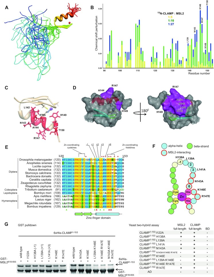
A family of 20 NMR structures of CLAMP87–153 was calculated (Figure 1A) using a set of 357 distance, 40 dihedral angle and 2 hydrogen bond restraints (see Supplementary Table S1 for details) and restrained molecular dynamics (MD) protocol as described in Supplementary Information. Solution structure reveals a well-defined zinc-finger domain of C2H2 type on the C-terminus of CLAMP87–153 (from F127 to E153), whereas the N-terminal portion (from N87 to S126) is disordered (Figure 1A).
Figure 1.
Drosophila protein CLAMP interacts with MSL2 through its N-terminal zinc-finger domain. (A) NMR structure of CLAMP87–153 (PDB: 7NF9). (B) Relative perturbation of CLAMP87–153 residues’ chemical shifts upon interaction with MSL2. Full spectra are shown in the Supplementary Figure S6. (C) Residues with the strongest chemical shifts are shown in red at the CLAMP N-terminal zinc finger structure. (D) The MSL2-contact surface of the CLAMP N-terminal zinc finger. (E) Multiple sequence alignment of CLAMP N-terminal zinc-fingers from various insects. Typical DNA-binding residues are shown, and red asterisks mark the residues displaying the largest chemical shift perturbations. Triangles depict residues subjected to mutagenesis, red represents a negative effect on binding, and green represents no detectable effect. (F) Schematic representation of zinc-finger structure showing the secondary structure, DNA- and MSL2-binding residue positions are depicted. Residues subjected to mutagenesis are shown in red circles. (G) GST-pulldown (left) and yeast two-hybrid assays (right) of the interaction between GST-tagged MSL2618–655 and 6xHis-thioredoxin-tagged CLAMP1–153 bearing point mutations within the zinc-finger domain. AD, activation domain; BD, DNA-binding domain of GAL4 protein. + or – denotes the ability of yeasts to grow on the media without histidine; assay plates are shown in the Supplementary Figure S9.
The CLAMP C2H2 domain structure details are described in Supplementary Figures S2 and S3. The region preceding the zinc finger is conserved in most insects except Hymenopterans (Supplementary Figure S4). CLAMP1–153 was found to be able to interact with full-length CLAMP in a yeast two-hybrid assay (Figure 1G) suggesting the presence of oligomerization domain. However, the CLAMP40–153 itself does not have dimerization activity in vitro as determined by NMR relaxation studies (Supplementary Figure S5). The superposition of the spectra of CLAMP87–153 and CLAMP1–153 indicates that the signals of the 126−153 region have almost the same positions regardless of the lengths of the upstream peptide sequence, while all signals of the 1−125 region are present in the spectral area corresponding to the non-structured polypeptide chain (Supplementary Figure S3).
To identify critical MSL2-binding residues within CLAMP, we performed NMR chemical shift perturbation experiments with 15N-labeled CLAMP40–153 titrated with an excess of unlabeled MSL2618–655 (Figure 1B and C, Supplementary Figure S6). This experiment showed that only the residues within the C2H2 zinc-finger domain exhibit the perturbation of the chemical shifts (Figure 1C and D). The most significant changes were found for the residues located within the alpha-helix of the C2H2 domain: N143 (+5 relative to the alpha-helix), T150 (+12), positively charged residues K146 (+8) and R147 (+9) and less for L142 (+4) and A144 (+6). Most of these residues are highly conserved in insects (Figure 1E, Supplementary Figure S4). We also observed chemical shift changes for H149, involved in zinc-ion binding. In this case, the chemical shift perturbation may reflect the slight conformation change of this residue upon binding the MSL2 peptide sequence. Interestingly, most amino acids involved in MSL2 binding differ from those commonly involved in DNA binding by C2H2 zinc fingers (relative to the alpha-helix: –1, +2, +3, +6 and in some cases +1 (19,20)). We compared amino acid residues of the CLAMP N-terminal zinc finger at DNA-binding positions with their average abundance in C2H2 zinc fingers (Supplementary Figures S7 and S8). Results of the analysis suggest that the CLAMP N-terminal zinc-finger has quite an atypical pattern of DNA-binding residues. It is very likely, that this zinc-finger is not involved in DNA binding (details provided in Supplementary Information). Previous study (12) showed that the cluster of zinc-fingers 2–7 is responsible for CLAMP binding to (GA)n motif.
To confirm the role of the identified amino acids in MSL2 binding, we tested mutant variants of the C2H2 domain for interaction with MSL2618–655 in the GST-pulldown and full-length MSL2 in yeast two-hybrid (Y2H) assays; the ability of CLAMP1–153 to interact with full-length CLAMP served as a control (Figure 1G and Supplementary Figure S9). We engineered single amino acid substitutions of K146 and R147 to oppositely charged glutamate to induce electrostatic repulsion and N143 to alanine. Also, we mutated conserved residues at DNA-binding positions H138 (–1), L139 (+1) and L141 (+3) to alanines (Figure 1F). Residues +2 and +6 are not conserved in CLAMP proteins (Figure 1E). Correct folding of mutant proteins was confirmed with 1D and 2D NMR spectra (Figure S10).
Among seven single amino acid substitutions tested, only L139A and K146E considerably affected the interaction of CLAMP1–153 and MSL2 in Y2H (Figure 1G). In GST-pulldown, only double and triple mutations in CLAMP1–153 displayed strong effects on the interaction with MSL2618–655 (Figure 1G). K146E attenuated the in vitro interaction and adding R147E enhanced this effect. However, adding N143A did not have an additive weakening effect on the interaction in combination with other mutations (Figure 1G). The inability of double mutant CLAMP to interact with MSL2 was also confirmed by 2D NMR demonstrating that MSL2 titrated with CLAMP41–153 L139A/K146E no longer exhibits the same chemical shift perturbations as it does in case of wild-type CLAMP41–153 construct (Supplementary Figure S11).
The N-terminal zinc finger of CLAMP is preceded by sequence NTISNIS conserved in most insects, which may contribute to protein-protein binding; however, no changes in chemical shifts were observed for these residues. We introduced an alanine substitution for conservative isoleucine 122 (I122A), but this mutation did not affect the interaction efficiency (Figure 1G).
Altogether, our results suggest that residues in the alpha-helix of CLAMP N-terminal C2H2 domain display redundancy in MSL2 interactions. Only the substitution of several residues involved in the interaction considerably affects the MSL2 binding.
The CLAMP contact area of the MSL2 protein
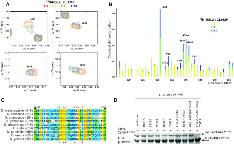
We performed a reverse experiment to understand further the mechanism of CLAMP N-terminal C2H2 domain binding to the unfolded MSL2 peptide. We produced stable isotope-labeled MSL2618–655 protein and performed a backbone resonance assignment using a set of 3D NMR spectra on 15N- and 13C-labelled protein samples. Molecular modeling suggests a possibility to form β-hairpins at V634−N638 and G641−N647, but according to the chemical shift values, MSL2618–655 is mostly unstructured in solution (Supplementary Figure S12), which is also supported by the lack of NOE signals. Upon titration with unlabeled CLAMP40–153 we observed chemical shift perturbation of the certain residues of MSL2618–655 (Figure 2A, relative perturbation is shown in Figure 2B, full spectra in Supplementary Figure S13). We did not observe a significant change in chemical shifts of MSL2618–655 upon binding to CLAMP, which indicates that it does not acquire ordered conformation in the process of complex formation.
Figure 2.
The small unfolded sequence of MSL2 interacts with CLAMP. (A) Sections of 1H–15N MSL2618–655 spectra showing perturbations of chemical shifts of MSL2 residues upon CLAMP binding. Full spectra are shown in the Supplementary Figure S13. (B) Relative perturbation of MSL2 residues’ chemical shifts upon interaction with CLAMP. (C) Multiple sequence alignment of CLAMP-interacting MSL2 region from Drosophila species. Red asterisks show the residues with the largest chemical shift perturbations upon binding to CLAMP. Triangles mark residues subjected for mutagenesis, red represents a negative effect on binding, and green represents no detectable effect. (D) GST-pulldown assay of interaction between GST-tagged MSL2618–655 (represented by *) and 6xHis-thioredoxin-tagged CLAMP1–153.
The NMR spectra measured during titration experiments indicate a fast exchange between free and bound states, which is a consequence of relatively weak protein-protein interaction. For fast exchange between bound and free states (koff >> the frequency change), resonances of labeled protein moved from free to bound positions upon the increase of concentration of unlabeled interacting protein. Analysis of chemical shift perturbation during the titration allowed to estimate the Kd of CLAMP–MSL2 interaction as 0.2 ± 0.1 mM at 25°C (see Supplementary Figure S14 and Supplementary Information for details).
Largest chemical shift changes were observed for both hydrophobic (L633, V634, Y643) and charged clusters of residues (E639, K640), with the most significant change for H631. To further demonstrate the influence of these residues on CLAMP binding, we designed substitutions for conserved residues H631A, L633A, V634A, E639A and Y643A. We utilized combinations of these substitutions and introduced them simultaneously. Unexpectedly, H631A did not affect the interaction even though it displayed the strongest chemical shift perturbation. L633A and V634A had little effect, but E639A, Y643A, and their combination significantly weakened binding of MSL2 to CLAMP in vitro. Furthermore, simultaneous substitutions of four residues (L633A, V634A, E639A and Y643A) resulted in almost complete loss of interaction in the pulldown assay (Figure 2D). These results suggest that the interaction between MSL2 and CLAMP is an additive effect of multiple contacts and allows in the course of evolution to gradually select the most effective combination of amino acids in MSL2 for interaction with the CLAMP N-terminal C2H2 domain.
Almost all residues responsible for the interaction with CLAMP are highly conserved in the MSL2 proteins of various Drosophilids (Figure 2C). However, the full motif is not conserved even in the closest Dipterans (Supplementary Figure S15). This low conservation contrasts with the CLAMP C2H2 domain, which remains highly conserved in all insects. Bees and other social Hymenopterans have a completely different mechanism of sex determination (males are haploid) and thus might not utilize dosage compensation similar to Drosophilids (21). At the same time, the N-terminal C2H2 domain has a high level of homology between CLAMP proteins from Drosophila melanogaster and Apis mellifera. We studied CLAMP–MSL2 interaction in Apis mellifera (hereafter amCLAMP and amMSL2). The amCLAMP and amMSL2 do not interact directly (Supplementary Figure S16A). At the same time, amCLAMP can interact with D. melanogaster MSL2618–655, which is not surprising since most protein-interacting residues are conserved (Figure 1E, Supplementary Figure S16A).
Mutations of the MSL2–CLAMP interface affect DCC recruitment in vivo
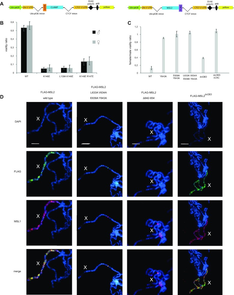
To assess the effect of mutations disrupting the CLAMP:MSL2 contact surface in vivo, we generated transgenic flies expressing 3xHA-tagged CLAMPWT, CLAMPK146E, CLAMPK146E;R147E, and CLAMPL139A;K146E under strong ubiquitin (Ubi63E) promoter (Figure 3A). The transgenes were inserted into the same 86Fb region on the third chromosome, using a φC31 integrase-based integration system (22). Immunoblot analysis showed that all CLAMP variants were expressed at similar levels (Supplementary Figure S17A). To understand the functional roles of the mutations in the C2H2 domain, we used a previously described null mutation in the clamp gene, named clamp2 (23). We examined the ability of transgenes expressing wild-type and mutant proteins to complement the clamp2 mutation (Figure 3B, Supplementary Table S2). Expression of the CLAMPWT protein restored to a greater extent the survival rate of clamp2 flies. At the same time, males and females expressing any of the CLAMP mutants displayed low viability. Thus, the N-terminal domain of C2H2 is required for CLAMP activity, which is not related to its role in dosage compensation.
Figure 3.
The CLAMP–MSL2 interaction is required for the correct recruitment of the dosage compensation complex. (A) Schematic representation of rescue constructs expressing 3xHA-tagged CLAMP 3xFLAG-tagged MSL2 proteins under the control of ubiquitin-p63E promoter; SV40 poly(A)–SV40 polyadenylation signal; attB is the site for φC31-mediated recombination used for site-specific insertion of the construct; yellow represents the intronless yellow gene used as a reporter. (B) Comparison of the viability (relative to clamp2/+ mutant background) of males and females upon the rescue of the clamp2/clamp2 mutant background with CLAMP proteins expressed in heterozygous (k/TM6) and homozygous (k/k) transgenic constructs. Complete data are shown in the Supplementary Table S2. (C) Relative viability of females with ectopic expression of the MSL2 variants. (D) Effect of single amino-acid substitutions and replacement of the CLAMP binding domain (CBD) in the FLAG-tagged MSL2 variants on DCC recruitment shown by immunostaining of polytene chromosomes with anti-FLAG and anti-MSL1 antibodies in females. All staining's are shown in Supplementary Figures S18A and S19. Polytene chromosomes stained with MSL2 antibodies are shown in Supplementary Figure S18B. Scale bar is 20 μm.
To define the contribution in vivo of key amino acids in the CLAMP-interacting region of MSL2, we created transgenic lines expressing different FLAG-tagged MSL2 mutants under control of the Ubi63E promoter in the 86Fb region (Figure 3A). We tested the mutations in the MSL2 that affect interaction with CLAMP in vitro: MSL2Y643A, MSL2E639A;Y643A, and MSL2L633A;V634A;E639A;Y643A. The previously obtained transgenic lines (12) expressing MSL2WT and MSL2Δ642–654 were used as positive and negative controls. Immunoblot analysis showed that all MSL2 variants were present in the transgenic flies at nearly equivalent levels as MSL2WT (Supplementary Figure S17A).
Ectopic expression of MSL2 in females resulted in the assembly of functional DCC (24,25) that led to a strong increase of gene expression, and as a consequence, decreased viability. This model is most sensitive for identifying mutations in MSL2 that affect the specific binding of the MSL complex to the X chromosome. Females carrying homozygous MSL2wt transgenes had low viability (Figure 3C). At the same time, females carrying homozygous transgenes expressing MSL2Δ642–654 or any of the tested mutant versions of MSL2 (MSL2Y643A, MSL2E639A;Y643A and MSL2L633A;V634A;E639A;Y643A) displayed normal viability suggesting that the functional activity of MSL2 is impaired in all mutants (Figure 3C). These results indicate that all mutations in MSL2 negatively influence the assembly of functional DCC in females.
The CLAMP interacting regions in the MSL2 proteins from D. melanogaster and D. virilis (dvMSL2) differ in 10 aa (Figure 2C). However, it was shown that the dvMSL2 interacts with CLAMP more strongly than MSL2 in D. melanogaster (26). To test how the strength of MSL2–CLAMP interaction affects the dosage compensation, we made transgenic line expressing MSL2 variant (MSL2dvCBD) in which the 618−655 aa region is substituted the similar region from dvMSL2. Both MSL2 peptides interact strongly with CLAMP in vitro (Figure S16B). We also constructed transgenic lines expressing MSL2dvCBD+ΔCXC variant with the deletion of CXC domain. Immunoblot analysis showed that both CLAMP variants were expressed at levels equivalent to MSL2wt (Figure S17A). Females carrying homozygous MSL2dvCBD transgenes had low viability suggesting that the chimeric MSL2 is functional (Figure 3C). In contrast MSL2dvCBD+ΔCXC females displayed normal viability, consistent with the important role of the CXC domain in recruitment of the MSL complex on the X chromosome.
Polytene chromosomes in the nuclei of salivary glands are a well-established model system for studying the recruitment and spreading of the MSL complex along the X chromosome (27–30). We used anti-FLAG mouse antibodies to identify tagged MSL2 variants and anti-MSL1 and anti-MSL2 rabbit antibodies to confirm the recruitment of endogenous components of the DCC (Figure 3D, Supplementary Figure S18A). Transgenic expression of MSL2WT in females led to localization to the X-chromosomes of both the MSL2 and MSL1 proteins (12). The deletions in the CLAMP interacting region resulted in an almost complete absence of binding sites for the MSL2Δ642–654 mutant and MSL1 on the female X chromosomes. The Y643A and E639A;Y643A mutations in MSL2 only slightly decreased the binding of the mutant MSL2 variants and MSL1 to the female X chromosomes. Simultaneous mutation of four amino acids (L633A;V634A;E639A;Y643A) led to the same effect as the deletion of the CLAMP interacting region. These results confirm that several residues in MSL2 additively interact with the C2H2 domain of CLAMP. The effectiveness of such interaction depends on the number of residues involved in the formation of a specific CLAMP–MSL2 contact.
Transgenic expression of the chimeric MSL2dvCBD protein in females led to recruiting of the MSL complex on the polytene X chromosome like in females expressing MSL2WT (Figure 3D, Supplementary Figure S19). Thus, the MSL2dvCBD protein retains complete functional activity. Similar to results obtained previously with MSL2ΔCXC (12), in MSL2dvCBD+ΔCXC females, we did not observe binding of the MSL complex on the X chromosome. Taken together the results suggest that the CLAMP interaction regions in the MSL2 proteins from two Drosophila species have the same properties and cannot substitute the absence of the CXC domain.
Recent study (31) has shown that the C-terminal portion of MSL2 is involved in a specific interaction with roX RNAs, which is critical for the specific recruitment of the MSL complex on the X chromosome. In Drosophila, roX2 RNA expression improves the efficiency of binding of the MSL complex to the X chromosome (26,31). Therefore, we asked whether strong expression of roX2 could compensate for the inactivation of the CLAMP interaction region or of the CXC domain upon specific recruitment of the MSL complex on the X chromosome. We generated transgenic line (roX2), in which the roX2 gene under control of the ubiquitin promoter was inserted into the 86Fb region. In roX2/MSL2ΔCXC and roX2/MSL2Δ642–654 females, the expression level of roX2 was nearly similar to WT males (Figure S17B). Next, we examined binding of MSL1 and MSL2 to polytene chromosomes in females roX2/MSL2ΔCXC, roX2/MSL2Δ642–654 and roX2/MSL2L633A;V634A;E639A;Y643A (Figure S20). As it was shown above, expression of both MSL2 variants in females did not result in binding of the MSL proteins on the X chromosome. Simultaneous expressing of these mutants with roX2 RNA only partially restored the recruitment of MSL complex on the X chromosome. These results suggest that roX2 overexpression cannot fully compensate for inactivation of the CXC domain or CLAMP binding region of MSL2. Expression of roX2 improves the binding of all studied MSL2 mutants to the X chromosome to the same extent. Since the CXC domain is not required for the MSL2-roX interaction, it seems likely that the CLAMP interacting region is also not critical for roX2 binding to MSL2.
DISCUSSION
For the first time, this study reveals the features of the highly specific interaction of the C2H2 zinc-finger with an intrinsically disordered polypeptide chain – part of MSL2. According to NMR data this fragment of MSL2 does not become structured upon binding to CLAMP. The N-terminal C2H2 domain of CLAMP at DNA-binding positions contains residues that differ significantly from those typical to the C2H2 domain involved in DNA binding. Single amino-acid substitutions have little effect on the CLAMP–MSL2 interaction suggesting an additive role of multiple bonds in forming a stable and specific interaction. Both hydrophilic and hydrophobic residues are involved in the interaction; notably, strong responses were shown for E639 in MSL2 and K146 and R147 in CLAMP, suggesting electrostatic interactions and possible salt bridge formation. Unexpectedly, whereas MSL2 peptide displayed significant chemical shifts for multiple hydrophobic residues after binding to CLAMP, in the CLAMP we can see perturbations mostly for the polar and charged residues. One possible explanation is that the hydrophobic core of the zinc finger did not undergo structural rearrangements after binding the MSL2, but the same residues formed stable hydrophobic pockets involved in the interaction with hydrophobic MSL2 residues.
All residues involved in the interaction are highly conserved in CLAMP proteins but almost non-conserved in family of MSL2 proteins outside the Drosophila genus, suggesting that the CLAMP N-terminal zinc finger has an important role and most likely is involved in interactions with other factors besides MSL2. Expression of the mutant CLAMP proteins has an equal effect on male and female viability, further supporting this hypothesis. Even Apis mellifera CLAMP can interact with Drosophila MSL2, supporting the assumption of strong evolutionary conservation of the CLAMP N-terminal C2H2 domain. However, we did not observe any interaction between amMSL2 and amCLAMP. Thus, it seems likely that the interaction between CLAMP and MSL2 occurs only in Drosophilidae and closely related species since we were unable to identify MSL2 motifs that can interact with the CLAMP C2H2 domain outside the Drosophila genus (Supplementary Figure S15).
Previously it was suggested that the interaction between MSL2 and the CLAMP C2H2 domain is essential for recruiting of the DCC to the male X chromosome (12). It was also shown that the roX RNAs bind to the C-terminal portion of MSL2, including the CLAMP interaction region (31,32). The roX RNAs and the MSL2 CTD form a stably condensed state that allows specific recruitment of the MSL complex on the X chromosome. Here, we have demonstrated the additive contribution of CLAMP and CXC interacting domains of MSL2 and roX2 RNA to the specific recruitment of the MSL complex to the X chromosome.
CLAMP is a key early developmental pioneer transcription factor involved in maintaining open chromatin and recruiting major transcription factors (10,23,33,34). During early embryogenesis, CLAMP preferentially binds to genomic regions outside HAS (35). The MSL complex initially binds nonspecifically to CLAMP-rich regions throughout the genome. Subsequently, preferential enrichment of the MSL complex and CLAMP occurs at HAS (11,35). Cooperative interaction with DNA and CLAMP allows MSL to compete more effectively with nucleosomes when binding to chromatin (11). Improving the interaction between MSL2 and CLAMP has an evolutionary advantage as it results in more efficient specific binding of the MSL complex to the male X chromosome. It can be assumed that in the course of evolution, there was a gradual increase in the strength of interaction between CLAMP and MSL2 as a result of mutations in the MSL2 region, which is not conservative.
In D. virilis, a species separated from D. melanogaster by 40 million years of evolution, orthologs of MSL2 and CLAMP (dvMSL2 and dvCLAMP) interact much more strongly than in D. melanogaster (32). We have demonstrated that CLAMP interacting regions in MSL2 proteins from both Drosophila species have approximately the same activity in specific recruiting of MSL complex on the X chromosome. Unlike D. melanogaster MSL2, the CXC domain in dvMSL2 does not specifically recognize HAS sites and binds X chromosome and autosomes with the same efficiency (26). It seems likely that in D. virilis, the loss of specific binding of CXC on HAS is compensated by additional domain in dvMSL2 that is involved in specific recruitment of MSL complex on the X chromosome.
In addition to recruiting the MSL complex, MSL2 binds to autosomal promoters of genes involved in patterning and morphogenesis and is required for the proper development of males (36). It can be assumed that the lack of specificity of the interaction between the CXC domain and the HAS allows dvMSL2 to more efficiently bind to autosomal promoters and participate in their regulation in D. virilis.
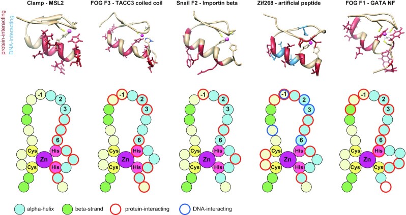
Our results suggest that interactions between C2H2 domains and intrinsically disordered regions may be widespread for creating new protein-protein interactions. However, the availability of structural data supporting the protein binding potential of C2H2 domains is limited. We have summarized the available data for protein-protein interactions mediated by the classical C2H2 domains (Figure 4 and Supplementary Figure S21). The interactions between the C2H2 and CCHC FOG domains with folded protein domains (coiled-coil domain or GATA-type zinc finger) are formed by the alpha-helix, and residues in such interfaces significantly overlap with those involved in DNA-binding (37,38). Zinc fingers of Snail protein also use the DNA-binding side of the alpha-helix for interaction with Importin beta (39), but the set of residues significantly differs from that normally used for nucleic acid binding (Figure 4).
Figure 4.
Comparison of the zinc-finger residues used for protein binding. Cartoons were drawn according to the following structures (PDB ID): 1F2I (Zif268-artificial peptide (40)), 1Y0J (FOG F1–GATA (38)), 1SRK (FOG F3–TACC3 coiled-coil (37)), 3W5K (Snail F2–importin beta (39)). The cartoons for intramolecular interactions mediated by zinc fingers are shown in the Supplementary Figure S21.
In contrast to these complexes, protein-binding residues of the CLAMP N-terminal C2H2 domain are located closer to the C-terminus and at the opposite side of the alpha-helix. NMR spectra demonstrate that the CLAMP-interacting sequences of MSL2 are unfolded. There is only one example of a similar interaction in which an artificial unfolded peptide contacted with the surface of the alpha-helix opposite to that used for DNA binding in the C2H2 domain of Zif268 (40) (Figure 4). This interface of C2H2 also was implicated in multiple contacts with structured protein domains. For example, this interface participates in intermolecular interactions between neighbor C2H2 domains of the GLI protein (41,42) and between the first and third C2H2 domains of Kaiso with their additional C-terminal beta-strands (43) (Supplementary Figure S21). A similar interface is used by some closely related UBZ-type C2H2 fingers for ubiquitin recognition, while other domains of this type use only the distal part of alpha-helix (44,45) (Supplementary Figure S21). These examples demonstrate that protein interactions mediated by non-DNA-binding interfaces of classical zinc fingers can be a common property of these domains. Most of the described interactions involve structured protein domains. Therefore, the recognition of an intrinsically disordered MSL2 peptide by a classical C2H2 zinc finger of CLAMP is the only described naturally occurring interaction of this type. Many other classical C2H2 zinc-finger domains can be involved in such interactions.
DATA AVAILABILITY
Atomic coordinates and structure factors for the reported NMR structure have been deposited with the Protein Data Bank under accession number 7NF9. The 1H, 15N and 13C chemical shifts of CLAMP87–153 and the backbone assignments of MSL2618–655 have been deposited into the BioMagResBank (www.bmrb.wisc.edu) under the accession numbers BMRB-34600 and BMRB-51286, correspondingly.
Supplementary Material
ACKNOWLEDGEMENTS
We thank Erica Larschan for the clamp2 mutation, Farhod Hasanov and Aleksander Parshikov for fly injections, and Olga Kyrchanova for assistance with fly genetics. This study was performed using the equipment of the IGB RAS Core Facilities Centre and Center for shared facilities of Kazan federal university.
Contributor Information
Evgeniya Tikhonova, Department of the Control of Genetic Processes, Institute of Gene Biology, Moscow 119334, Russia.
Sofia Mariasina, Center for Magnetic Tomography and Spectroscopy, Faculty of Fundamental Medicine, M.V. Lomonosov Moscow State University, Moscow 119991, Russia.
Sergey Efimov, NMR Laboratory, Institute of Physics, Kazan Federal University, Kazan 420008, Russia.
Vladimir Polshakov, Center for Magnetic Tomography and Spectroscopy, Faculty of Fundamental Medicine, M.V. Lomonosov Moscow State University, Moscow 119991, Russia.
Oksana Maksimenko, Center for Precision Genome Editing and Genetic Technologies for Biomedicine, Institute of Gene Biology, Moscow 119334, Russia.
Pavel Georgiev, Department of the Control of Genetic Processes, Institute of Gene Biology, Moscow 119334, Russia.
Artem Bonchuk, Department of the Control of Genetic Processes, Institute of Gene Biology, Moscow 119334, Russia; Center for Precision Genome Editing and Genetic Technologies for Biomedicine, Institute of Gene Biology, Moscow 119334, Russia.
SUPPLEMENTARY DATA
Supplementary Data are available at NAR Online.
FUNDING
Russian Foundation for Basic Research [19-04-00933 (NMR experiments), in part]; Russian Science Foundation [21-14-00211 to O.M., E.T. for genetic and immunostaining experiments, 19-74-10099 to A.B. for expression and purification of proteins and their mutants]; Ministry of Science and Higher Education of the Russian Federation [075-15-2019-1661 (analysis of protein-protein interactions)]; Interdisciplinary Scientific and Educational School of Moscow University “Molecular Technologies of the Living Systems and Synthetic Biology” (NMR data analysis). Funding for open access charge: Russian Science Foundation, Ministry of Science and Higher Education of the Russian Federation.
Conflict of interest statement. None declared.
REFERENCES
- 1. Samata M., Akhtar A.. Dosage compensation of the X chromosome: a complex epigenetic assignment involving chromatin regulators and long noncoding RNAs. Annu. Rev. Biochem. 2018; 87:323–350. [DOI] [PubMed] [Google Scholar]
- 2. Kuroda M.I., Hilfiker A., Lucchesi J.C.. Dosage compensation in Drosophila—a model for the coordinate regulation of transcription. Genetics. 2016; 204:435–450. [DOI] [PMC free article] [PubMed] [Google Scholar]
- 3. Lucchesi J.C. Transcriptional modulation of entire chromosomes: dosage compensation. J. Genet. 2018; 97:357–364. [PubMed] [Google Scholar]
- 4. Beckmann K., Grskovic M., Gebauer F., Hentze M.W.. A dual inhibitory mechanism restricts msl-2 mRNA translation for dosage compensation in Drosophila. Cell. 2005; 122:529–540. [DOI] [PubMed] [Google Scholar]
- 5. Straub T., Grimaud C., Gilfillan G.D., Mitterweger A., Becker P.B.. The chromosomal high-affinity binding sites for the Drosophila dosage compensation complex. PLos Genet. 2008; 4:e1000302. [DOI] [PMC free article] [PubMed] [Google Scholar]
- 6. Alekseyenko A.A., Peng S., Larschan E., Gorchakov A.A., Lee O.K., Kharchenko P., McGrath S.D., Wang C.I., Mardis E.R., Park P.J.et al.. A sequence motif within chromatin entry sites directs MSL establishment on the Drosophila X chromosome. Cell. 2008; 134:599–609. [DOI] [PMC free article] [PubMed] [Google Scholar]
- 7. Fauth T., Muller-Planitz F., Konig C., Straub T., Becker P.B.. The DNA binding CXC domain of MSL2 is required for faithful targeting the dosage compensation complex to the X chromosome. Nucleic Acids Res. 2010; 38:3209–3221. [DOI] [PMC free article] [PubMed] [Google Scholar]
- 8. Zheng S., Villa R., Wang J., Feng Y., Becker P.B., Ye K.. Structural basis of X chromosome DNA recognition by the MSL2 CXC domain during Drosophila dosage compensation. Genes Dev. 2014; 28:2652–2662. [DOI] [PMC free article] [PubMed] [Google Scholar]
- 9. Villa R., Schauer T., Smialowski P., Straub T., Becker P.B.. PionX sites mark the X chromosome for dosage compensation. Nature. 2016; 537:244–248. [DOI] [PubMed] [Google Scholar]
- 10. Soruco M.M., Chery J., Bishop E.P., Siggers T., Tolstorukov M.Y., Leydon A.R., Sugden A.U., Goebel K., Feng J., Xia P.et al.. The CLAMP protein links the MSL complex to the X chromosome during drosophila dosage compensation. Genes Dev. 2013; 27:1551–1556. [DOI] [PMC free article] [PubMed] [Google Scholar]
- 11. Albig C., Tikhonova E., Krause S., Maksimenko O., Regnard C., Becker P.B.. Factor cooperation for chromosome discrimination in drosophila. Nucleic Acids Res. 2019; 47:1706–1724. [DOI] [PMC free article] [PubMed] [Google Scholar]
- 12. Tikhonova E., Fedotova A., Bonchuk A., Mogila V., Larschan E.N., Georgiev P., Maksimenko O.. The simultaneous interaction of MSL2 with CLAMP and DNA provides redundancy in the initiation of dosage compensation in drosophila males. Development. 2019; 146:dev179663. [DOI] [PubMed] [Google Scholar]
- 13. Brayer K.J., Kulshreshtha S., Segal D.J.. The protein-binding potential of C2H2 zinc finger domains. Cell Biochem. Biophys. 2008; 51:9–19. [DOI] [PubMed] [Google Scholar]
- 14. Wheeler T.J., Clements J., Finn R.D.. Skylign: a tool for creating informative, interactive logos representing sequence alignments and profile hidden markov models. BMC Bioinf. 2014; 15:7. [DOI] [PMC free article] [PubMed] [Google Scholar]
- 15. Marley J., Lu M., Bracken C.. A method for efficient isotopic labeling of recombinant proteins. J. Biomol. NMR. 2001; 20:71–75. [DOI] [PubMed] [Google Scholar]
- 16. Zolotarev N., Fedotova A., Kyrchanova O., Bonchuk A., Penin A.A., Lando A.S., Eliseeva I.A., Kulakovskiy I.V., Maksimenko O., Georgiev P.. Architectural proteins pita, Zw5,and ZIPIC contain homodimerization domain and support specific long-range interactions in drosophila. Nucleic Acids Res. 2016; 44:7228–7241. [DOI] [PMC free article] [PubMed] [Google Scholar]
- 17. Sabirov M., Kyrchanova O., Pokholkova G.V., Bonchuk A., Klimenko N., Belova E., Zhimulev I.F., Maksimenko O., Georgiev P.. Mechanism and functional role of the interaction between CP190 and the architectural protein pita in drosophila melanogaster. Epigenet. Chromatin. 2021; 14:16. [DOI] [PMC free article] [PubMed] [Google Scholar]
- 18. Shen Y., Delaglio F., Cornilescu G., Bax A.. TALOS plus : a hybrid method for predicting protein backbone torsion angles from NMR chemical shifts. J. Biomol. NMR. 2009; 44:213–223. [DOI] [PMC free article] [PubMed] [Google Scholar]
- 19. Vandevenne M., Jacques D.A., Artuz C., Nguyen C.D., Kwan A.H., Segal D.J., Matthews J.M., Crossley M., Guss J.M., Mackay J.P.. New insights into DNA recognition by zinc fingers revealed by structural analysis of the oncoprotein ZNF217. J. Biol. Chem. 2013; 288:10616–10627. [DOI] [PMC free article] [PubMed] [Google Scholar]
- 20. Gupta A., Christensen R.G., Bell H.A., Goodwin M., Patel R.Y., Pandey M., Enuameh M.S., Rayla A.L., Zhu C., Thibodeau-Beganny S.et al.. An improved predictive recognition model for cys(2)-his(2) zinc finger proteins. Nucleic Acids Res. 2014; 42:4800–4812. [DOI] [PMC free article] [PubMed] [Google Scholar]
- 21. Heimpel G.E., de Boer J.G.. Sex determination in the hymenoptera. Annu. Rev. Entomol. 2008; 53:209–230. [DOI] [PubMed] [Google Scholar]
- 22. Bischof J., Maeda R.K., Hediger M., Karch F., Basler K.. An optimized transgenesis system for drosophila using germ-line-specific phiC31 integrases. Proc. Natl. Acad. Sci. U.S.A. 2007; 104:3312–3317. [DOI] [PMC free article] [PubMed] [Google Scholar]
- 23. Urban J.A., Doherty C.A., Jordan W.T., Bliss J.E., Feng J., Soruco M.M., Rieder L.E., Tsiarli M.A., Larschan E.N.. The essential drosophila CLAMP protein differentially regulates non-coding roX RNAs in male and females. Chromosome Res. 2017; 25:101–113. [DOI] [PMC free article] [PubMed] [Google Scholar]
- 24. Kelley R.L., Wang J., Bell L., Kuroda M.I.. Sex lethal controls dosage compensation in drosophila by a non-splicing mechanism. Nature. 1997; 387:195–199. [DOI] [PubMed] [Google Scholar]
- 25. Kelley R.L., Solovyeva I., Lyman L.M., Richman R., Solovyev V., Kuroda M.I.. Expression of msl-2 causes assembly of dosage compensation regulators on the X chromosomes and female lethality in drosophila. Cell. 1995; 81:867–877. [DOI] [PubMed] [Google Scholar]
- 26. Villa R., Jagtap P.K.A., Thomae A.W., Campos Sparr A., Forne I., Hennig J., Straub T., Becker P.B.. Divergent evolution toward seX chromosome-specific gene regulation in drosophila. Genes Dev. 2021; 35:1055–1070. [DOI] [PMC free article] [PubMed] [Google Scholar]
- 27. Zhou S., Yang Y., Scott M.J., Pannuti A., Fehr K.C., Eisen A., Koonin E.V., Fouts D.L., Wrightsman R., Manning J.E.et al.. Male-specific lethal 2, a dosage compensation gene of drosophila, undergoes sex-specific regulation and encodes a protein with a RING finger and a metallothionein-like cysteine cluster. EMBO J. 1995; 14:2884–2895. [DOI] [PMC free article] [PubMed] [Google Scholar]
- 28. Li F., Schiemann A.H., Scott M.J.. Incorporation of the noncoding roX RNAs alters the chromatin-binding specificity of the drosophila MSL1/MSL2 complex. Mol. Cell. Biol. 2008; 28:1252–1264. [DOI] [PMC free article] [PubMed] [Google Scholar]
- 29. Morra R., Yokoyama R., Ling H., Lucchesi J.C.. Role of the ATPase/helicase maleless (MLE) in the assembly, targeting, spreading and function of the male-specific lethal (MSL) complex of drosophila. Epigenet. Chromatin. 2011; 4:6. [DOI] [PMC free article] [PubMed] [Google Scholar]
- 30. Kelley R.L., Meller V.H., Gordadze P.R., Roman G., Davis R.L., Kuroda M.I.. Epigenetic spreading of the drosophila dosage compensation complex from roX RNA genes into flanking chromatin. Cell. 1999; 98:513–522. [DOI] [PubMed] [Google Scholar]
- 31. Valsecchi C.I.K., Basilicata M.F., Georgiev P., Gaub A., Seyfferth J., Kulkarni T., Panhale A., Semplicio G., Manjunath V., Holz H.et al.. RNA nucleation by MSL2 induces selective X chromosome compartmentalization. Nature. 2021; 589:137–142. [DOI] [PubMed] [Google Scholar]
- 32. Villa R., Jagtap P.K.A., Thomae A.W., Campos Sparr A., Forne I., Hennig J., Straub T., Becker P.B.. Divergent evolution toward seX chromosome-specific gene regulation in drosophila. Genes Dev. 2021; 35:1055–1070. [DOI] [PMC free article] [PubMed] [Google Scholar]
- 33. Urban J.A., Urban J.M., Kuzu G., Larschan E.N.. The drosophila CLAMP protein associates with diverse proteins on chromatin. PLoS One. 2017; 12:e0189772. [DOI] [PMC free article] [PubMed] [Google Scholar]
- 34. Rieder L.E., Koreski K.P., Boltz K.A., Kuzu G., Urban J.A., Bowman S.K., Zeidman A., Jordan W.T., Tolstorukov M.Y., Marzluff W.F.et al.. Histone locus regulation by the drosophila dosage compensation adaptor protein CLAMP. Genes Dev. 2017; 31:1494–1508. [DOI] [PMC free article] [PubMed] [Google Scholar]
- 35. Rieder L.E., Jordan W.T., Larschan E.N.. Targeting of the dosage-compensated male X-Chromosome during early Drosophila development. Cell Rep. 2019; 29:4268–4275. [DOI] [PMC free article] [PubMed] [Google Scholar]
- 36. Valsecchi C.I.K., Basilicata M.F., Semplicio G., Georgiev P., Gutierrez N.M., Akhtar A.. Facultative dosage compensation of developmental genes on autosomes in drosophila and mouse embryonic stem cells. Nat. Commun. 2018; 9:3626. [DOI] [PMC free article] [PubMed] [Google Scholar]
- 37. Simpson R.J., Yi Lee S.H., Bartle N., Sum E.Y., Visvader J.E., Matthews J.M., Mackay J.P., Crossley M.. A classic zinc finger from friend of GATA mediates an interaction with the coiled-coil of transforming acidic coiled-coil 3. J. Biol. Chem. 2004; 279:39789–39797. [DOI] [PubMed] [Google Scholar]
- 38. Liew C.K., Simpson R.J.Y., Kwan A.H.Y., Crofts L.A., Loughlin F.E., Matthews J.M., Crossley M., Mackay J.P.. Zinc fingers as protein recognition motifs: structural basis for the GATA-1/Friend of GATA interaction. Proc. Natl. Acad. Sci. U.S.A. 2005; 102:583–588. [DOI] [PMC free article] [PubMed] [Google Scholar]
- 39. Choi S., Yamashita E., Yasuhara N., Song J., Son S.Y., Won Y.H., Hong H.R., Shin Y.S., Sekimoto T., Park I.Y.et al.. Structural basis for the selective nuclear import of the C2H2 zinc-finger protein snail by importin beta. Acta. Crystallogr. D Biol. Crystallogr. 2014; 70:1050–1060. [DOI] [PubMed] [Google Scholar]
- 40. Wang B.S., Grant R.A., Pabo C.O.. Selected peptide extension contacts hydrophobic patch on neighboring zinc finger and mediates dimerization on DNA. Nat. Struct. Biol. 2001; 8:589–593. [DOI] [PubMed] [Google Scholar]
- 41. Dutnall R.N., Neuhaus D., Rhodes D.. The solution structure of the first zinc finger domain of SWI5: a novel structural extension to a common fold. Structure. 1996; 4:599–611. [DOI] [PubMed] [Google Scholar]
- 42. Pavletich N.P., Pabo C.O.. Crystal structure of a five-finger GLI-DNA complex: new perspectives on zinc fingers. Science. 1993; 261:1701–1707. [DOI] [PubMed] [Google Scholar]
- 43. Buck-Koehntop B.A., Stanfield R.L., Ekiert D.C., Martinez-Yamout M.A., Dyson H.J., Wilson I.A., Wright P.E.. Molecular basis for recognition of methylated and specific DNA sequences by the zinc finger protein kaiso. Proc. Natl. Acad. Sci. U.S.A. 2012; 109:15229–15234. [DOI] [PMC free article] [PubMed] [Google Scholar]
- 44. Bomar M.G., Pai M.T., Tzeng S.R., Li S.S., Zhou P.. Structure of the ubiquitin-binding zinc finger domain of human DNA Y-polymerase eta. EMBO Rep. 2007; 8:247–251. [DOI] [PMC free article] [PubMed] [Google Scholar]
- 45. Suzuki N., Rohaim A., Kato R., Dikic I., Wakatsuki S., Kawasaki M.. A novel mode of ubiquitin recognition by the ubiquitin-binding zinc finger domain of WRNIP1. FEBS J. 2016; 283:2004–2017. [DOI] [PubMed] [Google Scholar]
Associated Data
This section collects any data citations, data availability statements, or supplementary materials included in this article.
Supplementary Materials
Data Availability Statement
Atomic coordinates and structure factors for the reported NMR structure have been deposited with the Protein Data Bank under accession number 7NF9. The 1H, 15N and 13C chemical shifts of CLAMP87–153 and the backbone assignments of MSL2618–655 have been deposited into the BioMagResBank (www.bmrb.wisc.edu) under the accession numbers BMRB-34600 and BMRB-51286, correspondingly.


