Abstract
Background
Esophageal microbiota may influence esophageal squamous cell carcinoma (ESCC) pathobiology. Therefore, we investigated the characteristics and interplay of the esophageal microbiota in ESCC.
Methods
We performed 16S ribosomal RNA sequencing on paired esophageal tumor and tumor-adjacent samples obtained from 120 primarily ESCC patients. Analyses were performed using quantitative insights into microbial 2 (QIIME2) and phylogenetic investigation of communities by reconstruction of unobserved states 2 (PICRUSt2). Species found to be associated with ESCC were validated using quantitative PCR.
Results
The microbial diversity and composition of ESCC tumor tissues significantly differed from tumor-adjacent tissues; this variation between subjects beta diversity is mainly explained by regions and sampling seasons. A total of 56 taxa were detected with differential abundance between the two groups, such as R. mucilaginosa, P. endodontalis, N. subflava, H. Pylori, A. Parahaemolyticus, and A. Rhizosphaerae. Quantitative PCR confirmed the enrichment of the species P. endodontalis and the reduction of H. Pylori in tumor-adjacent tissues. Compared with tumor tissue, a denser and more complex association network was formed in tumor-adjacent tissue. The above differential taxa, such as H. Pylori, an unclassified species in the genera Sphingomonas, Haemophilus, Phyllobacterium, and Campylobacter, also participated in both co-occurrence networks but played quite different roles. Most of the differentially abundant taxa in tumor-adjacent tissues were negatively associated with the epidermal growth factor receptor (EGFR), erb-b2 receptor tyrosine kinase 2 (ERBB2), erb-b2 receptor tyrosine kinase 4 (ERBB4), and fibroblast growth factor receptor 1 (FGFR1) signaling pathways, and positively associated with the MET proto-oncogene, receptor tyrosine kinase (MET) and phosphatase and tensin homolog (PTEN) signaling pathways in tumors.
Conclusion
Alterations in the microbial co-occurrence network and functional pathways in ESCC tissues may be involved in carcinogenesis and the maintenance of the local microenvironment for ESCC.
Supplementary Information
The online version contains supplementary material available at 10.1186/s12885-022-09771-2.
Keywords: Esophageal squamous cell carcinoma, Microbiota, Co-occurrence network, PICRUSt2
Introduction
Esophageal cancer is a malignant cancer and contributes to disease burden, especially in developing countries. In 2020, there were 604,100 new cases of esophageal cancer globally [1]: China had the highest number of incident cases, accounting for up to 53% of new cases [2]. There are two main histological subtypes of esophageal cancer, esophageal squamous cell carcinoma (ESCC) and esophageal adenocarcinoma (EAC), among which ESCC accounts for more than 85% of global esophageal cancer cases [3]. ESCC develops from the squamous epithelial cells that make up the inner lining of the esophagus. The etiology of ESCC is multifactorial and still not well elucidated, which restricts the effective prevention of this disease. Thus, there is an urgent need to clarify the pathogenesis of ESCC and to explore new diagnostic and therapeutic possibilities.
The microbiome is an emerging area in the etiological exploration of various diseases. A balance of the microbiome is strongly associated with a variety of human diseases: the gastrointestinal microbiota plays an indispensable role in maintaining the health of humans [4]. The normal microenvironment is conducive to maintaining the steady state of the esophagus, and dysbiosis of the microbiota can lead to the occurrence and development of ESCC [5, 6]. However, there are insufficient studies on the changes in esophageal microbiota in ESCC, and the findings of these studies are also inconsistent [7, 8]. The earliest findings are that lower microbial richness in the upper digestive tract is independently associated with esophageal diseases [9]. However, these upper digestive microbiota may not colonize the esophagus. Then, Yang investigated the differences in esophageal microbiota in ESCC (n = 18) and patients with physiologically normal esophagus (n = 11) by 16S rRNA profiling [8]. In our previous studies, we detected the esophageal microbiota associated with alcohol consumption (a well-known risk factor for ESCC) [10] and prognosis [11] in ESCC. Shao characterized the microbial communities of paired tumor and nontumor samples from only 67 patients with ESCC in northern China [7]. Though the above studies explored the esophageal microbiota, the sample sizes were too small to find differences in diversity with adequate power [12] and did not take other factors that influenced the microbiota into consideration, including different regions and sampling seasons.
Recently, factors, including the region [13], sampling season [14], and interactions between microbes [15], have been shown to influence microbiota composition. He et al. determined that host location showed the strongest associations with microbiota variations [13]. Moreover, there are significant shifts in human microbiome composition across different seasons [14]. In addition, microbes involved in a variety of behaviors involving complex cooperation and communication may be involved in the development of human diseases, and no studies have reported the interaction network of the esophageal microbiota thus far. Hence, it is essential to consider these factors in microbiome studies.
Therefore, to identify the impact of different regions and sampling seasons on the esophageal mucosal microbiota of ESCC, we conducted a study and performed high-throughput profiling of the esophageal mucosal microbiota in paired tumor and tumor-adjacent tissues in the same ESCC cases. Additionally, we detected the microbial interactions by co-occurrence networks and their functional effects on the host.
Methods
Study population
We performed a hospital-based retrospective study of 120 patients pathologically diagnosed with primary ESCC between February 2013 and October 2017 at Fujian Provincial Cancer Hospital and Zhangzhou Municipal Hospital (Supplementary file 1). Subjects were chosen according to the following criteria. Inclusion criteria: (a) underwent esophagectomy surgery; (b) pathologically diagnosed with primary ESCC; (c) tumor stage clarified with number of harvested lymph nodes (HLN) ≥ 20; (d) undergoing neither preoperative radiotherapy nor chemotherapy; (e) no antibiotic use through preoperative two months; (g) no record of other infectious diseases; and (h) resident of Fujian Province for more than 10 years. Exclusion criteria: (a) incomplete clinicopathological data and nonavailability of tissue samples; (b) metastatic malignancy or recurrent esophageal cancer; (c) received pharmacotherapy (such as oral, intramuscular, and intravenous antibacterial drugs, various probiotics or other drugs affecting the microbiota) within a month. Written informed consent was obtained from all the patients. The study was approved by the Ethics Committee of Fujian Medical University (approval no. 201495).
Demographic and clinical information
Basic information for all the participants was collected through a detailed questionnaire that included sociodemographic status, dietary habits, daily physical activity, smoking status, alcohol consumption, family history of cancer and gastrointestinal symptoms. Clinicopathological features including tumor location, and tumor, node, and metastasis (TNM) stage for each patient were also collected from their respective medical records.
Sample collection and preservation
Paired tumor and tumor-adjacent tissue samples were obtained from each patient immediately after surgical resection in the operating room. The tumor-adjacent tissue samples were from an area at a distance of 3 cm from the tumor tissue. The tissue samples were cut into small pieces, placed in autoclaved cryovials, stored in liquid nitrogen for 1 h, and, then transferred to a − 80 °C refrigerator for storage. All samples were evaluated by pathological hematoxylin-eosin (HE) staining.
Bacterial DNA extraction and 16S rRNA sequencing
The sodium dodecyl sulfate (SDS) method was used to extract bacterial DNA from the samples. The extracted DNA was quantitatively detected by a Qubit fluorometer (Invitrogen, America), and the results were acceptable. Each extraction was performed with a blank buffer control to detect contaminants from either reagents or other unintentional sources. However, the negative controls detected too little DNA to prepare the library and hence were not sequenced.
Amplification of the 16S ribosomal RNA (rRNA) gene used primers targeting regions V3–V4, which included a forward primer (341F: 5′-CCTAYGGGRBGCASCAG-3′) and reverse primer (806R: 5′-GGACTACNNGGGTATCTAAT-3′). The sequencing platform was the HiSeq2500 PE250 (Illumina, America).
Quantitative PCR (qPCR) validation
DNA from samples was diluted 10- and 100-fold, respectively, in PCR grade water to facilitate downstream analysis. Relative abundance was calculated by the ΔCt method. The cycle threshold (Ct) values for P. endodontalis and H. pylori were normalized using a primer set for the total bacteria. The primer sequences for each assay were as follows: P. endodontalis forward primer, 5′-CGGTAATACGGAGGATACG-3′; P. endodontalis reverse primer, 5′-TTCAACGGCAAGACTACA-3′; H. pylori forward primer, 5′-GGTGAGTAACGCATAGGT-3′; H. pylori reverse primer, 5′-CTGATAGGACATAGGCTGAT-3′; Eubacteria 16S forward primer, 5′-GGTGAATACGTTCCCGG-3′; Eubacteria 16S reverse primer, 5′- TACGGCTACCTTGTTACGACTT-3′. The fold difference (2-ΔΔCt) in P. endodontalis and H. pylori abundance in tumor versus tumor-adjacent tissue was calculated by subtracting ΔCttumor from ΔCttumor-adjacent, where ΔCt is the difference in threshold cycle number for the test and reference assay. Amplification was performed with the ABI QuantStudio™ 5 RealTime PCR system (Applied Biosystems) using the following reaction conditions: 30 sec at 95 °C, and 40 cycles of 5 sec at 95 °C and 30 sec at 60 °C.
Total RNA was extracted using TRIzol total RNA isolation reagent (ACCURATE BIOTECHNOLOGY, HUNAN,Co.,Ltd). RNA was used to reverse transcribe cDNA using an Evo M-MLV RT Kit with gDNA Clean for qPCR (ACCURATE BIOTECHNOLOGY, HUNAN,Co.,Ltd). Phosphatase and tensin homolog (PTEN) (forward primer, 5′- GAGGGCCAGGTCATAAATAA-3′, reverse primer, 5′- ACCATAAAATGTAAGCAAGGC-3′) and glyceraldehyde-3-phosphate dehydrogenase (GAPDH) (forward primer, 5′-GCACCGTCAAGGCTGAGAAC-3′, reverse primer, 5′-TGGTGAAGACGCCAGTGGA-3′) mRNA levels were determined using quantitative reverse transcription PCR (qRT-PCR).
Sequence data processing
Raw sequencing data from patients with ESCC were imported into quantitative insights into microbial ecology (QIIME2–2020.02) [16] and processed using the DEBLUR algorithm to denoise and then to infer exact amplicon sequence variants (ASVs). The detailed analysis workflow is presented in Supplementary file 1. To provide better resolution and limit the false discovery rate (FDR) penalty on statistical tests, the low abundance features (with total ASV counts less than 100 or detected in less than 20 samples) were filtered before the differential abundance analysis (955 features left) (Table S1).
Statistical analysis
Questionnaires and clinicopathological data were double-entered into EpiData (version 3.1, Denmark). The demographic and baseline clinical features were displayed using n (%). The individuals’ risk index of ESCC was calculated by variables including age, smoking, drinking, eating speed, hot food, pickled food, and fruit from the questionnaire (Supplementary file 2). All statistical analyses were evaluated using R software (R version 4.0.2), and a two-tailed P < 0.050 was considered statistically significant.
The detailed analyses of microbial diversity are shown in Supplementary file 1. The analysis of composition of microbiomes 2 (ANCOM2) tests [17] were performed to detect the differential abundance in different tissue groups. For the functional prediction, the phylogenetic investigation of communities by reconstruction of unobserved states 2 (PICRUSt2) [18] pipeline was used to generate predictions for EC numbers and MetaCyc pathways. The strength of the edges of the microbial co-occurrence network was assessed by the SparCC algorithm [19], and the interaction network diagram was visualized with Cytoscape [20]. The top hub taxa were assessed by the plugin cytoHubba [21] in Cytoscape. Then, we selected four well-established pathogenesis enzyme genes in ESCC according to Lin [22] from the Kyoto Encyclopedia of Genes and Genomes (KEGG) database [23]. Then, the Spearman correlation was performed to explore the association between the differential taxa and four enzyme genes. The DESeq algorithm [24] was applied to calculate the differential MetaCyc pathways and visualized by volcano plot. Then, the Spearman correlation between differential taxa and differential MetaCyc pathways was calculated.
Results
Basic characteristics of all participants
The demographic and clinical data of the study population are reported in Table S2. A total of 120 ESCC patients were recruited from two independent clinical centers. There were 89 males and 31 females. The median age was 61 years. The majority of the tumors originated from the middle (n = 57) and lower (n = 52) esophagus. Most of the patients had advanced tumors (n = 72). Approximately 85 cases had a high risk index.
Esophageal microbial diversity analysis
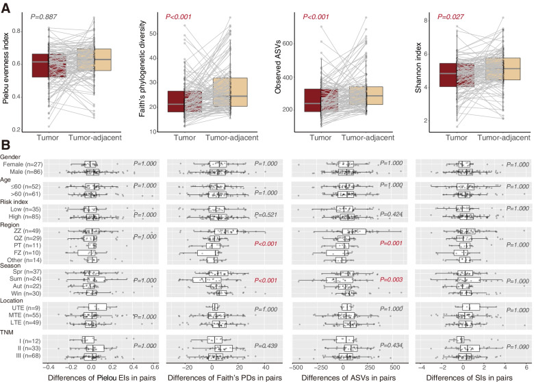
For alpha diversity, four characteristic metrics were evaluated. The alpha diversity in tumor-adjacent tissue was significantly higher than that in tumor tissue, except for Pielou evenness index (Faith’s phylogenetic diversity P < 0.001, observed ASVs P < 0.001, Shannon index P = 0.027) (Fig. 1A). The difference in alpha diversity between paired tumor and tumor-adjacent tissue was only observed in regions (Faith’s phylogenetic diversity P < 0.001, observed ASVs P < 0.001) and sampling seasons (Faith’s phylogenetic diversity P < 0.001, Observed ASVs P = 0.003) (Fig. 1B).
Fig. 1.
Microbial comparison for alpha diversity between tumor and tumor-adjacent tissues. A Alpha diversity based on the Pielou evenness index (P = 0.887), Faith’s phylogenetic diversity (P < 0.001), observed ASVs (P < 0.001), and Shannon index (P = 0.027). B General linear regression analysis to detect pairwise differences in alpha diversity after adjusting for gender, age, risk index, region, sampling season, tumor location, and TNM stage
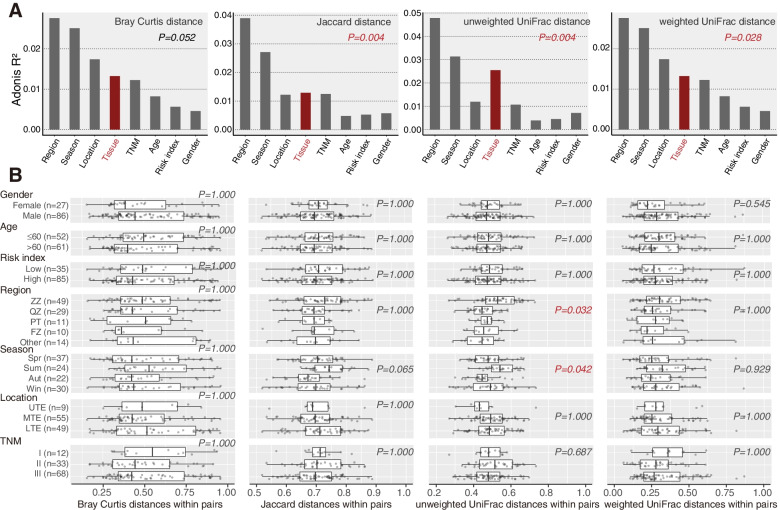
In the multivariate Adonis test, the beta diversity of tumor tissue was significantly different from that of tumor-adjacent tissue, as measured by Jaccard distance (P = 0.004), unweighted UniFrac distance (P = 0.004) or weighted UniFrac distance (P = 0.028) (Fig. 2A). About 1–2% of the variance in beta diversity was explained by tissue type. Moreover, the differences in beta diversity between within paired tumor and tumor-adjacent tissue were associated with regions (P = 0.032) and sampling seasons (P = 0.042) based on unweighted UniFrac distance (Fig. 2B).
Fig. 2.
Microbial comparison for beta diversity using the multivariate Adonis test between tumor and tumor-adjacent tissues. A Beta diversity based on Bray Curtis distance (P = 0.052), Jaccard distance (P = 0.004), unweighted UniFrac distance (P = 0.004), and weighted UniFrac distance (P = 0.028). B General linear regression analysis to detect within-pair differences in beta diversity after adjusting for gender, age, risk index, region, sampling season, tumor location, and TNM stage
Microbial composition analysis
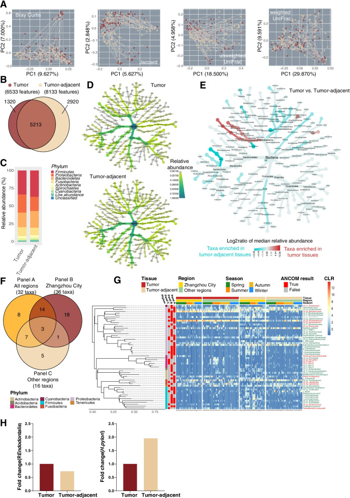
Pairwise Principal coordinate analysis (PCoA) results are displayed in Fig. 3A. Based on Bray Curtis, Jacarrd, and unweighted UniFrac distance, the microbiota from the two tissue types were separated into clusters. A total of 9453 features were found after the sequence was denoised in all samples (Supplementary file 1). More taxa were observed in tumor-adjacent tissue than in tumor tissue (8133 vs. 6533) and approximately 5213 taxa were detected in both tissues (Fig. 3B). The composition of the esophageal microbiota between tumor and tumor-adjacent tissue at the phylum and genus levels is shown in Fig. 3C and Fig. S1, respectively. To elucidate the phylogenetic relationship of the tumor and tumor-adjacent microbiota, the heat trees of microbiota (relative abundance > 0.1%) were plotted, as shown in Fig. 3D. The relative abundance of corresponding branches of bacteria in the phyla Proteobacteria, Bacteroidetes, Fusobacteria and Firmicutes was similar between tumor and tumor-adjacent tissue. However, some bacteria were enriched in different tissues (Fig. 3E). In class Alphaproteobacteria, the branches of bacteria of order Rhizobiales and Sphingomonadales were enriched in tumor tissue, while order Rhodospirillles was higher in tumor-adjacent tissue. Most bacteria of the class Alphaproteobacteria and its branch were enriched in tumor tissue, except for the family Klebsiella. Moreover, the microbiota from phylum TM7 and its branches of bacteria, class Bacilli, family Helicobacteraceae and its genus Helicobacter and its species pylori were higher in tumor-adjacent tissue.
Fig. 3.
The profile of esophageal microbiota between tumor and tumor-adjacent tissues. A PCoA plots based on four distances between tumor and tumor-adjacent tissues. B The overlap of microbiota features between tumor and tumor-adjacent tissues. C Microbial relative abundances at the phylum level in tumor and tumor-adjacent tissues. D Heat tree plot of the relative abundance (higher than 0.1%) of microbiota in tumor and tumor-adjacent tissues. E The heat trees of microbiota for log2 ratio of median relative abundance between tumor and tumor-adjacent tissues by univariate Wilcoxon rank sum test. F The differentially abundant taxa between cancer and para-cancer in all regions (Panel A), Zhangzhou city (Panel B) and other regions (Panel C) after multivariate adjustment using ANCOM2. G Heatmap of the relative abundance of differential microbiota (the red words on the right side of the heatmap are enriched differential taxa in tumor tissue, and the green words are enriched that in tumor-adjacent tissue)
Differential abundance analysis
A total of 56 differential taxa were selected by the ANCOM2 algorithm (Fig. 3G). As the host region exerted the strongest effect on the microbiota, we divided all participants into the Zhangzhou group and other regions group (Table S3). There were 32, 36, and 16 differentially abundant taxa between tumor and tumor-adjacent tissue in the all regions group, the Zhangzhou group and other regions group, respectively (Fig. 3F). There were only three shared differential bacteria in tumor and tumor-adjacent tissue from different regions, named family Enterobacteriaceae, unclassified species from genus Sphingomonas and genus Phyllobacterium (Fig. 3G). It was clearly ascertained that differential bacteria in the phyla Firmicutes and Proteobacteria were enriched in tumor-adjacent tissue from Zhangzhou city. Moreover, we observed an interesting alteration in which the unclassified species in the genus Mycoplane was enriched in tumor tissue from other regions but enriched in tumor-adjacent tissue from Zhangzhou city. Sampling seasons were another powerful factor that influenced host microbiota (Fig. 3G). The microbiota in the phylum Cyanobacteria in tumor tissue sampled in summer had a higher relative abundance. The relative abundance of bacteria in the phylum Proteobacteria was significantly enriched in tumor-adjacent tissue when sampled in spring and summer from Zhangzhou city.
Hence, the dominant candidate differential taxa (Table 1) between tumor and tumor-adjacent tissue were selected according to the grand means of relative abundance that exceeded 0.1% from the above 56 differential taxa. They were species R. mucilaginosa, P. endodontalis, unclassified species in the genus Leptotrichia, unclassified species in the genus Phyllobacterium, and unclassified species in the genus Sphingomonas, which were enriched in tumor tissue. On the other hand, class Bacilli, N. subflava, H. pylori, A. parahaemolyticus, A. rhizosphaerae, unclassified species in the genus Campylobacter and unclassified species in the genus Haemophilus were enriched in tumor-adjacent tissue. Next, to explore the confounding effect of regions, sampling seasons, tumor location and risk index on host microbiota, we added these four covariates into the ANCOM2 analysis. The results showed that the relative abundance of unclassified species in the genus Leptotrichia, unclassified species in the genus Sphingomonas, and A. rhizosphaerae could be influenced by region; the relative abundance of unclassified species in the genus Campylobacter could be influenced by sampling season and tumor location. However, none of the differential taxa were significantly different in either the low- or high-risk indices in ESCC.
Table 1.
The candidate taxa with differential abundance between tumor and tumor-adjacent tissues in ESCCa
| Phylum | Class | Order | Family | Genus | Species | Relative abundance (%)b | FCc | ANCOM’s W (normalized W)d |
Association with other covariatese | ||||
|---|---|---|---|---|---|---|---|---|---|---|---|---|---|
| T | TA | Region | Season | Location | RI | ||||||||
| Actinobacteria | Actinobacteria | Actinomycetales | Micrococcaceae | Rothia | Mucilaginosa | 0.291 | 0.043 | 6.767 | 644 (0.850) | FALSE | FALSE | FALSE | FALSE |
| Bacteroidetes | Bacteroidia | Bacteroidales | Porphyromonadaceae | Porphyromonas | Endodontalis | 0.286 | 0.027 | 10.593 | 465 (0.613) | FALSE | FALSE | FALSE | FALSE |
| Firmicutes | Bacilli | – | – | – | – | 0.009 | 0.482 | 0.019 | 608 (0.802) | FALSE | FALSE | FALSE | FALSE |
| Fusobacteria | Fusobacteriia | Fusobacteriales | Leptotrichiaceae | Leptotrichia | UC | 0.482 | 0.148 | 3.257 | 147 (0.194) | TRUE | FALSE | FALSE | FALSE |
| Fusobacteria | Fusobacteriia | Fusobacteriales | Leptotrichiaceae | Leptotrichia | UC | 0.287 | 0.037 | 7.757 | 686 (0.905) | FALSE | FALSE | FALSE | FALSE |
| Proteobacteria | α-Proteobacteria | Rhizobiales | Phyllobacteriaceae | Phyllobacterium | UC | 8.805 | 0.953 | 9.239 | 678 (0.894) | FALSE | FALSE | FALSE | FALSE |
| Proteobacteria | α-Proteobacteria | Sphingomonadales | Sphingomonadaceae | Sphingomonas | UC | 2.736 | 0.412 | 6.641 | 532 (0.702) | TRUE | FALSE | FALSE | FALSE |
| Proteobacteria | β-Proteobacteria | Neisseriales | Neisseriaceae | Neisseria | Subflava | 0.458 | 0.950 | 0.482 | 608 (0.802) | FALSE | FALSE | FALSE | FALSE |
| Proteobacteria | ε-Proteobacteria | Campylobacterales | Campylobacteraceae | Campylobacter | UC | 0.253 | 0.555 | 0.456 | 686 (0.905) | FALSE | TRUE | TRUE | FALSE |
| Proteobacteria | ε-Proteobacteria | Campylobacterales | Helicobacteraceae | Helicobacter | Pylori | 0.322 | 0.904 | 0.356 | 732 (0.966) | FALSE | FALSE | FALSE | FALSE |
| Proteobacteria | γ-Proteobacteria | Pasteurellales | Pasteurellaceae | Actinobacillus | Parahaemolyticus | 0.685 | 1.452 | 0.472 | 678 (0.894) | FALSE | FALSE | FALSE | FALSE |
| Proteobacteria | γ-Proteobacteria | Pasteurellales | Pasteurellaceae | Haemophilus | UC | 0.068 | 0.171 | 0.398 | 532 (0.702) | FALSE | FALSE | FALSE | FALSE |
| Proteobacteria | γ-Proteobacteria | Pseudomonadales | Moraxellaceae | Acinetobacter | Rhizosphaerae | 0.057 | 0.233 | 0.245 | 644 (0.850) | TRUE | FALSE | FALSE | FALSE |
UC unclassified, T tumor tissue, TA tumor-adjacent tissue, FC fold change, RI risk index
a The candidate differential taxa were selected according to the following two conditions: 1) pass the ANCOM tests conducted in patients from Zhangzhou City (50 pairs) or the other regions (70 pairs), or the pooled population (120 pairs) (Fig. 4G); 2) the grand means of relative abundance were exceeded 0.1%
b Mean relative abundance were presented
c Fold change (FC) = mean relative abundance in tumor/ mean relative abundance in tumor-adjacent tissue
d The differential abundant taxa between tumor and tumor-adjacent tissues were selected by the ANCOM2 algorithm under detected cut-off at 0.7, and were adjusted for sex, age, risk index, TNM, season, tumor location and regions. The normalized ANCOM’s W statistics were calculated by divided the W over the number of total taxa which were identified as none-structural zero
e The ANCOM2 algorithm was applied for association detection. The detected cut-off at 0.7 was adopt for all analyses. The variables included in ANCOM2 comparisons were in line with those included in differential abundant taxa detection. TRUE or FALSE indicated that the relative abundance of candidate taxa could be or not be influenced by specific factors, respectively
qPCR validation of esophageal species differential abundance
To validate the species we identified as differentially abundant in ESCC from our 16S rRNA-based microbiome analyses, we used qPCR to quantify the presence of P. endodontalis and H. pylori. Fold changes of qPCR abundances in tumor versus tumor-adjacent tissue are shown in Fig. 3H. We measured an increase for P. endodontalis (P = 0.320) and a decrease for H. pylori (P = 0.160) in tumor-adjacent samples, demonstrating consistency with the result of the altered abundance of these species based on 16S rRNA-based analyses.
Microbial co-occurrence networks
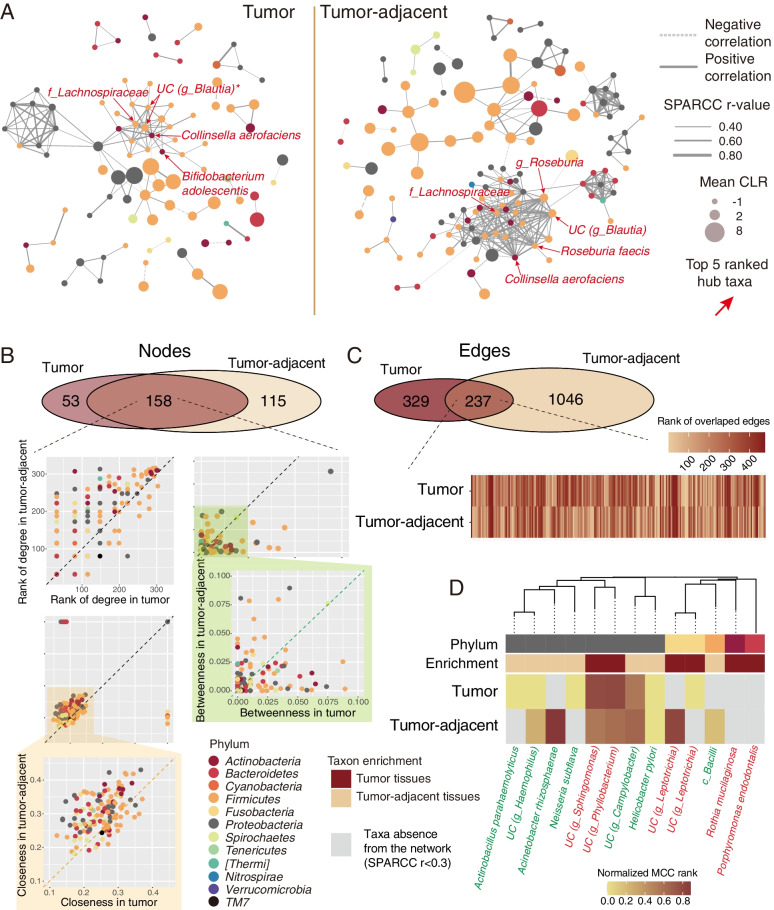
To understand the interaction among esophageal microbiota in tumor and tumor-adjacent tissue, we illustrated the microbial co-occurrence networks of the two groups. There were 3089 positive and 348 negative correlations in the tumor tissue, while the tumor-adjacent samples had 3761 positive and 355 negative correlations. Obviously, the microbial co-occurrence networks were distinct between the tumor and tumor-adjacent tissue (Fig. 4A). However, wide correlations were investigated in the family Lachnospiraceae, species C. aerofaciens and unclassified species in the genus Blautia in both tissue types.
Fig. 4.
The analyses of microbial co-occurrence networks. A Co-occurrence network of the microbiota in tumor and tumor-adjacent tissue (only Sparcc absolute r > =0.4 is shown). B Discrepancies of co-occurrence network nodes between tumor and tumor-adjacent tissue and its measurement dimensions included degree, betweenness, and closeness centrality. C Discrepancies of co-occurrence edges between tumor and tumor-adjacent tissues. D Discrepancies in the importance of differential taxa in the co-occurrence network
To quantify such differences, we counted the number of nodes and their centrality in the microbial networks in different tissue types. As expected, the numbers of interacting microbiota constituents in tumor-adjacent tissue (273 nodes) were higher than those in tumor tissue (201 nodes) (Fig. 4A and B). The centrality of co-occurrence networks was described with three dimensions: degree, betweenness, and closeness centrality. Interestingly, the degree and closeness of shared nodes between tumor and tumor-adjacent tissue were quite different. Next, the edges of the networks were evaluated (Fig. 4C). Despite having a few overlapping edges, the distribution of the rank of overlapping edges varies in the tumor and tumor-adjacent tissue.
To show the importance of the above candidate differential taxa in the network, the heatmap is shown in Fig. 4D. Most of the differentially abundant taxa in the network were from the phylum Proteobacteria, and certain differentially abundant taxa did not emerge in the co-occurrence networks. The roles of differential taxa in different networks (tumor vs. tumor-adjacent tissue) also varied. The discrepancies in microbial co-occurrence networks in the two groups may be attributed to the specific metabolism in different tissue types.
The association between esophageal microbiota and predicted function
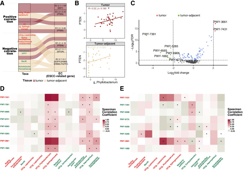
Most of the differentially abundant taxa in tumor-adjacent tissue were negatively associated with EC 2.7.10.1, which regulated the epidermal growth factor receptor (EGFR), erb-b2 receptor tyrosine kinase 2 (ERBB2), erb-b2 receptor tyrosine kinase 4 (ERBB4), and fibroblast growth factor receptor 1 (FGFR1) signaling pathways. In contrast, the differentially abundant taxa in tumor were positively associated with EC2.1.1.107 and EC3.1.3.16 in the MET proto-oncogene, receptor tyrosine kinase (MET) and PTEN signaling pathways respectively (Fig. 5A). Moreover, the expression of PTEN by qRT-PCR was positively correlated with the abundance of unclassified species in genus Phyllobacterium in both tumor (R = 0.22, P = 0.180) and tumor-adjacent tissues (R = 0.37, P = 0.029), which was in line with the predicted results (Fig. 5B). Among the eight MetaCyc metabolic pathways that were significantly different between tumor and tumor-adjacent tissues (Fig. 5C, Table S4), the relative abundance of PWY-3661 and PWY-7431 was increased in the tumor tissue, and six other pathways (PWY-1882, PWY-5265, PWY-6565, PWY-6731, PWY-6906, and PWY-7391) were enriched in tumor-adjacent tissue. Notably, the differentially abundant taxa played various roles in different tissues (Fig. 5D-E). For example, unclassified species from the genera Phyllobacterium and Sphingomonas were positively associated with PWY-6906 and PWY-1882 in tumors but negatively with PWY-6731 and PWY-6565 in tumor-adjacent tissue. Similarly, the correlation was demonstrated between taxa that were A. parahaemolyticus and A. rhizosphaerae and pathways including PWY-7431, PWY-7391, PWY-6731, and PWY-1882 in tumor tissue, but PWY-6565 in tumor-adjacent tissue. In addition, the unclassified species in the genus Haemophilus was only associated with the pathways in tumors.
Fig. 5.
The association between esophageal microbiota and predicted function. A The association between the differential microbiota and ESCC-related functional enzymes. B The correlation between the expression of PTEN by qRT-PCR and the abundance of unclassified species in the genus Phyllobacterium. C Volcano plot shows the differential MetaCyc metabolic pathways between tumor and tumor-adjacent tissues. The X-axis indicates the log2(fold change), and the Y-axis indicates -log10(FDR). The significant pathways enriched in tumor tissue (FDR < 0.05 and log2 fold change> 2) are colored as red dots, that are increased in tumor-adjacent tissue (FDR < 0.05 and log2 fold change<− 2) are colored as green dots. D, E The heatmap indicates the association between the differential MetaCyc metabolic pathways and microbiota in tumor tissue (D) and tumor-adjacent tissue (E)
Discussion
To characterize the esophageal microbiota in ESCC, we compared the microbial diversity and composition of paired tumor and tumor-adjacent tissues for ESCC in this study. Moreover, 56 candidate differential taxa were identified, of which 13 taxa were more abundant. In addition, we suggested that the microbial co-occurrence network was another critical aspect of the microbial community. Furthermore, the relationship between differential taxa and predicted functional ESCC-related gene pathways was investigated.
We observed differences in microbial diversity between the tumor and tumor-adjacent tissues for ESCC, which was similar to the findings of Yang [8] and Yu [9]. The microbial structure is modified due to the altered esophageal microenvironment in the progression of carcinogenesis. Interestingly, in the general linear regression model, the within-pair differences in microbial diversity were only impacted by regions and sampling seasons, which was possibly attributed to regions and sampling seasons explaining most of the observed variations in the diversity measures of esophageal microbiota (Fig. 2A). This is in agreement with many studies that environmental factors including sampling regions or seasons are the relevant factors affecting microbiota [13, 25]. In addition, the microbiota composition varies in response to other factors [26], such as age, sex, diet, nutritional status, environmental factors, lifestyles, and medications. According to Gupta [27], the important role of regions and seasons in microbiota diversity has been attributed to differences in host genetics or other host-specific factors such as innate/adaptive immunity, diet, and environmental exposure. Therefore, further studies involving microbiomics are needed to consider the effect of these factors on the microbiota to ensure generalizability.
Several differential bacteria were detected in tumor-adjacent tissue for ESCC. Of note, it has been reported that R. mucilaginosa upregulated TNF-α, and the upregulation of CD36 in all cell lines in cocultures [28] and the increased abundance of R. mucilaginosa exhibits the ability to produce acetaldehyde [29, 30]. The above results could explain the findings of the present study of a higher R. mucilaginosa in esophageal tumor tissue than that in tumor-adjacent tissue. In accordance with our results, Richard et al. [31] and Liu [32] identified that the tumor mucosal genus Sphingomonas had a particularly high prevalence in colitis-associated and bladder cancer. If the microbiota is involved in carcinogenesis, we can expect that it will be enriched in the tumor-adjacent tissue according to Wang’s hypothesis [33]. Many studies have indicated N. subflava is significantly more abundant in healthy controls, as found in our study [34, 35]. In addition, similar observations previously reported that H. pylori was increased in adjacent compared to tumor tissues for gastric cancer, which is in line with our results [36, 37]. Sandra investigated Campylobacter species that appear to be more prevalent and abundant in the esophagus of patients in Barrett’s esophagus [38], but their abundance decreased with the progression to EAC [39]. Our current study proved this finding in ESCC. Furthermore, we used species-specific qPCR to validate our 16S rRNA-based findings of enrichment of P. endodontalis and reduction of H. pylori in tumor-adjacent tissue, thus providing confidence for our results. At present, there is no other study of esophageal microbiota in ESCC that has taken this additional step of qPCR to corroborate 16S rRNA-based results.
A disease-associated microenvironment could be affected by a specific microbial network. Several studies have shown ecological interaction networks of microbiota in tumor and adjacent mucosal tissues in gastric [37] and colorectal cancer [33]. The esophagus is a part of the gastrointestinal tract, and we highlight the esophageal microbial co-occurrence network for ESCC to extend digestive research. Our microbial co-occurrence network analysis suggests that bacterial compositions in different sample types show specific correlation patterns. Interestingly, strong co-occurrence interactions formed by the family Lachnospraceae, C. aerofaciens, and undefined species from the genus Blautia showed the centralities of these taxa in both networks. This suggests that hub taxa may have a predominant impact on the structure of the microbiota in ESCC patients, which deserves further investigation. Notably, the above differentially abundant taxa also participated in the co-occurrence network and had various degrees of importance. It is implied that the microbial interaction is complex, and full consideration of the microbial interaction network should be taken in further microbiome studies.
We have preliminarily revealed differences in the predicted microbiota functions in tumor and tumor-adjacent tissues. Interestingly, regardless of the presence or absence of mutations in these genes in ESCC, their products (ERBB2 and MET) are often overexpressed in tumor tissues, indicating their vital carcinogenic role in ESCC [22]. It is possible that the abnormal profiled abundance of differential bacteria causes changes in the expression of PTEN, ERBB2, ERBB4, and MET and stimulates tumor growth. Additionally, the enriched differential MetaCyc pathways in tumors are linked to amino acid degradation. It has been implicated that microbiota in dysplasia tissues perturbed amino acid metabolism that is probably involved in the process of tumor development [40]. On the other hand, a large proportion of increased pathways in tumor-adjacent tissue are associated with bacteria-related biomolecule synthesis. This could explain why the more abundant microbiota were detected in tumor-adjacent samples. We found that H. pylori was negatively associated with polyamine biosynthesis, which was in line with Takashima [41], who found investigation that H. pylori significantly inhibited the proliferation of polyamine biosynthesis enzymes in cells. Moreover, Rousseau [42] found that Haemophilus in animals created changes in peptidoglycan metabolism, which is also consistent with our results. To develop a deeper understanding of esophageal carcinogenesis, further studies are needed to examine the significance of microbial functional variations in the ESCC microenvironment.
Association-based research provides limited insight into the role of microbiota in cancer. The microbial community in patients with cancer interacts with their hosts and influences tumor development by a variety of mechanisms. Unraveling the causal relationships between microbiota and cancer, as well as understanding the underlying mechanisms, has become the focus of future research.
In this study, esophageal mucosa samples were obtained from ESCC patients undergoing surgery, thus avoiding possible oral microbial contamination that may occur during upper digestive endoscopy sampling. Our work provides insights into the composition, function and interaction network of the mucosa-associated bacterial community in the tumor microenvironment in ESCC. However, our study had certain limitations. First, our experimental protocol for extracting the DNA of bacteria did not include a bead-beating step. This could lead to an overestimation of the proportion of Gram-negative bacteria in the results. Additionally, this study did not include esophageal tissues from individuals without ESCC for comparison, and the application of these results is limited. In addition, 16S rRNA sequencing only amplified the 16S rRNA-specific region and was less sophisticated than metagenomic sequencing. This is another limitation of the current study.
Conclusion
Compared with tumor-adjacent tissues, the microbiota in tumor tissues showed significant differences in diversity and composition. Alterations in the microbial co-occurrence network and functional pathways in ESCC tissues may be involved in carcinogenesis and the maintenance of the local microenvironment for ESCC. These discoveries of the esophageal microbiota for ESCC patients may contribute to the etiology for ESCC prevention, diagnosis, early intervention, and treatment.
Supplementary Information
Additional file 1: Supplementary File 1. The details of method
Additional file 2: Supplementary File 2. Risk index of esophageal squamous cell carcinoma (ESCC)
Additional file 3: Table S1. The relative abundance of 955 features in ESCC tumor and tumor-adjacent tissues.
Additional file 4: Table S2. Basic information of 120 ESCCs.
Additional file 5: Table S3. The characteristics of ESCC patients from Zhangzhou City (n = 50) and other regions (n = 70).
Additional file 6: Table S4. The corresponding description of the differential MetaCyc metabolic pathways.
Additional file 7: Fig. S1. Microbial relative abundances at the genus level in tumor and tumor-adjacent tissues.
Acknowledgments
This work was supported by a grant from Nature Science Foundation of Fujian Province (No.2021 J01726), Central government-led local science and technology development special project (No.2019 L3006, No.2020 L3009) and Startup Fund for scientific research, Fujian Medical University (No.2020QH2008). There was no potential financial or personal conflict of interest. We are grateful thank to the Fujian Provincial Cancer Hospital Affiliation to Fujian Medical University for data collection.
Abbreviations
- ESCC
Esophageal squamous cell carcinoma
- EAC
Esophageal adenocarcinoma
- HLN
Harvested lymph nodes
- TNM
Tumor, node, and metastasis
- HE
Haematoxylin-eosin
- SDS
Sodium dodecyl sulfate
- QIIME
Quantitative insights into microbial ecology
- ASVs
Amplicon sequence variants
- FDR
False discovery rate
- PICRUSt2
Phylogenetic investigation of communities by reconstruction of unobserved states 2
- qPCR
Quantitative PCR
- Ct
Cycle threshold
- qRT-PCR
quantitative reverse transcription PCR
- ANCOM2
Analysis of composition of microbiomes 2
- KEGG
Kyoto Encyclopedia of Genes and Genomes
- EGFR
Epidermal growth factor receptor;
- ERBB2
Erb-b2 receptor tyrosine kinase 2
- ERBB4
Erb-b2 receptor tyrosine kinase 4
- FGFR1
Fibroblast growth factor receptor 1
- MET
MET proto-oncogene, receptor tyrosine kinase
- PTEN
Phosphatase and tensin homolog
- GAPDH
Glyceraldehyde-3-phosphate dehydrogenase
Authors’ contributions
Zheng Lin: Writing – Review and Editing, Formal analysis, Conceptualization, Methodology and Software. Wenqing Rao: Writing – Original Draft Preparation, Writing – Review and Editing, Conceptualization, Investigation and Methodology. Zhisheng Xiang: Formal analysis, Conceptualization, Investigation and Methodology. Qiaoyan Zeng: Data Curation, Methodology and Investigation. Shuang Liu: Software, Validation and Visualization. Kaili Yu: Investigation and Visualization. Jinsong Zhou: Data Curation and Investigation. Jianwen Wang: Sofware. Weilin Chen: Software. Yuanmei Chen: Resources. Xiane Peng: Methodology and Validation. Zhijian Hu: Methodology, Validation, Writing - Review and Editing, Supervision, Project administration and Funding. All authors read and approved the final manuscript.
Funding
Funding was obtained from Nature Science Foundation of Fujian Province (No.2021 J01726), Central government-led local science and technology development special project (No.2019L3006, No.2020 L3009) and Startup Fund for scientific research, Fujian Medical University (No.2020QH2008).
Availability of data and materials
The datasets analysed during the current study are available from the corresponding author on reasonable request.
Declarations
Ethics approval and consent to participate
All procedures performed in studies involving human participants were carried out by the ethical standards of the institutional and/or national research committee, and with the 1964 Helsinki declaration and its later amendments or comparable ethical standards. Informed consent was obtained from participants, and the study was approved by the Institutional Review Board of Fujian Medical University (number: 201495).
Consent for publication
NO
Competing interests
The author(s) declare that there are no conflicts of interest.
Footnotes
Publisher’s Note
Springer Nature remains neutral with regard to jurisdictional claims in published maps and institutional affiliations.
Zheng Lin and Wenqing Rao have contributed to the work equally.
References
- 1.Sung H, Ferlay J, Siegel RL, Laversanne M, Soerjomataram I, Jemal A, et al. Global cancer statistics 2020: GLOBOCAN estimates of incidence and mortality worldwide for 36 cancers in 185 countries. CA Cancer J Clin. 2021;71(3):209–249. doi: 10.3322/caac.21660. [DOI] [PubMed] [Google Scholar]
- 2.Cao W, Chen H-D, Yu Y-W, Li N, Chen W-Q. Changing profiles of cancer burden worldwide and in China: a secondary analysis of the global cancer statistics 2020. Chin Med J. 2021;134(7):783–791. doi: 10.1097/CM9.0000000000001474. [DOI] [PMC free article] [PubMed] [Google Scholar]
- 3.Arnold M, Laversanne M, Brown LM, Devesa SS, Bray F. Predicting the future burden of esophageal Cancer by histological subtype: international trends in incidence up to 2030. Am J Gastroenterol. 2017;112(8):1247–1255. doi: 10.1038/ajg.2017.155. [DOI] [PubMed] [Google Scholar]
- 4.Fischbach MA. Microbiome: focus on causation and mechanism. Cell. 2018;174(4):785–790. doi: 10.1016/j.cell.2018.07.038. [DOI] [PMC free article] [PubMed] [Google Scholar]
- 5.Yang L, Lu X, Nossa CW, Francois F, Peek RM, Pei Z. Inflammation and intestinal metaplasia of the distal esophagus are associated with alterations in the microbiome. Gastroenterology. 2009;137(2):588–597. doi: 10.1053/j.gastro.2009.04.046. [DOI] [PMC free article] [PubMed] [Google Scholar]
- 6.Liu N, Ando T, Ishiguro K, Maeda O, Watanabe O, Funasaka K, et al. Characterization of bacterial biota in the distal esophagus of Japanese patients with reflux esophagitis and Barrett’s esophagus. BMC Infect Dis. 2013;13:130. doi: 10.1186/1471-2334-13-130. [DOI] [PMC free article] [PubMed] [Google Scholar]
- 7.Shao D, Vogtmann E, Liu A, Qin J, Chen W, Abnet CC, et al. Microbial characterization of esophageal squamous cell carcinoma and gastric cardia adenocarcinoma from a high-risk region of China. Cancer. 2019;125(22):3993–4002. doi: 10.1002/cncr.32403. [DOI] [PMC free article] [PubMed] [Google Scholar]
- 8.Yang W, Chen C-H, Jia M, Xing X, Gao L, Tsai H-T, et al. Tumor-associated microbiota in esophageal squamous cell carcinoma. Front Cell Dev Biol. 2021;9:641270. doi: 10.3389/fcell.2021.641270. [DOI] [PMC free article] [PubMed] [Google Scholar]
- 9.Yu G, Gail MH, Shi J, Klepac-Ceraj V, Paster BJ, Dye BA, et al. Association between upper digestive tract microbiota and cancer-predisposing states in the esophagus and stomach. Cancer Epidemiol Biomark Prev. 2014;23(5):735–741. doi: 10.1158/1055-9965.EPI-13-0855. [DOI] [PMC free article] [PubMed] [Google Scholar]
- 10.Rao W, Lin Z, Liu S, Zhang Z, Xie Q, Chen H, et al. Association between alcohol consumption and oesophageal microbiota in oesophageal squamous cell carcinoma. BMC Microbiol. 2021;21(1):73. doi: 10.1186/s12866-021-02137-x. [DOI] [PMC free article] [PubMed] [Google Scholar]
- 11.Liu Y, Lin Z, Lin Y, Chen Y, Peng X-E, He F, et al. Streptococcus and Prevotella are associated with the prognosis of oesophageal squamous cell carcinoma. J Med Microbiol. 2018;67(8):1058–1068. doi: 10.1099/jmm.0.000754. [DOI] [PubMed] [Google Scholar]
- 12.Casals-Pascual C, González A, Vázquez-Baeza Y, Song SJ, Jiang L, Knight R. Microbial diversity in clinical microbiome studies: sample size and statistical power considerations. Gastroenterology. 2020;158(6):1524–1528. doi: 10.1053/j.gastro.2019.11.305. [DOI] [PubMed] [Google Scholar]
- 13.He Y, Wu W, Zheng H-M, Li P, McDonald D, Sheng H-F, et al. Regional variation limits applications of healthy gut microbiome reference ranges and disease models. Nat Med. 2018;24(10):1532–1535. doi: 10.1038/s41591-018-0164-x. [DOI] [PubMed] [Google Scholar]
- 14.Davenport ER, Mizrahi-Man O, Michelini K, Barreiro LB, Ober C, Gilad Y. Seasonal variation in human gut microbiome composition. PLoS One. 2014;9(3):e90731. doi: 10.1371/journal.pone.0090731. [DOI] [PMC free article] [PubMed] [Google Scholar]
- 15.West SA, Diggle SP, Buckling A, Gardner A, Griffin AS. The social lives of microbes. Annu Rev Ecol Evol Syst. 2007;38(1):53–77. doi: 10.1146/annurev.ecolsys.38.091206.095740. [DOI] [Google Scholar]
- 16.Bolyen E, Rideout JR, Dillon MR, Bokulich NA, Abnet CC, Al-Ghalith GA, et al. Reproducible, interactive, scalable and extensible microbiome data science using QIIME 2. Nat Biotechnol. 2019;37(8):852–857. doi: 10.1038/s41587-019-0209-9. [DOI] [PMC free article] [PubMed] [Google Scholar]
- 17.Mandal S, van Treuren W, White RA, Eggesbø M, Knight R, Peddada SD. Analysis of composition of microbiomes: a novel method for studying microbial composition. Microb Ecol Health Dis. 2015;26:27663. doi: 10.3402/mehd.v26.27663. [DOI] [PMC free article] [PubMed] [Google Scholar]
- 18.Douglas GM, Maffei VJ, Zaneveld JR, Yurgel SN, Brown JR, Taylor CM, et al. PICRUSt2 for prediction of metagenome functions. Nat Biotechnol. 2020;38(6):685–688. doi: 10.1038/s41587-020-0548-6. [DOI] [PMC free article] [PubMed] [Google Scholar]
- 19.Friedman J, Alm EJ. Inferring correlation networks from genomic survey data. PLoS Comput Biol. 2012;8(9):e1002687. doi: 10.1371/journal.pcbi.1002687. [DOI] [PMC free article] [PubMed] [Google Scholar]
- 20.Smoot ME, Ono K, Ruscheinski J, Wang P-L, Ideker T. Cytoscape 2.8: new features for data integration and network visualization. Bioinformatics. 2011;27(3):431–432. doi: 10.1093/bioinformatics/btq675. [DOI] [PMC free article] [PubMed] [Google Scholar]
- 21.Chin C-H, Chen S-H, Wu H-H, Ho C-W, Ko M-T, Lin C-Y. cytoHubba: identifying hub objects and sub-networks from complex interactome. BMC Syst Biol. 2014;8(Suppl 4):S11. doi: 10.1186/1752-0509-8-S4-S11. [DOI] [PMC free article] [PubMed] [Google Scholar]
- 22.Lin D-C, Wang M-R, Koeffler HP. Genomic and Epigenomic aberrations in esophageal squamous cell carcinoma and implications for patients. Gastroenterology. 2018;154(2):374–389. doi: 10.1053/j.gastro.2017.06.066. [DOI] [PMC free article] [PubMed] [Google Scholar]
- 23.Kyoto Encyclopedia of Genes and Genomes database. Kanehisa Laboratories. https://www.kegg.jp
- 24.Love MI, Huber W, Anders S. Moderated estimation of fold change and dispersion for RNA-seq data with DESeq2. Genome Biol. 2014;15(12):550. doi: 10.1186/s13059-014-0550-8. [DOI] [PMC free article] [PubMed] [Google Scholar]
- 25.Rothschild D, Weissbrod O, Barkan E, Kurilshikov A, Korem T, Zeevi D, et al. Environment dominates over host genetics in shaping human gut microbiota. Nature. 2018;555(7695):210–215. doi: 10.1038/nature25973. [DOI] [PubMed] [Google Scholar]
- 26.El-Sayed A, Aleya L, Kamel M. Microbiota's role in health and diseases. Environ Sci Pollut Res Int. 2021;28(28):36967–36983. doi: 10.1007/s11356-021-14593-z. [DOI] [PMC free article] [PubMed] [Google Scholar]
- 27.Gupta VK, Paul S, Dutta C. Geography, ethnicity or subsistence-specific variations in human microbiome composition and diversity. Front Microbiol. 2017;8:1162. doi: 10.3389/fmicb.2017.01162. [DOI] [PMC free article] [PubMed] [Google Scholar]
- 28.Baraniya D, Jain V, Lucarelli R, Tam V, Vanderveer L, Puri S, et al. Screening of health-associated Oral Bacteria for anticancer properties in vitro. Front Cell Infect Microbiol. 2020;10:575656. doi: 10.3389/fcimb.2020.575656. [DOI] [PMC free article] [PubMed] [Google Scholar]
- 29.Moritani K, Takeshita T, Shibata Y, Ninomiya T, Kiyohara Y, Yamashita Y. Acetaldehyde production by major oral microbes. Oral Dis. 2015;21(6):748–754. doi: 10.1111/odi.12341. [DOI] [PubMed] [Google Scholar]
- 30.Amer A, Galvin S, Healy CM, Moran GP. The microbiome of potentially malignant Oral leukoplakia exhibits enrichment for Fusobacterium, Leptotrichia, Campylobacter, and Rothia species. Front Microbiol. 2017;8:2391. doi: 10.3389/fmicb.2017.02391. [DOI] [PMC free article] [PubMed] [Google Scholar]
- 31.Richard ML, Liguori G, Lamas B, Brandi G, da Costa G, Hoffmann TW, et al. Mucosa-associated microbiota dysbiosis in colitis associated cancer. Gut Microbes. 2018;9(2):131–142. doi: 10.1080/19490976.2017.1379637. [DOI] [PMC free article] [PubMed] [Google Scholar]
- 32.Liu F, Liu A, Lu X, Zhang Z, Xue Y, Xu J, et al. Dysbiosis signatures of the microbial profile in tissue from bladder cancer. Cancer Med. 2019;8(16):6904–6914. doi: 10.1002/cam4.2419. [DOI] [PMC free article] [PubMed] [Google Scholar]
- 33.Wang Y, Zhang C, Hou S, Wu X, Liu J, Wan X. Analyses of potential driver and passenger Bacteria in human colorectal Cancer. Cancer Manag Res. 2020;12:11553–11561. doi: 10.2147/CMAR.S275316. [DOI] [PMC free article] [PubMed] [Google Scholar]
- 34.Al-Hebshi NN, Abdulhaq A, Albarrag A, Basode VK, Chen T. Species-level core oral bacteriome identified by 16S rRNA pyrosequencing in a healthy young Arab population. J Oral Microbiol. 2016;8:31444. doi: 10.3402/jom.v8.31444. [DOI] [PMC free article] [PubMed] [Google Scholar]
- 35.Barbadoro P, Ponzio E, Coccia E, Prospero E, Santarelli A, Rappelli GGL, et al. Association between hypertension, oral microbiome and salivary nitric oxide: a case-control study. Nitric Oxide. 2021;106:66–71. doi: 10.1016/j.niox.2020.11.002. [DOI] [PubMed] [Google Scholar]
- 36.Liu X, Shao L, Liu X, Ji F, Mei Y, Cheng Y, et al. Alterations of gastric mucosal microbiota across different stomach microhabitats in a cohort of 276 patients with gastric cancer. EBioMedicine. 2019;40:336–348. doi: 10.1016/j.ebiom.2018.12.034. [DOI] [PMC free article] [PubMed] [Google Scholar]
- 37.Chen X-H, Wang A, Chu A-N, Gong Y-H, Yuan Y. Mucosa-associated microbiota in gastric Cancer tissues compared with non-cancer tissues. Front Microbiol. 2019;10:1261. doi: 10.3389/fmicb.2019.01261. [DOI] [PMC free article] [PubMed] [Google Scholar]
- 38.Macfarlane S, Furrie E, Macfarlane GT, Dillon JF. Microbial colonization of the upper gastrointestinal tract in patients with Barrett’s esophagus. Clin Infect Dis. 2007;45(1):29–38. doi: 10.1086/518578. [DOI] [PubMed] [Google Scholar]
- 39.Kaakoush NO, Castaño-Rodríguez N, Man SM, Mitchell HM. Is Campylobacter to esophageal adenocarcinoma as Helicobacter is to gastric adenocarcinoma? Trends Microbiol. 2015;23(8):455–462. doi: 10.1016/j.tim.2015.03.009. [DOI] [PubMed] [Google Scholar]
- 40.Ilhan ZE, Łaniewski P, Thomas N, Roe DJ, Chase DM, Herbst-Kralovetz MM. Deciphering the complex interplay between microbiota, HPV, inflammation and cancer through cervicovaginal metabolic profiling. EBioMedicine. 2019;44:675–690. doi: 10.1016/j.ebiom.2019.04.028. [DOI] [PMC free article] [PubMed] [Google Scholar]
- 41.Takashima T, Fujiwara Y, Watanabe T, Tominaga K, Oshitani N, Higuchi K, et al. High molecular protein of Helicobacter pylori responsible for inhibition of ornithine decarboxylase activity of human gastric cultured cells. Aliment Pharmacol Ther. 2002;16(Suppl 2):167–173. doi: 10.1046/j.1365-2036.16.s2.20.x. [DOI] [PubMed] [Google Scholar]
- 42.Rousseau N, Dargis M, Gourde P, Beauchamp D, Malouin F. Effect of beta-lactams on peptidoglycan metabolism of Haemophilus influenzae grown in animals. Antimicrob Agents Chemother. 1992;36(10):2147–2155. doi: 10.1128/AAC.36.10.2147. [DOI] [PMC free article] [PubMed] [Google Scholar]
Associated Data
This section collects any data citations, data availability statements, or supplementary materials included in this article.
Supplementary Materials
Additional file 1: Supplementary File 1. The details of method
Additional file 2: Supplementary File 2. Risk index of esophageal squamous cell carcinoma (ESCC)
Additional file 3: Table S1. The relative abundance of 955 features in ESCC tumor and tumor-adjacent tissues.
Additional file 4: Table S2. Basic information of 120 ESCCs.
Additional file 5: Table S3. The characteristics of ESCC patients from Zhangzhou City (n = 50) and other regions (n = 70).
Additional file 6: Table S4. The corresponding description of the differential MetaCyc metabolic pathways.
Additional file 7: Fig. S1. Microbial relative abundances at the genus level in tumor and tumor-adjacent tissues.
Data Availability Statement
The datasets analysed during the current study are available from the corresponding author on reasonable request.


