PLOS ONE logoLink to PLOS ONE
. 2022 Jun 24;17(6):e0269525. doi: 10.1371/journal.pone.0269525

Eradication of avian leukosis virus subgroups J and K in broiler cross chickens by selection against infected birds using multilocus PCR

Alexander M Borodin 1,2, Zhanna V Emanuilova 1, Sergei V Smolov 1, Olga A Ogneva 1, Nina V Konovalova 3, Elena V Terentyeva 3, Natalia Y Serova 4, D N Efimov 5, V I Fisinin 5, Anthony J Greenberg 6,*, Yakov I Alekseev 3,7,*
Editor: Michelle Wille8
PMCID: PMC9231750  PMID: 35749432

Abstract

The avian leukosis virus (ALV) is a serious threat to sustainable and economically viable commercial poultry management world-wide. Active infections can result in more than 20% flock loss, resulting in significant economic damage. ALV detection and elimination from flocks and breeding programs is complicated by high sequence variability and the presence of endogenous virus copies which show up as false positives in assays. Previously-developed approaches to virus detection are either too labor-intensive to implement on an industrial scale or suffer from high false negative or positive rates. We developed a novel multi-locus multiplex quantitative real-time PCR system to detect viruses belonging to the J and K genetic subgroups that are particularly prevalent in our region. We used this system to eradicate ALV from our broiler breeding program comprising thousands of individuals. Our approach can be generalized to other ALV subgroups and other highly genetically diverse pathogens.

Introduction

As the global population continues to grow, agricultural systems must increase production while maintaining sustainable levels of input consumption. To achieve this goal, we must control disease outbreaks in agricultural animal production facilities. Infections cause significant economic harm and contribute to global food insecurity. An important pathogen in poultry is the avian leukosis virus (ALV), a diploid single-stranded RNA virus that belongs to the genus Alpharetrovirus of the Retroviridae family [1]. ALV leads to lymphoid and myeloid leukosis, as well as neoplasia in other tissues. Active infections result in over 20% mortality, lower productivity, and cause significant harm to industrial poultry management [1]. Even subclinical manifestations of ALV lead to decreased productivity and serious revenue loss [2, 3]. Spread of ALV presents significant problems worldwide. Several national eradication programs exist, for example in China, Australia, and Iran.

ALV types are classified into subgroups based on the GP85 envelope protein structure [4, 5]. Viruses from subgroups A through E [2], J [4], and K [5] are specific to chicken. Subgroup F and G viruses are specific to pheasants and are thus not relevant to the present study [6]. With the exception of the endogenous E group they are highly pathogenic. Endogenous virus genomes integrate into host chromosomes in the germline and are transmitted vertically via Mendelian inheritance [2]. ALV can integrate into somatic cell DNA and be induced by stress, leading to reversion of viremia [7]. Viruses belonging to the A, B, and J subgroups are common, while the C and D subgroups are encountered only rarely [2]. The K subgroup has been discovered relatively recently [5, 811]. Some of its variants are significantly pathogenic [1215]. Studying the K subgroup emergence may lead to novel insights into the modes of virus spread and virulence evolution. Eradication programs typically focus on the J subgroup viruses. Russian poultry industry is severely affected by ALV belonging to this subgroup, with 70% of 223 production sites, surveyed in 46 regions, testing positive for anti-subgroup J (and 90% for all subgroups) antibodies [16]. There is evidence that susceptibility to infection has a genetic component, with a major susceptibility locus linked to the ev21 locus. ev12 and ev21 produce complete endogenous viruses and associated with significant reductions in antibody response to ALV. These genes also predispose individuals to ALV shedding [17].

Fundamentally, the virus eradication problem in a breeding program must be solved by preventing vertical transmission to subsequent generations. This has been the foundation of previous approaches [18]. However, the high horizontal transmission rates exhibited by subgroup J viruses necessitates a more intensive screening [19]. The current standard involves serological tests of cloacal and vaginal swabs at 20 weeks, followed by viremic and blood serum ELISA at 22 weeks, a serological test of the first two eggs at 23 weeks, meconium of new-born chicks at 26 weeks, and finally sero-tests of egg albumen at 40 weeks [20]. Previous studies suggest that raising poultry in small groups and identifying infected chicks before hatching prevents horizontal transmission of ALV subgroup A [21] in layers and ALV J in broilers [22]. However, there is evidence that vertical transmission can occur even when specific viral antigens are undetectable [22, 23]. Furthermore, broiler chickens appear to be particularly susceptible to ALV subgroup J horizontal transmission [2], complicating the eradication process.

ALV identification via propagation in CEFs or DF-1 cells is currently the golden standard. This method suffers from severe drawbacks: it takes seven to nine days and requires significant investment in specialized equipment and laboratory space. An ELISA assay against a group-specific ALV p27 antigen is much more widely used. However, it also has several disadvantages, chiefly a high false positive rate due to endogenous virus p27 expression [24] and low sensitivity [2, 18].

DNA-based detection is possible because the RNA genome of the virus is reverse-transcribed upon cell entry, with subsequent host genome integration [25]. However, identifying ALV positive birds is complicated by the background noise coming from endogenous viruses and is compounded by exogenous virus genome variability. Several of published PCR-based protocols suffer from endogenous virus driven false positives. Nevertheless, PCR-based approaches are 15 to 20% more sensitive than culture or ELISA-based protocols [26]. The major difficulty in implementing a PCR protocol is the high variability of the ALV subgroup J target sequence. For example, there is up to 5.1% divergence between gp85 DNA sequences isolated from the same individual [27]. Amino-acid identity among isolates can be as low as 86.2% [28], implying and even higher DNA sequence diversity. We describe a PCR-based system for identification of ALV subgroups A, B, J, and K. We overcame the DNA diversity problem by using multiple loci and probes. We demonstrate the effectiveness of our method by using it as a screening method to identify infected individuals and thus eradicate ALV from our Smena8 broiler cross. Our approach can be easily deployed in typical industrial settings.

Materials and methods

Ethical statement

All samples included in this study were obtained from birds with official records. Sample collection did not involve animal killing and was performed in accordance with national and European regulations. No ethical approval was necessary.

Test system development

We used BLAST (http://www.ncbi.nlm.nih.gov/BLAST) and ClustalW (http://www.genome.jp/tools-bin/clustalw) on sequences obtained from GenBank to find regions of the ALV genome that are conserved within but divergent between subgroups. GenBank entries used as reference sequences for region selection are M37980, HM452341(ALV-A); AF052428, JF826241 (ALV-B), J02342 (ALV-C), D10652 (ALV-D), EF467236, AY013303, AY013304, KC610517 (ALV-E); Z46390, JF951728, JQ935966, HM776937, JX855935, JF932002, KX058878, DQ115805, KX034517, KU997685, HM235668, HM582657(ALV-J), KF746200, KP686143, and GD14LZ (ALV K). Primers and probes for real-time PCR (Table 1) were developed based on published sequences [10, 29, 30] and synthesized by Syntol (Moscow, Russia). We took care to pick primers that did not amplify known endogenous ALV sequences. In particular, EAV-HP elements contain parts of ALV-J envelope gene. We chose primers outside of the common region, thus eliminating EAV-HP amplification.

Table 1. Primers and probes used in this study.

Name* Sequence (5’–3’) Amplicon length, bp Subgroup Purpose
ALVAF GCCACACGGTTCCTCCTTAGA 114 ALV A Multiplex primer
ALVAR CGCAGTACTCACTCCCCATGAA 114 ALV A Multiplex primer
APL (5R6G)TACGGTGG(dT-BHQ1)GACAGCGGATAG-P ALV A Multiplex probe
ALBF1 GGCCGAGGCCTCCCCGAAA 77 ALV B Multiplex primer
ALVBR GTCTCATTAATTTCCTTTGATTGA 77 ALV B Multiplex primer
BPL1G (Cy5)CCCATGTACC(dT-BHQ2)CCCGTGCCTTG-P ALV B Multiplex probe
JFF1F GCCCTGGGAAGGTGAGCAAGA 139 ALV J Multiplex primer
JJR GGAAATAATAACCACGCACACGA 139 ALV J Multiplex primer
JNP (ROX)TCCTCTCGA(dT-BHQ2)GGCAGCAAGGGTGTC-P ALV J Multiplex probe
JJPLN (ROX)CAGCA AGGGTG(dT-BHQ2)CTTCTCCG-P ALV J Multiplex probe
ALVKF CGGAGCATTGACAAGCTTTCAGA 72 ALV K Multiplex primer
ALVKR GTGATTGCGGCGGAGGAGGA 72 ALV K Multiplex primer
KPL (Cy5.5)CCACCTCGTGAG(dT-BHQ2)TGCGGCC-P ALV K Multiplex probe
ALV-JNF [9] TTGCAGGCATTTCTGACTGG 214 ALV J Published primer
ALV-JNR [9] ACACGTTTCCTGGTTGTTGC 214 ALV J Published primer
JCP [9] (FAM)CCTGGGAAGGTGAGCAAGAAGGA-BHQ1 ALV J Published probe
H5 [10, 11] GGATGAGGTGACTAAGAAAG 545 ALV J Published primer
H7 [10, 11] CGAACCAAAGGTAACACACG 545 ALV J Published primer
Probe [10, 11] (FAM)CTCTTTGCAGGCATTTCTGACTGGGC(TAMRA) ALV J Published probe
SEQA-KR CGCGATCCCCACAAATGAGGAAA 443 ALV A Sequencing primer
SEQA-KR CGCGATCCCCACAAATGAGGAAA 253 ALV B Sequencing primer
SEQJF CCCTGGGAAGGTGAGCAAGAA 498 ALV J Sequencing primer
SEQJR CCTTTATAGCACACCGAACCGAA 498 ALV J Sequencing primer
SEQA-KR CGCGATCCCCACAAATGAGGAAA 466 ALV K Sequencing primer
JEF CCTATTCAAGTTGCCTCTGTGGA 72 ALV J LTR primer
JER GCTTGCTCTATTTGGCCGTCAGA 72 ALV J LTR primer
JEP (Cy5)CCATCCGAGC(dT-BHQ2)GCCTCCAGTCC-P ALV J LTR probe

*References provided for previously published primers

DNA isolation and quantitative real-time PCR

We isolated DNA from feathers (one feather per sample) using the M-Sorb-OOM kit from Syntol (catalog # OOM-502). Calamus fragments 0.3 to 0.5 cm long or chick cloacal smears (all birds from the Smena8 cross) were placed in 1.5 mL tubes and treated with 0.4 mL of the lysis buffer at 70°C for 15 minutes while stirring. We separated debris by a three-minute centrifugation at 13 000 rpm. We transferred the supernatant to a fresh tube and continued the isolation protocol according the manufacturer’s instructions. We used 1.5 μL of the resulting DNA solution in each PCR reaction. While screening the breeding population, individuals were re-tested up to eight times, unless determined to be positive and eliminated from the flock.

RNA isolation and reverse-transcription were performed using kits from Syntol (M-Sorb-OOM for extraction and OT-1 for reverse transcription). We used 1 feather from each bird. We confirmed quantitative PCR specificity by amplicon sequencing using primers ALVKF, SEQA-KR, SEQJF, SEQJR, ALVAF, and SEQA-KR on the Nanophore 05 genetic analyzer (Institute for Analytical Instrumentation RAS, St. Petersburg, Russia). Subsequent generations were tested for ALV-J and -K by pooling feathers from 10 birds in one sample.

We ran real-time PCR reactions on an ANK-48 machine (Institute for Analytical Instrumentation RAS, St. Petersburg, Russia). We ran 50 cycles, with the following steps: denaturation step (10 seconds, 93°C), annealing and elongation step (30 s, 55°C). We used 10 μL of the quantitative real-time PCR reaction mix from a commercial kit (M-428, Syntol, Moscow, Russia). Primer concentration was 450 nM and we used 100 nM of each probe per reaction.

ELISA

We used the IDEXX ALV-J Ab Test system (IDEXX Laboratories, Inc., USA) and the Avian leukosis virus antigen test kit from Synbiotics (USA) to perform ELISA on blood plasma samples from randomly selected birds according to the manufacturers’ instructions.

Results and discussion

Multiplex quantitative real-time PCR of common ALV subgroups

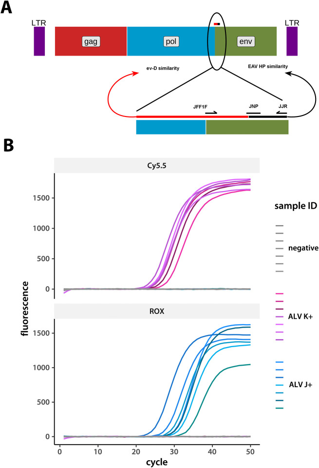
We designed our multiplex test system to identify all common ALV subgroups. We focused on the locus coding for the gp85 coat protein to target amplification of viruses of the A, B, J, and K subgroups (Table 1). Fig 1 shows primer positions and typical real-time fluorescence curves. We used synthetic DNA fragments corresponding to each subgroup, as well as field samples, as positive controls during test system development (Table 1). We performed serial ten- and subsequently two-fold dilutions of the positive control samples to estimate assay sensitivity and employed a previously described procedure [30, 31] to estimate sensitivity from our serial dilution data. Our essays can detect 25 genome-equivalents of A and B subgroup viruses, and 10 genome-equivalents of J and K.

Fig 1. Specificity of the developed real-time PCR system for detecting ALV J and K.

Fig 1

A. Primer positioning around the ev-D/EAV-HP (GenBank accession numbers DQ500016 and AC270426) similarity breakpoint in the ALV J gene that codes for the gp85 coat protein. A diagram of the full ALV genome is on top, with the region surrounding primer placement shown in detail below. B. A representative set of detection curves from the Cy5.5 and ROX channels (the KPL and JJPLN probes, respectively, in Table 1). Each curve is a sample from a separate bird. Eight ALV J positive, ALV K positive, and negative samples each were measured on the same 96-well plate and fluorescence values recorded simultaneously. Data for this graph are included as the S1 File.

To verify the performance of our system, we compared quantitative PCR threshold cycles obtained with the set of primers and probes specific to the J subgroup to previously published results (Table 1) [26, 29, 32]. We only used PCR methods for comparison since other approaches are unacceptably invasive for our application. Field isolates from poultry production facilities in the Moscow region were used for the latter estimates. We successfully verified our results using the Qin L system [26]. This system can detect fewer than 10 virus genome copies per reaction. We made the published primers ourselves (Table 1) and used them to replicate the previously described essays [26]. In our hands, the widely used primers H5 and H7 that detect the J subgroup genomes [29, 32] exhibit 100-fold lower sensitivity, consistent with previous reports [26]. Negative controls, either empty PCR buffer or specific pathogen free chicken DNA, gave no detectable signal after 50 cycles. We tested our identification system on a sample of 1200 individuals randomly picked from DNA isolates of four base lines used to initiate the Smena8 broiler cross. We found that up to 52% of all individuals in our flock were positive for the subgroup J ALV, while up to 10% carried subgroup K (some birds were double-infected). We did not detect any subgroup A or B cases. Previous studies [1] reported around 20% mortality in flocks affected by ALV. Given that not all infected birds die, our prevalence estimates appear to be in line with these previous observations.

We then moved on to analyze DNA from several tissues (feathers, liver, spleen, and blood) from the following regions in Russia: Moscow, Orenburg, Chelyabinsk, Kemerovo, Tyumen, Kaliningrad, Leningrad, Sverdlovsk, Novgorod, and Krasnodar. Samples collected in the Kaliningrad, Leningrad, Sverdlovsk, and Novgorod regions tested positive for K subgroup ALV. We found subgroup A infected individuals in the Leningrad region, while subgroup J positive samples were identified in areas surrounding Moscow, Sverdlovsk, and Leningrad. We did not detect any subgroup B positive samples, suggesting that this ALV variant was absent from Russia during the testing period. We confirmed our test system specificity by sequencing amplicons obtained from positive samples using primers described in Table 1. We did not see any subgroup misidentification.

Multi-locus multiplex quantitative PCR and its use in ALV-J and K eradication

Having established a robust assay, we set out to test its effectiveness in eradicating the subgroup J and K infections we uncovered in our Smena8 broiler cross. To increase assay reliability, we implemented a multiplex approach, adding an extra probe specific to the gp85 gene (JJPLN) and another that targets the long terminal repeat (LTR) sequence. Both probes are specific to the J subgroup viruses (Table 1). We succeeded in eradicating the virus infection by the 77th generation of the selection process.

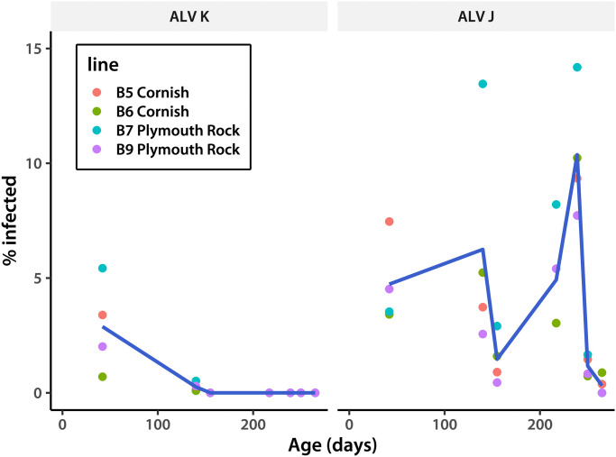
We started by employing our J-subgroup specific PCR system for four generations of the Smena8 broiler cross (Table 1). High variability of the J genomes led us to implement the multi-locus multiplex system mentioned above, containing an additional probe (JJPLN, Table 1). This resulted in an average 2.5% increase in virus identification rates. We also introduced an additional amplification of a subgroup J-specific LTR fragment (Table 1), further increasing identification rates. The latter step allows us to see additional J subgroup variants. Managing the time interval between successive tests proved crucial. Increasing the between-test duration to 62 days between cycle four and five resulted in a two-fold jump in infection rates. Therefore, from that point on testing was done no less frequently than once every two weeks. As a result, we no longer detected subgroup J viruses after cycle eight of the program in two Smena8 broiler cross lines (B7 and B9), while infection rates in two others were minimal (Table 2 and Fig 2). This in contrast to about 52% ALV subgroup J positive rate at the start of the program. Subgroup K viruses disappeared by cycle four, suggesting that they are easier to eradicate. Chickens from the next generation of the Smena8 broiler cross showed several ELISA-positive tests at day 267 after hatching. However, none of these were PCR-positive. We re-tested individuals from the generation after that at day 360 and found no positives by either PCR or ELISA. We saw no clinical manifestations of leukosis in the three years since. A handful of suspected cases proved to be PCR-negative.

Table 2. ALV eradication in the 77th generation of Smena8 broiler cross pure lines.

Pure line Age (days) Livestock Number infected ALV K/J ratio % infected
Line B5 Cornish 1 1886 208 N/A* 11.0
42 1032 112 35/77 10.9
140 804 31 1/30 3.9
155 775 7 0/7 0.9
217 757 23 0/23 3.0
239 610 57 0/57 9.3
250 548 8 0/8 1.5
265 531 2 0/2 0.4
Line B6 Cornish 1 2358 118 N/A* 5.0
42 1142 47 8/39 4.1
140 1088 58 1/57 5.3
155 939 15 0/15 1.6
217 923 28 0/28 3.0
239 772 79 0/79 10.2
250 687 5 0/5 0.7
265 682 6 0/6 1.0
Line B7 Plymouth Rock 1 2319 134 N/A* 5.8
42 1216 109 66/43 9.0
140 966 135 5/130 14.0
155 860 25 0/25 2.9
217 841 69 0/69 8.2
239 684 97 0/97 14.2
250 602 10 0/10 1.7
265 585 0 0/0 0.0
Line B9 Plymouth Rock 1 2466 67 N/A* 2.7
42 1438 94 29/65 6.5
140 1367 39 4/35 2.9
155 1118 5 0/5 0.4
217 1073 58 0/58 5.4
239 919 71 0/71 7.7
250 845 7 0/7 0.8
265 823 0 0/0 0.0

*N/A—data are not available

Fig 2. Prevalence of infected individuals over time.

Fig 2

Data for ALV K and J infections are shown on separate panels. Lines trace among-line means, calculated for each day separately. The data for this plot are in Table 2.

Conclusions

The avian leukosis virus is a serious threat to sustainable and economically viable commercial poultry management world-wide. As an RNA virus, it is highly variable, making reliable detection difficult. We developed a multi-locus multiplex quantitative real-time PCR system to identify individuals infected with subgroup J and K viruses with high sensitivity and specificity. We demonstrate the effectiveness of our approach by quickly eliminating both ALV J and K subgroups from our breeding program.

Our innovation is to couple quantitative PCR of multiple genes and the use of several probes per locus to increase sensitivity in the face of high genetic variation. In addition, using pulped feathers as the DNA source makes the assay non-invasive, cheap, and easy to implement [3335]. Preliminary data indicate that we can get an almost two order of magnitude sensitivity increase when we assay ALV J cDNA, suggesting that future assay development can improve on the current approach, albeit perhaps at the cost of increased labor. Our experience in successfully eliminating ALV from our population comprising thousands of individuals demonstrates that the proposed system has adequate sensitivity and specificity. The procedure may be further simplified by screening only parents selected for establishment of the next generation. This multilocus multiplex approach with multiple probes can also be used to detect retro- and coronaviruses, as well as any other highly variable microorganisms.

Supporting information

S1 File

(CSV)

Acknowledgments

The authors are grateful to the two anonymous reviewers for comments that helped improve the manuscript.

Data Availability

All relevant data are within the manuscript.

Funding Statement

ZhVE, AMB, SVS: Ministry of Science and Higher Education of the Russian Federation, the state assignment for Breeding and Genetic Center Smena No. 075-01297-20-00. https://minobrnauki.gov.ru/ The funders had no role in study design, data collection and analysis, decision to publish, or preparation of the manuscript.

References

  • 1. Gao L, Qin L, Pan W, Wang Y, Lee QX, Gao H, et al. Avian leukosis virus subgroup J in layer chickens, China. Emerg Infect Dis. 2010;16(10):1637–8. doi: 10.3201/eid1610.100780 [DOI] [PMC free article] [PubMed] [Google Scholar]
  • 2. Payne L. Retrovirus-induced disease in poultry. Poult Sci. 1998;77(8):1204–1212. doi: 10.1093/ps/77.8.1204 [DOI] [PubMed] [Google Scholar]
  • 3. Bacon LD, Fulton JE, Kulkarni GB. Methods for evaluating and developing commercial chicken strains free of endogenous subgroup E avian leukosis virus. Avian Pathol. 2004;33(2):233–243. doi: 10.1080/0307943042000195731 [DOI] [PubMed] [Google Scholar]
  • 4. Payne L, Brown S, Bumstead N, Howes K, Frazier J, Thouless M. A novel subgroup of exogenous avian leukosis virus in chickens. J Gen Virol. 1991;72:801–807. doi: 10.1099/0022-1317-72-4-801 [DOI] [PubMed] [Google Scholar]
  • 5. Li X, Lin W, Chang S, Zhao P, Zhang X, Liu Y, et al. Isolation, identification and evolution analysis of a novel subgroup of avian leukosis virus isolated from a local Chinese yellow broiler in South China. Arch Virol. 2016;161(10):2717–25. doi: 10.1007/s00705-016-2965-x [DOI] [PubMed] [Google Scholar]
  • 6. Fujita DJ, Chen YC, Friis RR, Vogt PK. RNA tumor viruses of pheasants: Characterization of avian leukosis subgroups F and G. Virology. 1974;60(2):558–571. doi: 10.1016/0042-6822(74)90350-X [DOI] [PubMed] [Google Scholar]
  • 7. Pandiri A, Gimeno I, Mays J, Reed W, Fadly A. Reversion to subgroup J avian leukosis virus viremia in seroconverted adult meat-type chickens exposed to chronic stress by adrenocorticotrophin treatment. Avian Dis. 2012;56(3):578–82. doi: 10.1637/9949-092611-ResNote.1 [DOI] [PubMed] [Google Scholar]
  • 8. Nakamura S, Ochiai K, Hatai H, Ochi A, Sunden U, Umemura T. Pathogenicity of avian leukosis viruses related to fowl glioma-inducing virus. Aian Pathol. 2011;40(5):499–505. [DOI] [PubMed] [Google Scholar]
  • 9. Cui N, Su S, Chen Z, Zhao X, Cui Z. Genomic sequence analysis and biological characteristics of a rescued clone of avian leukosis virus strain JS11C1, isolated from indigenous chickens. J Gen Virol. 2014;95(11):2512–22. doi: 10.1099/vir.0.067264-0 [DOI] [PubMed] [Google Scholar]
  • 10. Shao H, Wang L, Sang J, Li T, Liu Y, Wan Z, et al. Novel avian leukosis viruses from domestic chicken breeds in mainland China. Arch Virol. 2017;. doi: 10.1007/s00705-017-3344-y [DOI] [PubMed] [Google Scholar]
  • 11. Chang S, Hsu M, Wang C. Gene detection, virus isolation, and sequence analysis of avian leukosis viruses in Taiwan country chickens. Avian Dis. 2013;57(2):172–177. doi: 10.1637/10387-092612-Reg.1 [DOI] [PubMed] [Google Scholar]
  • 12. Lv L, Li T, Hu M, Deng J, Liu Y, Xie Q, et al. A recombination efficiently increases the pathogenesis of the novel K subgroup of avian leukosis virus. Vet Microbiol. 2019;231:214–217. doi: 10.1016/j.vetmic.2019.03.021 [DOI] [PubMed] [Google Scholar]
  • 13. Su Q, Li Y, Cui Z, Chang S, Zhao P. The emerging novel avian leukosis virus with mutations in the pol gene shows competitive replication advantages both in vivo and in vitro. Emerg Microbes Infect. 2018;7(1):117. doi: 10.1038/s41426-018-0111-4 [DOI] [PMC free article] [PubMed] [Google Scholar]
  • 14. Liang X, Gu Y, Chen X, Li T, Gao Y, Wang X, et al. Identification and characterization of a novel natural recombinant avian leucosis virus from Chinese indigenous chicken flock. Virus Genes. 2019;55(5):726–733. doi: 10.1007/s11262-019-01695-7 [DOI] [PubMed] [Google Scholar]
  • 15. Borodin AM, Alekseev YI, Konovalova NV, Terentyeva EV, Efimov DN, Emanuilova ZV, et al. Detection of avian leukosis virus subgroup K in Russia and its molecular genetic analysis. Agricultural Biology (in Russian). 2018;53(4):842–850. [Google Scholar]
  • 16. Plotnikov V, Grebennikova T, Dudnikova EK, Shulpin MI, Lazareva SP, Nikonova ZB, et al. About spreading the avian leukosis viruses in poultry farms in Russian Federation. Agricultural Biology (in Russian). 2013;6:36–42. [Google Scholar]
  • 17. Gavora J, Spencer J, Benkel B, Gagnon C, A Emsley A, Kulenkamp A. Endogenous viral genes influence infection with avian leukosis virus. Avian Pathol. 1995;24(4):653–664. doi: 10.1080/03079459508419105 [DOI] [PubMed] [Google Scholar]
  • 18. Lloyd Spencer J. Progress towards eradication of lymphoid leukosis viruses—A review. Avian Pathol. 1984;13(4):599–619. doi: 10.1080/03079458408418560 [DOI] [PubMed] [Google Scholar]
  • 19. Venugopal K. Avian leukosis virus subgroup J: a rapidly evolving group of oncogenic retroviruses. Res Vet Sci. 1999;67(2):113–119. doi: 10.1053/rvsc.1998.0283 [DOI] [PubMed] [Google Scholar]
  • 20. Payne LN. HPRS-103: A retrovirus strikes back. The emergence of subgroup J avian leukosis virus. Avian Pathol. 1998;27(S1):S36–S45. doi: 10.1080/03079459808419291 [DOI] [Google Scholar]
  • 21. Fadly AM, Okazaki W, Witter RL. Hatchery-related contact transmission and short-term small-group-rearing as related to lymphoid leukosis virus eradication programs. Avian Dis. 1981;25:667–677. doi: 10.2307/1589997 [DOI] [PubMed] [Google Scholar]
  • 22. Witter RL, M FA. Reduction of horizontal transmission of avian leukosis virus subgroup J in broiler breeder chickens hatched and reared in small groups. Avian Pathol. 2001;30:641–654. doi: 10.1080/03079450120092134 [DOI] [PubMed] [Google Scholar]
  • 23. Ignjatovic J. Congenital transmission of avian leukosis virus in the absence of detectable shedding of group specific antigen. Aust Vet J. 1990;67:299–301. doi: 10.1111/j.1751-0813.1990.tb07801.x [DOI] [PubMed] [Google Scholar]
  • 24. Peng H, Qin L, Bi Y, Wang P, Zou G, Li J, et al. Rapid detection of the common avian leukosis virus subgroups by real-time loop-mediated isothermal amplification. Virol J. 2015;12:195. doi: 10.1186/s12985-015-0430-1 [DOI] [PMC free article] [PubMed] [Google Scholar]
  • 25. Whitcomb J, Hughes S. Retroviral reverse transcription and integration: progress and problems. Annu Rev Cell Biol. 1992;8:275–306. doi: 10.1146/annurev.cb.08.110192.001423 [DOI] [PubMed] [Google Scholar]
  • 26. Qin L, Gao Y, Ni W, Sun M, Wang Y, Yin C, et al. Development and Application of Real-Time PCR for Detection of Subgroup J Avian Leukosis Virus. J Clin Microbiol. 2013;51(1):149–54. doi: 10.1128/JCM.02030-12 [DOI] [PMC free article] [PubMed] [Google Scholar]
  • 27. Meng F, Li X, Fang J, Gao Y, Zhu L, Xing G, et al. Genomic diversity of the Avian leukosis virus subgroup J gp85 gene in different organs of an infected chicken. J Vet Sci. 2016;17(4):497–503. doi: 10.4142/jvs.2016.17.4.497 [DOI] [PMC free article] [PubMed] [Google Scholar]
  • 28. Wang P, Lin L, Li H, Yang Y, Huang T, Wei P. Diversity and evolution analysis of glycoprotein GP85 from avian leukosis virus subgroup J isolates from chickens of different genetic backgrounds during 1989–2016: Coexistence of five extremely different clusters. Arch Virol. 2017;163(2):377–389. doi: 10.1007/s00705-017-3601-0 [DOI] [PubMed] [Google Scholar]
  • 29. Smith L, Brown S, Howes K, McLeod S, Arshad S, Barron G, et al. Development and application of polymerase chain reaction (PCR) tests for the detection of subgroup J avian leukosis virus. Virus Res. 1998;54(1):87–98. doi: 10.1016/S0168-1702(98)00022-7 [DOI] [PubMed] [Google Scholar]
  • 30. Yang S, Lin S, Kelen G, Quinn T, Dick J, Gaydos C, et al. Quantitative multiprobe PCR assay for simultaneous detection and identification to species level of bacterial pathogens. J Clin Microbiol. 2002;40(9):3449–54. doi: 10.1128/JCM.40.9.3449-3454.2002 [DOI] [PMC free article] [PubMed] [Google Scholar]
  • 31. Lamien C, Lelenta M, Goger W, Silber R, Tuppurainen E, Matijevic M, et al. Real time PCR method for simultaneous detection, quantitation and differentiation of capripoxviruses. J Virol Methods. 2011;171(1):134–40. doi: 10.1016/j.jviromet.2010.10.014 [DOI] [PubMed] [Google Scholar]
  • 32. Kim Y, Gharaibeh S, Stedman N, Brown T. Comparison and verification of quantitative competitive reverse transcription polymerase chain reaction (QC-RT-PCR) and real time RT-PCR for avian leukosis virus subgroup J. J Virol Methods. 2002;102(1–2):1–8. doi: 10.1016/S0166-0934(01)00372-X [DOI] [PubMed] [Google Scholar]
  • 33. Sung H, Reddy S, Fadly A. High virus titer in feather pulp of chickens infected with subgroup J avian leukosis virus. Avian Dis. 2002;46(2):281–286. doi: 10.1637/0005-2086(2002)046[0281:HVTIFP]2.0.CO;2 [DOI] [PubMed] [Google Scholar]
  • 34. Zavala G, Jackwood M, Hilt D. Polymerase chain reaction for detection of avian leukosis virus subgroup J in feather pulp. Avian Dis. 2002;46(4):971–8. doi: 10.1637/0005-2086(2002)046[0971:PCRFDO]2.0.CO;2 [DOI] [PubMed] [Google Scholar]
  • 35. Davidson I, Borenshtain R. The feather tips of commercial chickens are a favorable source of DNA for the amplification of Marek’s disease virus and avian leukosis virus, subgroup J. Avian Pathol. 2002;31(3):237–40. doi: 10.1080/03079450220136549 [DOI] [PubMed] [Google Scholar]

Decision Letter 0

Michelle Wille

Transfer Alert

This paper was transferred from another journal. As a result, its full editorial history (including decision letters, peer reviews and author responses) may not be present.

15 Dec 2021

PONE-D-20-34696Complete eradication of avian leukosis virus subgroups J and K using multilocus qRTPCR in broiler cross chickensPLOS ONE

Dear Dr. Greenberg,

Thank you for submitting your manuscript to PLOS ONE. After careful consideration, we feel that it has merit but does not fully meet PLOS ONE’s publication criteria as it currently stands. Therefore, we invite you to submit a revised version of the manuscript that addresses the points raised during the review process.

I would encourage the authors to pay careful attention to the comments provided by the reviewers. Specifically, one of the reviewers identified that an identical paragraph has appeared in both the introduction and discussion which should be rectified. Both reviewers also raised concerns about how PCR could eradicate this virus, so I would suggest the authors consider changing the title and modifying this in the manuscript. 

Please submit your revised manuscript by Jan 29 2022 11:59PM. If you will need more time than this to complete your revisions, please reply to this message or contact the journal office at plosone@plos.org. When you're ready to submit your revision, log on to https://www.editorialmanager.com/pone/ and select the 'Submissions Needing Revision' folder to locate your manuscript file.

Please include the following items when submitting your revised manuscript:

  • A rebuttal letter that responds to each point raised by the academic editor and reviewer(s). You should upload this letter as a separate file labeled 'Response to Reviewers'.

  • A marked-up copy of your manuscript that highlights changes made to the original version. You should upload this as a separate file labeled 'Revised Manuscript with Track Changes'.

  • An unmarked version of your revised paper without tracked changes. You should upload this as a separate file labeled 'Manuscript'.

If you would like to make changes to your financial disclosure, please include your updated statement in your cover letter. Guidelines for resubmitting your figure files are available below the reviewer comments at the end of this letter.

If applicable, we recommend that you deposit your laboratory protocols in protocols.io to enhance the reproducibility of your results. Protocols.io assigns your protocol its own identifier (DOI) so that it can be cited independently in the future. For instructions see: https://journals.plos.org/plosone/s/submission-guidelines#loc-laboratory-protocols. Additionally, PLOS ONE offers an option for publishing peer-reviewed Lab Protocol articles, which describe protocols hosted on protocols.io. Read more information on sharing protocols at https://plos.org/protocols?utm_medium=editorial-email&utm_source=authorletters&utm_campaign=protocols.

We look forward to receiving your revised manuscript.

Kind regards,

Michelle Wille

Academic Editor

PLOS ONE

Journal Requirements:

When submitting your revision, we need you to address these additional requirements.

1. Please ensure that your manuscript meets PLOS ONE's style requirements, including those for file naming. The PLOS ONE style templates can be found at 

https://journals.plos.org/plosone/s/file?id=wjVg/PLOSOne_formatting_sample_main_body.pdf and 

https://journals.plos.org/plosone/s/file?id=ba62/PLOSOne_formatting_sample_title_authors_affiliations.pdf.

2. Thank you for stating the following "The limited blood samples used in the study were taken during routine veterinary care. Infected animals were segregated from the general population but not culled." As part of your revision, we would ask that you include this within your Material and Methods section.

3. Please update your submission to use the PLOS LaTeX template. The template and more information on our requirements for LaTeX submissions can be found at http://journals.plos.org/plosone/s/latex.

4. We note that the grant information you provided in the ‘Funding Information’ and ‘Financial Disclosure’ sections do not match. 

When you resubmit, please ensure that you provide the correct grant numbers for the awards you received for your study in the ‘Funding Information’ section.

5. Thank you for stating the following in the Acknowledgments Section of your manuscript: 

[This study was supported financially by Ministry of Science and Higher Education

 of the Russian Federation, the state assignment for Breeding and Genetic Center

 Smena No. 075-01297-20-00.]

We note that you have provided funding information that is not currently declared in your Funding Statement. However, funding information should not appear in the Acknowledgments section or other areas of your manuscript. We will only publish funding information present in the Funding Statement section of the online submission form. 

Please remove any funding-related text from the manuscript and let us know how you would like to update your Funding Statement. Currently, your Funding Statement reads as follows: 

 [ZhVE, AMB, SVS: Ministry of Science and Higher Education of the Russian Federation, the state assignment for Breeding and Genetic Center Smena No. 075-01297-20-00. https://minobrnauki.gov.ru/ The funders had no role in study design, data collection and analysis, decision to publish, or preparation of the manuscript.]

Please include your amended statements within your cover letter; we will change the online submission form on your behalf.

6. We note that you have included the phrase “data not shown” in your manuscript. Unfortunately, this does not meet our data sharing requirements. PLOS does not permit references to inaccessible data. We require that authors provide all relevant data within the paper, Supporting Information files, or in an acceptable, public repository. Please add a citation to support this phrase or upload the data that corresponds with these findings to a stable repository (such as Figshare or Dryad) and provide and URLs, DOIs, or accession numbers that may be used to access these data. Or, if the data are not a core part of the research being presented in your study, we ask that you remove the phrase that refers to these data.

Additional Editor Comments:

I would encourage the authors to pay careful attention to the comments provided by the reviewers. Specifically, one of the reviewers identified that an identical paragraph has appeared in both the introduction and discussion which should be rectified. Both reviewers also raised concerns about how PCR could eradicate this virus, so I would suggest the authors consider changing the title and modifying this in the manuscript. 

[Note: HTML markup is below. Please do not edit.]

Reviewers' comments:

Reviewer's Responses to Questions

Comments to the Author

1. Is the manuscript technically sound, and do the data support the conclusions?

The manuscript must describe a technically sound piece of scientific research with data that supports the conclusions. Experiments must have been conducted rigorously, with appropriate controls, replication, and sample sizes. The conclusions must be drawn appropriately based on the data presented.

Reviewer #1: Partly

Reviewer #2: No

**********

2. Has the statistical analysis been performed appropriately and rigorously?

Reviewer #1: N/A

Reviewer #2: I Don't Know

**********

3. Have the authors made all data underlying the findings in their manuscript fully available?

The PLOS Data policy requires authors to make all data underlying the findings described in their manuscript fully available without restriction, with rare exception (please refer to the Data Availability Statement in the manuscript PDF file). The data should be provided as part of the manuscript or its supporting information, or deposited to a public repository. For example, in addition to summary statistics, the data points behind means, medians and variance measures should be available. If there are restrictions on publicly sharing data—e.g. participant privacy or use of data from a third party—those must be specified.

Reviewer #1: No

Reviewer #2: No

**********

4. Is the manuscript presented in an intelligible fashion and written in standard English?

PLOS ONE does not copyedit accepted manuscripts, so the language in submitted articles must be clear, correct, and unambiguous. Any typographical or grammatical errors should be corrected at revision, so please note any specific errors here.

Reviewer #1: Yes

Reviewer #2: Yes

**********

5. Review Comments to the Author

Please use the space provided to explain your answers to the questions above. You may also include additional comments for the author, including concerns about dual publication, research ethics, or publication ethics. (Please upload your review as an attachment if it exceeds 20,000 characters)

Reviewer #1: Borodin and colleagues present new PCR assays for the detection of typical exogenous Avian Leukosis Viruses present in their flocks of interest. The authors clearly present the need for PCR-based assays to detect particularly subgroups J and K, but limited data is presented as evidence for the improvements made by their tests. Tables contain redundant information, and graphical representation of reductions in overall infection rate would be most welcome. The authors need to ensure they are consistent and clear in the differences between exogenous and endogenous ALV, and that the distinction between particularly J and K needs to be made clear when discussing results.

Of most particular concern is the repeat of an entire section of the introduction in the conclusion. This makes me doubt the integrity of the entire manuscript.

Lines 60-2 – Make clearer the distinction between highly pathogenic exogenous viruses and the much less pathogenic endogenous group.

Lines 74-5 – ALVE21/ev21 is not a gene, but an ALVE integration site, so please change this sentence. I think “ERV” will then be superfluous here, but it is the first time used (and therefore not defined).

Line 75 – the indicated reference 16 doesn’t talk about ALV susceptibility – is there an error here? Other works by Smith, Levin, Fadly etc from the early 1990s do talk about the importance of ALVE sequences (and not just ALVE21, but also ALVE6, ALVE9 etc) for receptor interference, but the relevance of receptor interference depends on the subgroup-specific viral entry receptors.

Lines 112-113 – I don’t yet understand how you can eradicate an exogenous virus by PCR alone? Unless eradicate just means to identify infected individuals, rather than eradicating the impact from the flock entirely?

Line 123,125 – GenBank, rather than GeneBank

Line 133 – The ALV-J envelope is derived from an EAV-HP element, often numerous in the genome. What precautions were taken to ensure other, non-ALV chicken ERVs were not detected?

Lines 170-2 – to put these types of figures on detection is interesting, but could it have context? Can you compare this directly to existing PCR and non-PCR approaches? OK, lines 177-179 do some comparison, but you say “in your hands” – can you give context of what has previously been reported in terms of sensitivity?

Lines 181-185 – 52% seems very high! Is this what you expected? Or higher than you suspected? Also (line 183) – switch herd for flock.

Line 186 – were there any differences in detection prevalence or apparent sensitivity between tissues?

Line 204-5 – Is the 77th generation what the authors mean? 77th round of PCR testing? No data is shown to support generational crosses?

Lines 235-247 (in conclusion) are a direct repeat of the introduction, only skipping the week number in each part of the third sentence. This is a very very odd thing to do – why have the authors done this? It makes me doubt other parts of the manuscript, if copy and pasting is being used…

Lines 248-9 – the authors don’t appear to show at any point any difference in detection between multiple site probes? So how can this assertion be made? It could be that one probe set detects all the time?

Tables1-4 – lots of the primers are repeated, creating superfluous content. Better to have one large table showing how different primers are used together and for what purpose, than many separate tables with duplicated information. A single landscape orientation page would cover it. Table legends could be more informative.

Table 5 give lots of information, but it’s really had to get the message across. A graph depicting the infection rate would be much more informative, or proportional ratios as a graph.

Reviewer #2: In the manuscript entitled “Complete eradication of avian leukosis virus subgroups J and K using multilocus qRTPCR in broiler cross chickens”, the authors developed a novel qRT-PCR assay to ALV subgroups A, B, J, and K . The authors worked hardly to validate their assays but

the following points should be considered and addressed by the authors in the manuscript prior to being submitted for publication as detailed below:

Major:

#Materials and Methods

-The M&M is very short and the authors should expand their methods to show for example: alignment figure where they highlight the locations of primers/probe.

-In DNA isolation and qRTPCR: how many feather samples were used? How many samples were used for RNA isolation? How many field samples? only 10?

-Description for how the authors set their detection limit for each subgroup assay is unclear. How genome copies equivalent was calculated?

-As the virus can be detected in other sources than feathers (for example eggs), why the authors didn´t test their assays against egg sample, for example?

#Results and Discussion:

-Did the author validate their new assays on Egg samples?

-Did the author test their assays against mixed sample (different ALV subgroups in one sample)?

-Can the authors provide few PCR curves for the results of their assays?

Minor:

-Title: “complete eradication” can be removed. It is just an opinion of this reviewer that here the authors developed a tool which can be useful for control and prevention of ALV.

-Abstract and M&M: real-time quantitative reverse transcriptase PCR or quantitative real-time PCR?

-Line 31: “two genetic subgroups” please name them.

-Line 52: Please add reference.

-Line 60: where is group F circulating? In which species?

-Line 100, 117 and the rest of the manuscript: “animals” to birds

-Line 143: can move this paragraph after line 155.

-Line 145: 50 cycles? How could the authors exclude that this large number of cycles generate false positive results?

-Line 151: the abbreviations for the primers/probe should be clarified either at the M&M or below the tables.

-Line 179: “SPF” abbreviation was not mentioned in the manuscript.

-Line 187: “We then moved on to analyze DNA from several tissues (feathers, liver, spleen,

etc.)” what is etc? please name explicitly the tissue.

-Line 28: qRT-PCR?

-Table 1: why ALV B is after ALV J?

-Table 2: as those primers/probes are already published, why the authors mentioned them in a separate table? Did the authors make any modification?

**********

6. PLOS authors have the option to publish the peer review history of their article (what does this mean?). If published, this will include your full peer review and any attached files.

If you choose “no”, your identity will remain anonymous but your review may still be made public.

Do you want your identity to be public for this peer review? For information about this choice, including consent withdrawal, please see our Privacy Policy.

Reviewer #1: No

Reviewer #2: No

[NOTE: If reviewer comments were submitted as an attachment file, they will be attached to this email and accessible via the submission site. Please log into your account, locate the manuscript record, and check for the action link "View Attachments". If this link does not appear, there are no attachment files.]

While revising your submission, please upload your figure files to the Preflight Analysis and Conversion Engine (PACE) digital diagnostic tool, https://pacev2.apexcovantage.com/. PACE helps ensure that figures meet PLOS requirements. To use PACE, you must first register as a user. Registration is free. Then, login and navigate to the UPLOAD tab, where you will find detailed instructions on how to use the tool. If you encounter any issues or have any questions when using PACE, please email PLOS at figures@plos.org. Please note that Supporting Information files do not need this step.

PLoS One. 2022 Jun 24;17(6):e0269525. doi: 10.1371/journal.pone.0269525.r002

Author response to Decision Letter 0


1 Mar 2022

Reviewer #1: Borodin and colleagues present new PCR assays for the detection of typical exogenous Avian Leukosis Viruses present in their flocks of interest. The authors clearly present the need for PCR-based assays to detect particularly subgroups J and K, but limited data is presented as evidence for the improvements made by their tests. Tables contain redundant information, and graphical representation of reductions in overall infection rate would be most welcome. The authors need to ensure they are consistent and clear in the differences between exogenous and endogenous ALV, and that the distinction between particularly J and K needs to be made clear when discussing results.

Of most particular concern is the repeat of an entire section of the introduction in the conclusion. This makes me doubt the integrity of the entire manuscript.

Response: We thank the reviewer for the detailed comments on our manuscript. We address each point below.

R#1: Lines 60-2 – Make clearer the distinction between highly pathogenic exogenous viruses and the much less pathogenic endogenous group.

Response: We modified the text accordingly, see line 14 and below (from here on, line numbers reference the new version of the manuscript).

R#1: Lines 74-5 – ALVE21/ev21 is not a gene, but an ALVE integration site, so please change this sentence. I think “ERV” will then be superfluous here, but it is the first time used (and therefore not defined).

Response: We agree and have modified the text (line 29 and below).

R#1: Line 75 – the indicated reference 16 doesn’t talk about ALV susceptibility – is there an error here? Other works by Smith, Levin, Fadly etc from the early 1990s do talk about the importance of ALVE sequences (and not just ALVE21, but also ALVE6, ALVE9 etc) for receptor interference, but the relevance of receptor interference depends on the subgroup-specific viral entry receptors.

Response: We thank the reviewer for catching this mistake. We substituted the correct reference.

R#1: Lines 112-113 – I don’t yet understand how you can eradicate an exogenous virus by PCR alone? Unless eradicate just means to identify infected individuals, rather than eradicating the impact from the flock entirely?

Response: We identified and removed infected individuals. We endeavored to make this clearer in the text.

R#1: Line 123,125 – GenBank, rather than GeneBank

Response: Thank you, fixed.

R#1: Line 133 – The ALV-J envelope is derived from an EAV-HP element, often numerous in the genome. What precautions were taken to ensure other, non-ALV chicken ERVs were not detected?

Response: The ALV J envelope is only partially derived from the EAV-HP element. The other part contains a portion of ev-D. Our primers flank the breakpoint, eliminating potential non-ALV J and ERV amplicons. We now illustrate this in the new Figure 1A.

R#1: Lines 170-2 – to put these types of figures on detection is interesting, but could it have context? Can you compare this directly to existing PCR and non-PCR approaches?

OK, lines 177-179 do some comparison, but you say “in your hands” – can you give context of what has previously been reported in terms of sensitivity?

Response: We did not compare PCR and non-PCR methods for most of the experiment because the latter are unacceptably labor intensive for our large-scale applications (we added a statement to this effect on line 130). We synthesized the primers published by Qin et al. and repeated ALV detection according to their methods, but on our material (this is the meaning of "in our hands"). Our results are similar to those published by Qin et al. We added relevant clarification on line 132 and below. We did confirm PCR-negative birds using ELISA (line 179 and below) at the end of our eradication program.

R#1: Lines 181-185 – 52% seems very high! Is this what you expected? Or higher than you suspected?

Response: Given published estimates of ~20% overall flock mortality and the fact that not all infected birds die, our results seem in line with previous experience. We added discussion of this point on line 145. We were unable to find published PCR-based data on infection frequencies, however.

R#1: Also (line 183) – switch herd for flock.

Response: Done, thank you for catching this.

R#1: Line 186 – were there any differences in detection prevalence or apparent sensitivity between tissues?

Response: We did not do any among-tissue comparisons because our main goal is a non-invasive quick detection method. Since using feathers proved to be effective, we focused on them because they are the easiest to sample.

R#1: Line 204-5 – Is the 77th generation what the authors mean? 77th round of PCR testing? No data is shown to support generational crosses?

Response: Yes, we are referring to the 77th generation from the start of the breeding program. We made it clearer in the text (line 163).

R#1: Lines 235-247 (in conclusion) are a direct repeat of the introduction, only skipping the week number in each part of the third sentence. This is a very very odd thing to do – why have the authors done this? It makes me doubt other parts of the manuscript, if copy and pasting is being used

Response: We thank the reviewer for catching this unfortunate mistake. The second instance of the paragraph has been removed.

R#1: Lines 248-9 – the authors don’t appear to show at any point any difference in detection between multiple site probes? So how can this assertion be made? It could be that one probe set detects all the time?

Response: We discuss this in the paragraph starting from line 164. We conducted separate experiments, adding the JJPLN probe and the LTR locus on top of the initial JNP probe, with increased detection at each step.

R#1: Tables1-4 – lots of the primers are repeated, creating superfluous content. Better to have one large table showing how different primers are used together and for what purpose, than many separate tables with duplicated information. A single landscape orientation page would cover it. Table legends could be more informative.

Table 5 give lots of information, but it’s really had to get the message across. A graph depicting the infection rate would be much more informative, or proportional ratios as a graph.

Response: We consolidated all the primer information into one table (new Table 1) and added a figure with infection rates (Figure 2). We did, however, retain the eradication data table, in case some readers would like to see the raw numbers.

Reviewer #2: In the manuscript entitled “Complete eradication of avian leukosis virus subgroups J and K using multilocus qRTPCR in broiler cross chickens”, the authors developed a novel qRT-PCR assay to ALV subgroups A, B, J, and K . The authors worked hardly to validate their assays but the following points should be considered and addressed by the authors in the manuscript prior to being submitted for publication as detailed below:

Response: We thank the reviewer for taking the time to provide a detailed critique of our manuscript. We respond to each point below.

R#2: -The M&M is very short and the authors should expand their methods to show for example: alignment figure where they highlight the locations of primers/probe.

Response: We provide additional details in the M&M section. We added Figure 1, the A panel shows a schematic representation of our probe position.

R#2: -In DNA isolation and qRTPCR: how many feather samples were used? How many samples were used for RNA isolation? How many field samples? Only 10?

Response: We used 16 feathers for RNA isolation (now mentioned in M&M, line 98). All samples were field samples.

R#2: -Description for how the authors set their detection limit for each subgroup assay is unclear. How genome copies equivalent was calculated?

Response: We used cloned virus fragments from the relevant subtypes. In the case of ALV J, we also used the published test system (Qin et al., 2013) to verify our estimates.

R#2: -As the virus can be detected in other sources than feathers (for example eggs), why the authors didn´t test their assays against egg sample, for example?

Response: Our aim was to develop a non-invasive system, so we did not use eggs. We did, however, use several tissues in our regional survey (line 145 and below).

R#2: -Did the author validate their new assays on Egg samples?

Response: See above.

R#2: -Did the author test their assays against mixed sample (different ALV subgroups in one sample)?

Response: We used field samples, a portion of which were double infected by ALV J and K. This is now mentioned on line 140).

R#2: -Can the authors provide few PCR curves for the results of their assays?

Response: We added a representative set of curves in the B panel of Figure 1.

R#2: -Title: "complete eradication" can be removed. It is just an opinion of this reviewer that here the authors developed a tool which can be useful for control and prevention of ALV.

Response: Since we can no longer detect the virus in our flock, we believe "eradication" is an appropriate term. However, since our system has a finite precision, we agree with the reviewer that we cannot state that the eradication is complete. We dropped that word from the title.

R#2: -Abstract and M&M: real-time quantitative reverse transcriptase PCR or quantitative real-time PCR?

Response: It is quantitative real-time PCR. To avoid confusion, we no longer use the abbreviation in the text.

R#2: -Line 31: “two genetic subgroups” please name them.

Response: Changed the text accordingly.

R#2: -Line 52: Please add reference.

Response: Added (it is reference [1]).

R#2: -Line 60: where is group F circulating? In which species?

Response: This subgroup is specific to pheasants (we added a reference to that effect on line 17).

R#2: -Line 100, 117 and the rest of the manuscript: “animals” to birds

Response: Fixed, thank you.

R#2: -Line 143: can move this paragraph after line 155.

Response: We moved the paragraph as the reviewer suggests.

R#2: -Line 145: 50 cycles? How could the authors exclude that this large number of cycles generate false positive results?

Response: We used fresh reagents and negative controls, including specific pathogen-free chicken DNA (now mentioned on line 137), to check for false positives. We also note that false positives are less detrimental than false negatives when the goal is to eliminate infected individuals.

R#2: -Line 151: the abbreviations for the primers/probe should be clarified either at the M&M or below the tables.

Response: Primer and probe names are arbitrary and are not acronyms.

R#2: -Line 179: “SPF” abbreviation was not mentioned in the manuscript.

Response: "Specific Pathogen-Free." Now written in full (line 137).

R#2: -Line 187: “We then moved on to analyze DNA from several tissues (feathers, liver, spleen,

etc.)” what is etc? please name explicitly the tissue.

Response: All tissues now explicitly stated.

R#2: -Line 28: qRT-PCR?

Response: We no longer use the abbreviation.

R#2: -Table 1: why ALV B is after ALV J?

Response: Primers for all subgroups are now listed alphabetically in the table.

R#2: -Table 2: as those primers/probes are already published, why the authors mentioned them in a separate table? Did the authors make any modification?

Response: We re-synthesized the primers, using the exact published sequences. We list them here (new Table 1) so that the readers do not have to search through references for them and can verify that we in fact used the correct primers.

Attachment

Submitted filename: BorodinReviewResponce.txt

Decision Letter 1

Michelle Wille

29 Mar 2022

PONE-D-20-34696R1Eradication of avian leukosis virus subgroups J and K in broiler cross chickens by selection of infected birds using multilocus PCRPLOS ONE

Dear Dr. Greenberg,

Thank you for submitting your manuscript to PLOS ONE. We invite you to submit a revised version of the manuscript that addresses the points raised during the review process.

Please submit your revised manuscript by May 13 2022 11:59PM. If you will need more time than this to complete your revisions, please reply to this message or contact the journal office at plosone@plos.org. When you're ready to submit your revision, log on to https://www.editorialmanager.com/pone/ and select the 'Submissions Needing Revision' folder to locate your manuscript file.

Please include the following items when submitting your revised manuscript:

  • A rebuttal letter that responds to each point raised by the academic editor and reviewer(s). You should upload this letter as a separate file labeled 'Response to Reviewers'.

  • A marked-up copy of your manuscript that highlights changes made to the original version. You should upload this as a separate file labeled 'Revised Manuscript with Track Changes'.

  • An unmarked version of your revised paper without tracked changes. You should upload this as a separate file labeled 'Manuscript'.

If you would like to make changes to your financial disclosure, please include your updated statement in your cover letter. Guidelines for resubmitting your figure files are available below the reviewer comments at the end of this letter.

If applicable, we recommend that you deposit your laboratory protocols in protocols.io to enhance the reproducibility of your results. Protocols.io assigns your protocol its own identifier (DOI) so that it can be cited independently in the future. For instructions see: https://journals.plos.org/plosone/s/submission-guidelines#loc-laboratory-protocols. Additionally, PLOS ONE offers an option for publishing peer-reviewed Lab Protocol articles, which describe protocols hosted on protocols.io. Read more information on sharing protocols at https://plos.org/protocols?utm_medium=editorial-email&utm_source=authorletters&utm_campaign=protocols.

We look forward to receiving your revised manuscript.

Kind regards,

Michelle Wille

Academic Editor

PLOS ONE

Journal Requirements:

Please review your reference list to ensure that it is complete and correct. If you have cited papers that have been retracted, please include the rationale for doing so in the manuscript text, or remove these references and replace them with relevant current references. Any changes to the reference list should be mentioned in the rebuttal letter that accompanies your revised manuscript. If you need to cite a retracted article, indicate the article’s retracted status in the References list and also include a citation and full reference for the retraction notice.

[Note: HTML markup is below. Please do not edit.]

Reviewers' comments:

Reviewer's Responses to Questions

Comments to the Author

1. If the authors have adequately addressed your comments raised in a previous round of review and you feel that this manuscript is now acceptable for publication, you may indicate that here to bypass the “Comments to the Author” section, enter your conflict of interest statement in the “Confidential to Editor” section, and submit your "Accept" recommendation.

Reviewer #1: (No Response)

Reviewer #2: All comments have been addressed

**********

2. Is the manuscript technically sound, and do the data support the conclusions?

The manuscript must describe a technically sound piece of scientific research with data that supports the conclusions. Experiments must have been conducted rigorously, with appropriate controls, replication, and sample sizes. The conclusions must be drawn appropriately based on the data presented.

Reviewer #1: Partly

Reviewer #2: Yes

**********

3. Has the statistical analysis been performed appropriately and rigorously?

Reviewer #1: N/A

Reviewer #2: N/A

**********

4. Have the authors made all data underlying the findings in their manuscript fully available?

The PLOS Data policy requires authors to make all data underlying the findings described in their manuscript fully available without restriction, with rare exception (please refer to the Data Availability Statement in the manuscript PDF file). The data should be provided as part of the manuscript or its supporting information, or deposited to a public repository. For example, in addition to summary statistics, the data points behind means, medians and variance measures should be available. If there are restrictions on publicly sharing data—e.g. participant privacy or use of data from a third party—those must be specified.

Reviewer #1: Yes

Reviewer #2: Yes

**********

5. Is the manuscript presented in an intelligible fashion and written in standard English?

PLOS ONE does not copyedit accepted manuscripts, so the language in submitted articles must be clear, correct, and unambiguous. Any typographical or grammatical errors should be corrected at revision, so please note any specific errors here.

Reviewer #1: Yes

Reviewer #2: Yes

**********

6. Review Comments to the Author

Please use the space provided to explain your answers to the questions above. You may also include additional comments for the author, including concerns about dual publication, research ethics, or publication ethics. (Please upload your review as an attachment if it exceeds 20,000 characters)

Reviewer #1: Thank you to the authors for considering my previous review suggestions. Coupled with responses to the other reviewer, I feel this manuscript has been greatly improved and would be acceptable for publication with some additional minor revisions. These comments are directed at data presentation - the figures are very welcome, but executed at a standard below the level required for this journal.

Thank you also for removing the duplicated paragraph in the conclusions, though I reiterate to the authors how disconcerting this was in the original review. Care must be taken in future to not repeat this.

1. For clarity the title should probably say "against infected birds" rather than "of infected birds"

2. Table 2 - where no birds ever co-infected with both J and K? Given your numbers (particularly at early ages) that seems unlikely? (totals in the ratio column always add up to number infected)

3. Figure 1 - a schematic is very welcome, but 1A doesn't really show anything. It would be much better to present to-scale schematics for both ALV-J and -K showing where all primers go across the elements. This would allow you to address where the conserved regions are between subgroups, and where the unique regions are that you have targeted.

4. Figure 1 - similarly, inclusion of B is good but the low resolution graph and vague supporting text means you don't take much from the graph. Which lines specifically are the negative controls? Where is your cut-off for non-specific targeting? Did you check those with lowest fluorescence for homology with desired target?

5. Figure 2 - as flock sizes differ (slightly), % infected would be more informative, with some way of indicating the difference in drop-off rate for K and J. As you are describing the flock as a whole, you could use line graphs (with separate lines for each flock, and for K/J). Anything to improve the clarity. Similarly, a 6 word figure legend is not sufficient for understanding the graph in isolation.

Reviewer #2: (No Response)

**********

7. PLOS authors have the option to publish the peer review history of their article (what does this mean?). If published, this will include your full peer review and any attached files.

If you choose “no”, your identity will remain anonymous but your review may still be made public.

Do you want your identity to be public for this peer review? For information about this choice, including consent withdrawal, please see our Privacy Policy.

Reviewer #1: No

Reviewer #2: No

[NOTE: If reviewer comments were submitted as an attachment file, they will be attached to this email and accessible via the submission site. Please log into your account, locate the manuscript record, and check for the action link "View Attachments". If this link does not appear, there are no attachment files.]

While revising your submission, please upload your figure files to the Preflight Analysis and Conversion Engine (PACE) digital diagnostic tool, https://pacev2.apexcovantage.com/. PACE helps ensure that figures meet PLOS requirements. To use PACE, you must first register as a user. Registration is free. Then, login and navigate to the UPLOAD tab, where you will find detailed instructions on how to use the tool. If you encounter any issues or have any questions when using PACE, please email PLOS at figures@plos.org. Please note that Supporting Information files do not need this step.

PLoS One. 2022 Jun 24;17(6):e0269525. doi: 10.1371/journal.pone.0269525.r004

Author response to Decision Letter 1


13 May 2022

We thank the reviewer for the additional comments and reply point by point below.

1. For clarity the title should probably say "against infected birds" rather than "of infected birds"

> We changed the title as suggested

2. Table 2 - where no birds ever co-infected with both J and K? Given your numbers (particularly at early ages) that seems unlikely? (totals in the ratio column always add up to number infected)

> We did not see co-infections in this experiment. Lack of double infections is not that unlikely. For example, for the B7 Plymouth Rock line on day 140 (where the overall infection prevalence is highest), the prevalence of single infection is 0.5% (K) and 13.5% (J). Assuming independent infection, expected double-infection frequency is roughly 0.005 * 0.135 = 0.0007. This implies 0.0007*966 = 0.65 < 1 birds expected to be infected by both viruses.

3. Figure 1 - a schematic is very welcome, but 1A doesn't really show anything. It would be much better to present to-scale schematics for both ALV-J and -K showing where all primers go across the elements. This would allow you to address where the conserved regions are between subgroups, and where the unique regions are that you have targeted.

4. Figure 1 - similarly, inclusion of B is good but the low resolution graph and vague supporting text means you don't take much from the graph. Which lines specifically are the negative controls? Where is your cut-off for non-specific targeting? Did you check those with lowest fluorescence for homology with desired target?

> See the new Figure 1. We believe we addressed all these suggestions.

5. Figure 2 - as flock sizes differ (slightly), % infected would be more informative, with some way of indicating the difference in drop-off rate for K and J. As you are describing the flock as a whole, you could use line graphs (with separate lines for each flock, and for K/J). Anything to improve the clarity. Similarly, a 6 word figure legend is not sufficient for understanding the graph in isolation.

> See the new Figure 2. We implemented the changes the reviewer suggests.

Attachment

Submitted filename: BorodinReviewResponce.txt

Decision Letter 2

Michelle Wille

24 May 2022

Eradication of avian leukosis virus subgroups J and K in broiler cross chickens by selection against infected birds using multilocus PCR

PONE-D-20-34696R2

Dear Dr. Greenberg,

We’re pleased to inform you that your manuscript has been judged scientifically suitable for publication and will be formally accepted for publication once it meets all outstanding technical requirements.

Within one week, you’ll receive an e-mail detailing the required amendments. When these have been addressed, you’ll receive a formal acceptance letter and your manuscript will be scheduled for publication.

An invoice for payment will follow shortly after the formal acceptance. To ensure an efficient process, please log into Editorial Manager at http://www.editorialmanager.com/pone/, click the 'Update My Information' link at the top of the page, and double check that your user information is up-to-date. If you have any billing related questions, please contact our Author Billing department directly at authorbilling@plos.org.

If your institution or institutions have a press office, please notify them about your upcoming paper to help maximize its impact. If they’ll be preparing press materials, please inform our press team as soon as possible -- no later than 48 hours after receiving the formal acceptance. Your manuscript will remain under strict press embargo until 2 pm Eastern Time on the date of publication. For more information, please contact onepress@plos.org.

Kind regards,

Michelle Wille

Academic Editor

PLOS ONE

Additional Editor Comments (optional):

Reviewers' comments:

Acceptance letter

Michelle Wille

16 Jun 2022

PONE-D-20-34696R2

Eradication of avian leukosis virus subgroups J and K in broiler cross chickens by selection against infected birds using multilocus PCR

Dear Dr. Greenberg:

I'm pleased to inform you that your manuscript has been deemed suitable for publication in PLOS ONE. Congratulations! Your manuscript is now with our production department.

If your institution or institutions have a press office, please let them know about your upcoming paper now to help maximize its impact. If they'll be preparing press materials, please inform our press team within the next 48 hours. Your manuscript will remain under strict press embargo until 2 pm Eastern Time on the date of publication. For more information please contact onepress@plos.org.

If we can help with anything else, please email us at plosone@plos.org.

Thank you for submitting your work to PLOS ONE and supporting open access.

Kind regards,

PLOS ONE Editorial Office Staff

on behalf of

Dr. Michelle Wille

Academic Editor

PLOS ONE

Associated Data

    This section collects any data citations, data availability statements, or supplementary materials included in this article.

    Supplementary Materials

    S1 File

    (CSV)

    Attachment

    Submitted filename: BorodinReviewResponce.txt

    Attachment

    Submitted filename: BorodinReviewResponce.txt

    Data Availability Statement

    All relevant data are within the manuscript.


    Articles from PLoS ONE are provided here courtesy of PLOS

    RESOURCES