Extended Data Fig. 2:

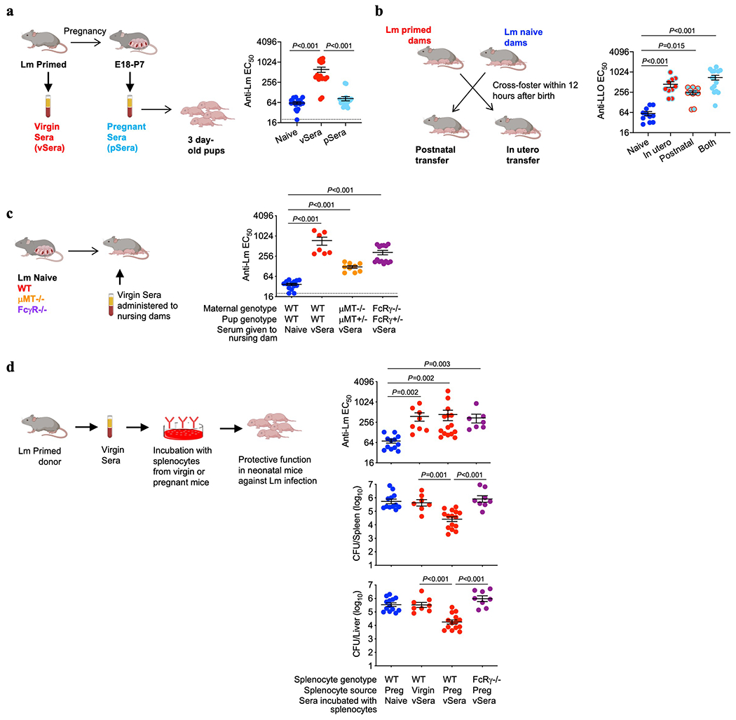
Anti-Lm antibodies acquire protective function during pregnancy through maternal FcγR-expressing cells. (a) Anti-Lm IgG titer in neonatal mice 72 hours after virulent Lm infection that were transferred sera from ΔActA Lm-primed virgin (vSera) or sera from late gestation/early post-partum (pSera) mice. (b) Cross-fostering schematic and anti-Lm IgG titer in each group of neonatal mice 72 hours after virulent Lm infection. (c) Anti-Lm IgG titer in neonatal mice nursed by WT, μMT−/−, or FcRγ−/− mice administered vSera on the day of delivery and 3 days later. (d) Anti-Lm IgG titers and bacterial burdens after virulent Lm infection in neonatal mice administered vSera that had been incubated with splenocytes from virgin or pregnant (E18) WT or FcRγ−/− mice for 48 hours. Pups were infected with virulent Lm 3-4 days after birth, 24 hours after antibody transfer, with enumeration of bacterial burden 72 hours post-infection. Each symbol represents an individual mouse, with graphs showing data combined from at least 2 independent experiments each with 3-5 mice per group per experiment. Bar, mean ± standard error. P values between key groups are shown as determined by one-way ANOVA adjusting for multiple comparisons. Dotted lines, limit of detection.
