Abstract
Aims
The aim of this study was to report a complete overview of both incidence, fracture distribution, mode of injury, and patient baseline demographics of paediatric distal forearm fractures to identify age of risk and types of activities leading to injury.
Methods
Population-based cohort study with manual review of radiographs and charts. The primary outcome measure was incidence of paediatric distal forearm fractures. The study was based on an average at-risk population of 116,950. A total number of 4,316 patients sustained a distal forearm fracture in the study period. Females accounted for 1,910 of the fractures (44%) and males accounted for 2,406 (56%).
Results
The overall incidence of paediatric distal forearm fractures was 738.1/100,000 persons/year (95% confidence interval (CI) 706/100,000 to 770/100,000). Female incidences peaked with an incidence of 1,578.3/100,000 persons/year at age ten years. Male incidence peaked at age 13 years, with an incidence of 1,704.3/100,000 persons/year. The most common fracture type was a greenstick fracture to the radius (48%), and the most common modes of injury were sports and falls from ≤ 1 m. A small year-to-year variation was reported during the five-year study period, but without any trends.
Conclusion
Results show that paediatric distal forearm fractures are very common throughout childhood in both sexes, with almost 2% of males aged 13 years sustaining a forearm fracture each year.
Cite this article: Bone Jt Open 2022;3(6):448–454.
Keywords: Incidence, Mode of trauma, Pediatric, Distal forearm fracture, distal forearm fractures, Fractures, epidemiology, forearm fractures, Radiographs, cohort study, trauma, ulna fractures, type I fracture, physicians
Introduction
Fracture of the distal forearm is a very common injury in children and adolescents, 1-8 and constitutes 74% of all paediatric fractures in the upper limb. 3 Despite attention increasing worldwide towards children’s safety during the last decades, 9 studies have reported an unexplained increase in incidence of paediatric distal forearm fractures. 7,8,10,11
Incidence of paediatric distal forearm fractures has previously been investigated in several studies reporting incidences between 263.3/100,000 persons/year and 801.0/100,000 persons/year. 2,8,11-16 However, most studies included a low number of patients, only specific fracture types (distal radius), and/or did not include an accurate at-risk paediatric population, making comparison and subgroup analysis in regard to safety issues difficult.
Furthermore, most population-based studies reporting on paediatric forearm fractures lack information regarding fracture classifications and/or information concerning the trauma mechanism. The reported change in children’s comorbidity and change in activity towards a more inactive lifestyle 17 may all be factors resulting in changes in the fracture incidence, trauma mechanism, fracture classification, and/or distribution of fractures among sexes and paediatric age groups.
At present, the reported incidence and epidemiology of paediatric distal forearm fractures are inconsistent, and the literature lacks a large-scale population-based study of all distal forearm fractures, based on an accurate at-risk population including all children and adolescents, reporting fracture classifications and associated mode of injury.
Such accurate data are essential to identify potential safety issues and develop potential prevention strategies to reduce the risk of distal forearm fractures in children. Furthermore, accurate data is essential in the allocation of healthcare resources in the emergency department and may be a strong predictor in determining cost of injury and associated consequences in society.
The aim of this study was to report a complete overview of both incidence, fracture distribution, mode of injury, and patient baseline demographics of paediatric distal forearm fractures to identify age of risk and types of activities prone to injury.
Methods
The present study was a retrospective population-based cohort study providing epidemiological data on distal forearm fractures in children between 1 January 2013 and 31 December 2017. The study was performed in the North Denmark Region served by Aalborg University Hospital (level 1 trauma centre) and five minor hospitals, representing all treatment facilities in the region.
All contacts with healthcare providers are registered digitally in the Danish Patient Registry, alongside updated medical charts, as required by Danish law. 18 All registrations are made and secured using a unique civil registration number (CPR) that all residents of Denmark are assigned at birth or on entry in to the country. 18 This provides researchers with accurate data on all health-related contact on both individual- and population-based levels.
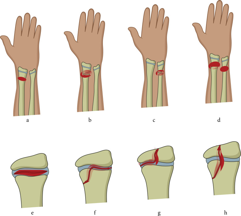
Included were children diagnosed with a distal forearm fracture in the five-year study period identified by the ICD-10 diagnosis: 19 DS525, DS525A, DS525B, DS525C, DS528, DS528C, and DS529 (Figure 1). Patients aged below 18 years and with open epiphyseal discs on radiograph were included. All patients’ records were validated by manual review of their charts and radiographs. Excluded were misclassified patients without a distal forearm fracture, patients with closed epiphyseal discs, and patients residing outside of North Denmark region.
Fig. 1.
Included children with a distal forearm fracture. legends: a) greenstick fracture (no distinguishing between greenstick and torus fractures); b) complete radius fracture; c) complete ulna fracture; d) complete radius and ulna fracture (antebrachium fracture); e) SH type 1; f) SH type 2; g) SH type 3; and h) SH type 4.
Patient chart data regarding age, injury/fracture type, and trauma mechanism were obtained. The trauma mechanism was divided into low- and high-energy trauma and mode of injury. Low-energy trauma includes falls, bicycle/vehicle accident, sports, and being hit by an object. High-energy trauma includes high-speed accident (> 65 km/h), significant damage to vehicle (> 0.5 m deformation), trapped in vehicle, getting hit by truck/train/bus as a pedestrian/cyclist/driver, passenger collision by other vehicle > 50 km/h, or falls from great height (> 2nd floor or 4 m).
Radiographs (anteroposterior (AP) and side radiograph) were used for classifying all fractures and to evaluate bone maturity (open/closed epiphyseal discs). Fractures were classified as: a) Salter Harris type I-V; 20 b) greenstick fracture to the radius and/or ulna; c) fracture through the radius with no epiphyseal contact; d) greenstick fracture to both radius and ulna; e) combination of fractures in both radius and ulna; and f) ulna fractures.
This study was conducted in accordance with the ethical principles of the 1975 Declaration of Helsinki. 21 The study was approved by the Danish Data Protection Agency (J nr: 2020 to 158). The reporting of the study complies with the Strengthening of the Reporting of Observational Studies in Epidemiology (STROBE) statement. 22
Statistical analysis
Percentages and frequencies are given for categorial data. Incidences were calculated by age, sex, and annual variation. Incidence was reported by incidence/100,000/persons year, including 95% confidence intervals (CIs). Incidence of age was reported by one-year increments. Prevalence of fractures are given for fracture classification, trauma mechanism, and monthly variation. The statistical analysis was performed by Stata 16 (StataCorp, USA).
Results
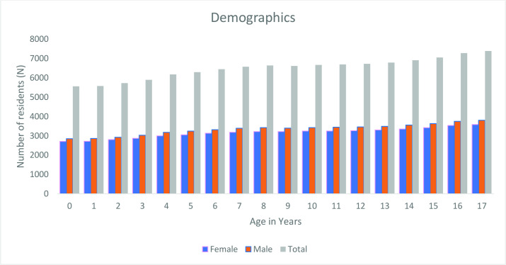
The present study was based on an average population of 116,950 people aged between zero and 17 years from 1 January 2013 to 31 December 2017 (Figure 2).
Fig. 2.
Average demographics of patients aged zero to 17 years during the study period.
A total number of 4,316 patients sustained distal forearm fractures in the study period. Females accounted for 1,910 of the fractures (44%), and males accounted for 2,406 (56%) (Table I).
Table I.
Distribution of the study population: children with fractures during the study period.
| Variable | Female, n (%) | Male, n (%) | Total, n (%) |
|---|---|---|---|
| Age, yrs | |||
| 0 | 0 (0.0) | 2 (0.0) | 2 (0.0) |
| 1 | 40 (0.9) | 25 (0.6) | 65 (1.5) |
| 2 | 28 (0.6) | 35 (0.8) | 63 (1.5) |
| 3 | 49 (1.1) | 44 (1.0) | 93 (2.2) |
| 4 | 58 (1.3) | 66 (1.5) | 124 (2.9) |
| 5 | 98 (2.3) | 103 (2.4) | 201 (4.7) |
| 6 | 139 (3.2) | 116 (2.7) | 255 (5.9) |
| 7 | 163 (3.8) | 137 (3.2) | 300 (7.0) |
| 8 | 175 (4.1) | 162 (3.8) | 337 (7.8) |
| 9 | 211 (4.9) | 188 (4.4) | 399 (9.2) |
| 10 | 256 (5.9) | 206 (4.8) | 462 (10.7) |
| 11 | 224 (5.2) | 263 (6.1) | 487 (11.3) |
| 12 | 201 (4.7) | 289 (6.7) | 490 (11.4) |
| 13 | 142 (3.3) | 297 (6.9) | 439 (10.2) |
| 14 | 75 (1.7) | 229 (5.3) | 304 (7.0) |
| 15 | 35 (0.8) | 138 (3.2) | 173 (4.0) |
| 16 | 11 (0.3) | 76 (1.8) | 87 (2.0) |
| 17 | 5 (0.1) | 30 (0.7) | 35 (0.8) |
| Total | 1,910 (44.3) | 2,406 (55.7) | 4,316 (100.0) |
| Side | |||
| Right | 795 (18.0) | 1,039 (24.0) | 1,834 (42.0) |
| Left | 1,114 (26.0) | 1,367 (32.0) | 2,481 (57.0) |
| Both | 1 (0.0) | 0 (0.0) | 1 (0.0) |
| Total | 1,910 (44.0) | 2,406 (56.0) | 4,316 (100.0) |
| Other fractures | |||
| No | 1,894 (44.0) | 2,356 (55.0) | 4,250 (98.0) |
| Another limb | 8 (0.0) | 31 (1.0) | 39 (1.0) |
| Same limb | 8 (0.0) | 18 (0.0) | 26 (1.0) |
| Missing data | 0 (0.0) | 1 (0.0) | 1 (0.0) |
| Total | 1,910 (44.0) | 2,406 (56.0) | 4,316 (100.0) |
Primary outcome
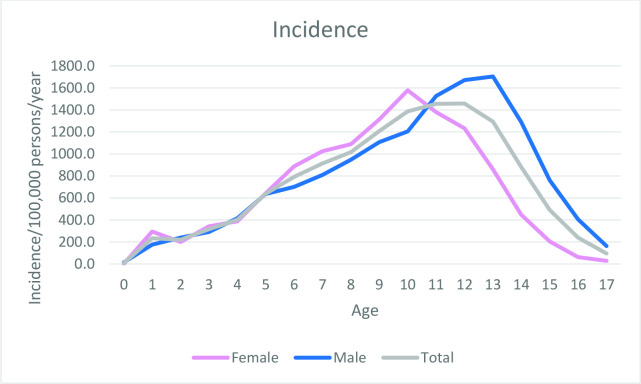
The overall incidence of distal forearm fractures in children was 738.1/100,000 persons/year (95% CI 706/100,000 to 770/100,000). Female incidence was 672.4/100,000 persons/year (95% CI 639/100,000 to 705/100,000) and male incidence was 800.2/100,000 persons/year (95% CI 768/100,000 to 833/100,000). Female incidences peaked between the ages of nine to 11 years, with the highest incidence of 1,578.3/100,000 persons/year at age ten years. Male incidence peaked between age 11 to 13 years with the highest incidence of 1,704.3/100,000 persons/year, at aged 13 years (Figure 3).
Fig. 3.
Incidence of fractures/100,000 persons/year for females, males, and in total.
Secondary outcomes
The sex-specific fracture classifications of forearm fractures in children are shown in Table II. The most common paediatric distal fracture of the forearm was a greenstick fracture to the radius representing 48% of all fractures, followed by a Salter Harris type I fracture, 20 representing 17% of all fractures.
Table II.
Classification of fractures, mode on injury, and energy of trauma.
| Variable | Female, n (‘%) | Male, n (‘%) | Total, n (‘%) |
|---|---|---|---|
| Fracture classification | |||
| SH 1 (radius) | 373 (8.6) | 363 (8.4) | 736 (17.1) |
| SH IIa (radius) | 116 (2.7) | 187 (4.3) | 303 (7.0) |
| SH IIb (radius) | 21 (0.5) | 32 (0.7) | 53 (1.2) |
| SH III (radius) | 5 (0.1) | 10 9 (0.2) | 15 (0.3) |
| SH IV (radius) | 2 (0.0) | 2 (0.0) | 4 (0.1) |
| SH V (radius) | 1 (0.0) | 2 (0.0) | 3 (0.1) |
| Greenstick radius | 958 (22.2) | 1,109 (25.7) | 2,067 (47.9) |
| Radius fracture* | 101 (2.3) | 218 (5.1) | 319 (7.4) |
| Greenstick radius and ulna | 237 (5.5) | 272 (6.3) | 509 (11.8) |
| Antebrachium fracture | 78 (1.8) | 177 (4.1) | 255 (5.9) |
| Ulna fracture | 15 (0.3) | 23 (0.5) | 38 (0.9) |
| Missing | 3 (0.1) | 11 (0.3) | 14 (0.3) |
| Total | 1,910 (44.3) | 2,406 (55.7) | 4,316 (100.0) |
| Mode of injury | |||
| Other | 56 (1.3) | 66 (1.5) | 122 (2.8) |
| Fall from > 1 m | 257 | 298 | 555 |
| Fall ≤ 1 m | 722 | 738 | 1460 |
| Bicycle accident | 125 | 159 | 284 |
| Sports | 687 | 1017 | 1704 |
| Motor vehicle† | 11 (0.3) | 58 (1.3) | 69 (1.6) |
| Hit by motor vehicle | 2 (0.0) | 4 (0.1) | 6 (0.1) |
| Hit by object | 48 (1.1) | 63 (1.5) | 111 (2.6) |
| Missing | 2 (0.0) | 3 (0.1) | 5 (0.1) |
| Total | 1,910 (44.3) | 2,406 (55.7) | 4,316 (100.0) |
| Energy level | |||
| High | 2 (0.0) | 8 (0.2) | 4 (0.1) |
| Low | 1,907 (44.2) | 2,397 (55.5) | 1,477 (34.2) |
| Missing | 1 (0.0) | 1 (0.0) | 2 (0.0) |
| Total | 1,910 (44.3) | 2,406 (55.7) | 4,316 (100.0) |
Radius fracture with no epiphyseal contact.
Driver or passenger.
SH, Salter Harris type fracture.
The modes of injury divided by sex are shown in Table II. The most common modes of injury were sports, represented by 39%, and falls from ≤ 1 m, representing 34% of all cases.
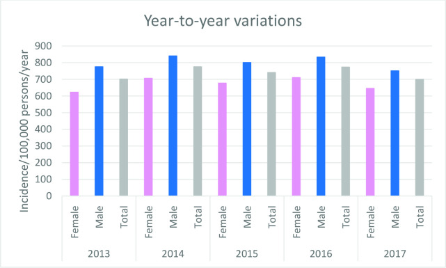
A small year-to-year variation (< 10%) in incidence of distal forearm fractures was observed between 2013 and 2017 with no apparent trend (Figure 4). The highest incidence of 775.5/100,000 persons/year was observed in 2014 and the lowest incidence of 699.7/100,000 persons/year was observed in 2017.
Fig. 4.
Year-to-year incidence of fractures during the study period.
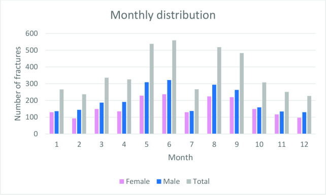
The monthly distribution of fractures is shown in Figure 5. The highest frequency of paediatric distal forearm fractures mainly occurred during the summer months, representing 31% of all fractures, whereas the winter months (December to February) represented only 17% of all fractures.
Fig. 5.
Monthly distribution of fractures.
Remarkably few fractures occurred during July compared to the other summer months (June and August). July represented 6% of all fractures, half of the fractures that occurred in June and August.
Discussion
This study presents a full epidemiological overview of paediatric distal forearm fractures. The overall incidence of paediatric distal forearm fractures was 738.1/100,000 persons/year. Female incidence was 672.4/100,000 persons/year, and male incidence was 800.2/100,000 persons/year. The most common fracture type was a greenstick fracture to radius (48%), and the most common modes of injury were sports and falls from ≤ 1 m. Results show that paediatric distal forearm fractures are very common throughout childhood in both sexes, with almost 2% of males aged 13 years sustaining a forearm fracture each year.
Incidence of paediatric distal forearm fractures has been investigated in several studies, reporting incidences between 393/100,000 persons/year and 801/100,000 persons/year. 12,13,15 Results from the present study show comparable results to Park et al, 12 reporting an overall incidence of 801/100,000 persons/year from a large-scale study. However, most studies available included low number of patients, and/or did not include an accurate at-risk paediatric population, making comparison to the present study difficult. Furthermore, the present study is the first study to report incidence based on a validated paediatric population investigated by manual review of all radiographs from the 4,316 fractures.
Both sexes showed a steady incline in incidences up to the ages of nine to 13 years, followed by a declining incidence. The highest female incidence was 1,140.6/100,000 persons/year at age ten years. The highest male incidence was 1,228.1/100,000 persons/year at age 13 years. Observed peak incidences correspond well with findings of other studies reporting peak incidences at age ten to 14 years. 7,8,12 This may be explained by children having almost the same sporting activities in different parts off the world, as well as the same bone-maturity and strength due to similarities in diets and overall health in the reported studies.
The most common fracture type was a greenstick fracture to the radius, representing 48% followed by Salter Harris type I fracture (17% of all paediatric distal fractures). These findings are in line with Kramhoeft et al, 7 reporting greenstick fractures in a paediatric population as the most common fracture (48%). This indicates that the distribution of children’s forearm fractures has not changed significantly in the last two decades. However, most studies available on paediatric distal forearm fractures lack information from manually-reviewed fracture classification, 12,13,15 and more research is needed to monitor the development of fracture distribution as a result of a change in children’s behaviour.
The most common trauma mechanisms reported were sports related injuries (39%) and falls ≤ 1 m (34%). These findings are supported by several authors, who have reported organized sports and recreational activities as the most common trauma mechanisms in paediatric distal forearm fractures. 1,7,8 Valid information regarding mode of injury is an essential to identify types of activities prone to injury.
Small annual and non-significant variations in incidence of fracture (699.7/100,000 persons/year to 775.5/100,000 persons/year) were observed in the present study. Results are comparable to Mamoowala et al, 2 who reported no overall change in incidence of paediatric distal radius fractures between 2007 and 2014. In contrast, several authors have reported an increase in incidence when comparing three or more historical decades. Furthermore, Khosla et al 8 reported an increase in incidence in childhood distal forearm fractures between 1969 to 1971 and 1989 to 1991, with a leveling off in 1999 to 2001.
The season with the highest frequency of paediatric distal forearm fractures was the summer period. The lowest frequency occurred during the winter period (December to February). These findings are supported by several authors with generally a small variation between studies. 1,2,12 Such small variations may be explained by seasonal differences between countries, schoolyear, and overall cultural differences.
A remarkable drop in fracture frequencies was observed in July. Many residents of North Denmark leave the region during summer vacation in July, reducing the number of residents in the region. We only included citizens with residency in the region and not visiting tourists. This might explain the remarkable drop in patients with paediatric distal forearm fractures. Moreover, in July, school and organized sports and many recreational activities are closed down as a consequence of summer vacation in Denmark.
Results from the present study are based on high-quality data from the Danish National Patient Registry (DNPR), which is one of the oldest nationwide health registries in the world and is used extensively for research. 18,23 Reporting to the DNPR is mandatory by law in Denmark, and the distribution of health resources is based to some extent on the reporting to the DNPR. The DNPR contains an overall high quality of data, and the positive predictive value of orthopaedic diseases is reported to be 89% to 91%. 23 Another major strength is inclusion of an accurate at-risk population used for calculations of incidence. 18 A further strength is the inclusion of all paediatric age groups manually evaluated by bone maturity. A limitation of our study is that medical charts and radiographs were manually reviewed by several physicians, which may result in differences in coding. However, a coding manual and meetings between physicians to discuss cases were initiated before study start to increase validity and reliability of data. All physicians were employed at the orthopaedic department and has the treatment of childrens forearm fractures as part of employment. The status of all physicians were postgraduate training.
In summary, this study showed the overall incidence of paediatric distal forearm fractures to be 738.1/100,000 persons/year. Female incidence was 672.4/100,000 persons/year and male incidence was 800.2/100,000 persons/year. The most common fracture type was a greenstick fracture to radius (48%), and the most common modes of injury were sports and falls from ≤ 1 m. Results show that paediatric distal forearm fractures are very common throughout childhood in both sexes, with almost 2% of males aged 13 years sustaining a forearm fracture each year.
Take home message
- The overall incidence of pediatric distal forearm fractures was 738.1/100,000 persons/year.
- Paediatric distal forearm fractures are very common throughout childhood in both sexes, with almost 2% of males aged 13 years sustaining a forearm fracture each year.
Acknowledgements
The authors thank Hanne Dalsgaard, Head of Hand Surgery at Aalborg University Hospital, Denmark, for supporting the study idea, study process, and comments to the final manuscript.
Footnotes
Author contributions: L. R. Korup: Conceptualization, Data curation, Analysis, Methodology, Validation, Writing original draft, Writing - review and editing.
P. Larsen: Conceptualization, Data curation, Analysis, Methodology, Validation, Writing - original draft, Writing - review and editing.
K. R. Nanthan: Data curation, Methodology, Validation, Writing - review and editing.
M. Arildsen: Data curation, Methodology, Validation, Writing - review and editing.
N. Warming: Data curation, Methodology, Validation, Writing - review and editing.
S. Sørensen: Conceptualization, Data curation, Analysis, Methodology, Validation, Writing - review and editing.
O. Rahbek: Conceptualization, Data curation, Methodology, Writing - original draft, Writing - review and editing.
R. Elsoe: Conceptualization, Data curation, Analysis, Methodology, Validation, Writing - original draft, Writing - review and editing.
Funding statement: The author(s) received no financial or material support for the research, authorship, and/or publication of this article.
ICMJE COI statement: The authors declare that they have no conflicts of interest to report.
Open access funding: The authors report that the open access funding for this manuscript was self-funded.
Twitter: Follow Aalborg University Hospital, Denmark @Trauma_Aalborg
Contributor Information
Lærke R. Korup, Email: l.korup@rn.dk.
Peter Larsen, Email: peter.larsen@rn.dk.
Kumanan R. Nanthan, Email: k.nanthan@rn.dk.
Marie Arildsen, Email: m.arildsen@rn.dk.
Nikolaj Warming, Email: n.warming@rn.dk.
Søren Sørensen, Email: soeren.soerensen@hotmail.com.
Ole Rahbek, Email: o.rahbek@rn.dk.
Rasmus Elsoe, Email: rae@rn.dk.
References
- 1. Ryan LM, Teach SJ, Searcy K, et al. Epidemiology of pediatric forearm fractures in Washington, DC. J Trauma. 2010;69(4 Suppl):S200-5. 10.1097/TA.0b013e3181f1e837 [DOI] [PubMed] [Google Scholar]
- 2. Mamoowala N, Johnson NA, Dias JJ. Trends in paediatric distal radius fractures: an eight-year review from a large UK trauma unit. Ann R Coll Surg Engl. 2019;101(4):297–303. 10.1308/rcsann.2019.0023 [DOI] [PMC free article] [PubMed] [Google Scholar]
- 3. Joeris A, Lutz N, Blumenthal A, Slongo T, Audigé L. The AO Pediatric Comprehensive Classification of Long Bone Fractures (PCCF). Acta Orthop. 2017;88(2):123–128. 10.1080/17453674.2016.1258532 [DOI] [PMC free article] [PubMed] [Google Scholar]
- 4. Joeris A, Lutz N, Wicki B, Slongo T, Audigé L. An epidemiological evaluation of pediatric long bone fractures - a retrospective cohort study of 2716 patients from two Swiss tertiary pediatric hospitals. BMC Pediatr. 2014;14:314. 10.1186/s12887-014-0314-3 [DOI] [PMC free article] [PubMed] [Google Scholar]
- 5. Naranje SM, Erali RA, Warner WC, Sawyer JR, Kelly DM. Epidemiology of pediatric fractures presenting to emergency departments in the United States. J Pediatr Orthop. 2016;36(4):e45-8. 10.1097/BPO.0000000000000595 [DOI] [PubMed] [Google Scholar]
- 6. Bailey DA, Wedge JH, McCulloch RG, Martin AD, Bernhardson SC. Epidemiology of fractures of the distal end of the radius in children as associated with growth. J Bone Joint Surg Am. 1989;71-A(8):1225–1231. 10.2106/00004623-198971080-00016 [DOI] [PubMed] [Google Scholar]
- 7. Kramhøft M, Bødtker S. Epidemiology of distal forearm fractures in Danish children. Acta Orthop Scand. 1988;59(5):557–559. 10.3109/17453678809148784 [DOI] [PubMed] [Google Scholar]
- 8. Khosla S, Melton LJ, Dekutoski MB, Achenbach SJ, Oberg AL, Riggs BL. Incidence of childhood distal forearm fractures over 30 years: a population-based study. JAMA. 2003;290(11):1479–1485. 10.1001/jama.290.11.1479 [DOI] [PubMed] [Google Scholar]
- 9. Sminkey L. World report on child injury prevention. Inj Prev. 2008;14(1):69. 10.1136/ip.2007.018143 [DOI] [PubMed] [Google Scholar]
- 10. Jónsson B, Bengnér U, Redlund-Johnell I, Johnell O. Forearm fractures in Malmö, Sweden. Changes in the incidence occurring during the 1950s, 1980s and 1990s. Acta Orthop Scand. 1999;70(2):129–132. 10.3109/17453679909011249 [DOI] [PubMed] [Google Scholar]
- 11. Hedström EM, Svensson O, Bergström U, Michno P. Epidemiology of fractures in children and adolescents. Acta Orthop. 2010;81(1):148–153. 10.3109/17453671003628780 [DOI] [PMC free article] [PubMed] [Google Scholar]
- 12. Park MS, Chung CY, Choi IH, et al. Incidence patterns of pediatric and adolescent orthopaedic fractures according to age groups and seasons in South Korea: a population-based study. Clin Orthop Surg. 2013;5(3):161–166. 10.4055/cios.2013.5.3.161 [DOI] [PMC free article] [PubMed] [Google Scholar]
- 13. Bell SW, McLaughlin D, Huntley JS. Paediatric forearm fractures in the west of Scotland. Scott Med J. 2012;57(3):139–143. 10.1258/smj.2012.012018 [DOI] [PubMed] [Google Scholar]
- 14. Brudvik C, Hove LM. Childhood fractures in Bergen, Norway: identifying high-risk groups and activities. J Pediatr Orthop. 2003;23(5):629–634. 10.1097/00004694-200309000-00010 [DOI] [PubMed] [Google Scholar]
- 15. Cooper C, Dennison EM, Leufkens HGM, Bishop N, van Staa TP. Epidemiology of childhood fractures in Britain: a study using the general practice research database. J Bone Miner Res. 2004;19(12):1976–1981. 10.1359/JBMR.040902 [DOI] [PubMed] [Google Scholar]
- 16. Lyons RA, Delahunty AM, Kraus D, et al. Children’s fractures: a population based study. Inj Prev. 1999;5(2):129–132. 10.1136/ip.5.2.129 [DOI] [PMC free article] [PubMed] [Google Scholar]
- 17. Lobstein T, Baur L, Uauy R. IASO international obesity taskforce. obesity in children and young people: a crisis in public health. Obes Rev. 2004;4–104. 10.1111/j.1467-789X.2004.00133.x [DOI] [PubMed] [Google Scholar]
- 18. Lynge E, Sandegaard JL, Rebolj M. The Danish National Patient Register. Scand J Public Health. 2011;39(7 Suppl):30–33. 10.1177/1403494811401482 [DOI] [PubMed] [Google Scholar]
- 19. World Health Organization . 2022. https://www.who.int/standards/classifications/classification-of-diseases (date last accessed 28 April 2022).
- 20. Cepela DJ, Tartaglione JP, Dooley TP, Patel PN. Classifications In brief: Salter-Harris classification of pediatric physeal fractures. Clin Orthop Relat Res. 2016;474(11):2531–2537. 10.1007/s11999-016-4891-3 [DOI] [PMC free article] [PubMed] [Google Scholar]
- 21. World Medical Association . Declaration of Helsinki. Law Med Health Care. 1991;19(3–4):264–265. 10.1111/j.1748-720X.1991.tb01824.x [DOI] [PubMed] [Google Scholar]
- 22. von Elm E, Altman DG, Egger M, et al. The Strengthening the Reporting of Observational Studies in Epidemiology (STROBE) Statement: guidelines for reporting observational studies. Int J Surg. 2014;12(12):1495–1499. 10.1016/j.ijsu.2014.07.013 [DOI] [PubMed] [Google Scholar]
- 23. Schmidt M, Schmidt SAJ, Sandegaard JL, Ehrenstein V, Pedersen L, Sørensen HT. The Danish National Patient Registry: a review of content, data quality, and research potential. Clin Epidemiol. 2015;7:449–490. 10.2147/CLEP.S91125 [DOI] [PMC free article] [PubMed] [Google Scholar]


