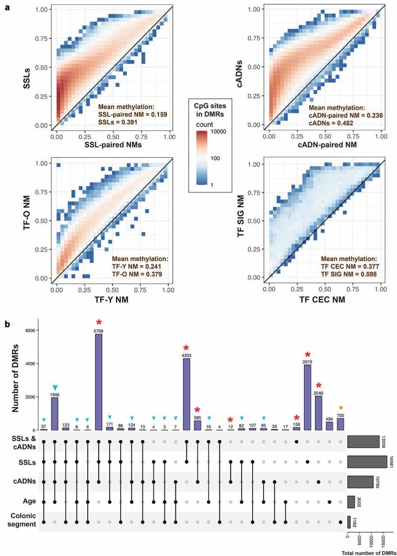
Figure 3.

Hypermethylated differentially methylated regions (DMRs) related to early colorectal tumorigenesis, normal mucosa ageing, and/or anatomical location of normal mucosa. a. Scatter plots showing the number of methylated CpG sites within the identified hypermethylated DMRs (colour gradient) and mean methylation levels at these sites in precancerous tumours (SSLs or cADNs) versus SSL-paired NM and cADN-paired NM, respectively; in TF-O NM samples vs. TF-Y NM samples; and in TF SIG NM vs. TF CEC NM. b. UpSet plot showing the tumorigenesis-, ageing-, and colon segment-specificities of the hypermethylated DMRs. The vast majority of the hypermethylated DMRs in tumours (in SSLs, cADNs, or both, red asterisks) were tumorigenesis-specific, i.e., regions that displayed no ageing-related or colon segment-related changes in the NM. In contrast, over 80% of the ageing-associated DMRs overlapped tumorigenesis-associated DMRs (blue arrowheads). Most regions that were differentially methylated in SIG vs. CEC NM samples displayed no methylation alterations associated with ageing or tumorigenesis (orange dots). (Abbreviations as defined in Figure 1.).
