Abstract
Analysis of stool offers simple, non-invasive monitoring for many gastrointestinal (GI) diseases and access to the gut microbiome, however adherence to stool sampling protocols remains a major challenge because of the prevalent dislike of handling one’s feces. We present a technology that enables individual stool specimen collection from toilet wastewater for fecal protein and molecular assay. Human stool specimens and a benchtop test platform integrated with a commercial toilet were used to demonstrate reliable specimen collection over a wide range of stool consistencies by solid/liquid separation followed by spray-erosion. The obtained fecal suspensions were used to perform occult blood tests for GI cancer screening and for microbiome 16S rRNA analysis. Using occult blood home test kits, we found overall 90% agreement with standard sampling, 96% sensitivity and 86% specificity. Microbiome analysis revealed no significant difference in within-sample species diversity compared to standard sampling and specimen cross-contamination was below the detection limit of the assay. Furthermore, we report on the use of an analogue turbidity sensor to assess in real time loose stools for tracking of diarrhea. Implementation of this technology in residential settings will improve the quality of GI healthcare by facilitating increased adherence to routine stool monitoring.
Subject terms: Gastrointestinal diseases, Biomedical engineering, Gastroenterology, Biochemical assays
Introduction
Noninvasive individual health monitoring conducted by smartphones, wearables and home sensors offers the advantages of objective and more frequent data collection, the convenience of remote monitoring, and holds promise for integration of this data into meaningful clinical assessments of disease progression and treatment response1,2. There is growing interest in remote health monitoring in gastrointestinal (GI) medicine for widely prevalent conditions such as functional GI diseases, autoimmune diseases, and colorectal cancer3,4. These conditions are estimated to affect as many as 1 in 10 people globally and significantly impact quality of life, ability to work, as well as healthcare costs5–8.
Traditionally underutilized, stool is an attractive biological specimen for remote GI disease monitoring because it can be collected non-invasively and longitudinally. Less invasive and costly than a colonoscopy, stool-based biochemical analysis allows for detection of many acute and chronic GI conditions such as GI cancer9,10, enteric infections such as Clostridium difficile11, response to treatment in inflammatory bowel disease12,13, and gluten consumption by celiac disease patients14. In addition to diagnosis of disease, access to fecal specimens coupled with molecular analysis and sequencing enables analysis of the gut microbiome, and pursuit of novel understanding of the etiology of disease and of novel therapeutic approaches in a number of intestinal and extra-intestinal conditions15–18. Time-dense stool analysis for the composition of the microbiota could provide an ability to discern the effect of pathophysiology from environmental factors such as dietary habits, drug treatments and intestinal motility19.
Despite being non-invasive and effective, implementation of stool-based analysis is severely limited in practice: patients are rarely able to produce a stool specimen during the clinical encounter20,22–27 and a number of studies from different geographies have found low uptake and adherence to GI disease surveillance based on regular stool testing20–24. Studies exploring barriers to surveillance by fecal tests have found that blood testing is preferred to fecal testing and suggest that only patients with active disease are likely to comply with a stool test20,25. Surveillance studies carried out when patients are stable consistently found adherence of only 30% for completing four or more repeated fecal tests21,22,26. Survey studies have revealed that a major reason for poor compliance on repeated fecal screening was avoidance/forgetfulness27,28 and over 60% of respondents stated that the reason for low acceptability of fecal tests was disgust/embarrassment in stool collection23,25,29.
Approaches to better leverage stool-based data and integrate stool analysis with routine use of the toilet have been recently proposed30,31. Emerging efforts by academia and toilet manufacturers for excreta monitoring within and around a toilet have focused on analysis of urine31–37.
Accessing fecal specimens is technically challenging due to the extremely sticky nature of the material and its heterogeneity38,39. Image capture of stool in the toilet bowl, either by the user40 or without user intervention by a camera in the toilet seat31, have been proposed to track stool appearance characteristics since significant disparities have been reported between patient assessment and stool properties such as consistency, particularly for diarrhea41–43. However, there are major social taboos and privacy concerns associated with operating a camera in a bathroom and particularly in the toilet seat.
Here, we report on an approach to isolating human feces just downstream from a flush toilet specifically designed for sample collection and analysis. Wastewater has been used for many years for molecular analysis-based surveillance of infectious disease at the community level for polio44, and since 2020, it has been extensively used for detection of SARS-CoV-2 to assess prevalence of COVID-19 infection at the community level45–51. In our approach, we have sought to improve the resolution of wastewater-based analysis to the level of a single toilet and individual stool being flushed through it. The purpose of this technology is to make the stool sampling process automatic and seamless with a daily routine for improved adherence to a testing regimen that enables better disease management and improved outcomes.
This work presents a proof-of-concept demonstration of hands-free human stool collection for continuous health monitoring. The approach separates stool from wastewater after the toilet is flushed for the purpose of analysis. A method based on buffer spray-erosion is used to collect a small portion of the stool for biochemical analysis in a manner that is amenable to automation and manages issues of cross-contamination. Two types of physiologically relevant data, occult blood measurements and 16S rRNA microbiome analysis, were conducted on fecal specimens obtained from our prototype system. The analytical integrity of the results was assessed by conducting pair-wise comparison against stool specimens sampled conventionally. For the clinically relevant cases of watery and loose stool, we demonstrate an inline analog turbidity sensor with the ability to discriminate loose stools from other types of wastewater for tracking of diarrhea.
Results
Design criteria
A foundational design criterion for our approach is to seamlessly integrate stool analysis with the routine use of the toilet without requiring any action nor habit change in users nor engendering discomfort. This criterion minimizes barriers to adoption and improves likelihood of long-term use for longitudinal data collection. We achieved this objective by leaving the toilet bowl appearance and function unchanged, and by operating in the plumbing where sampling and analysis occurs outside the purview of the user, after the specimen leaves the bowl.
Another key requirement is the ability to sample the stool in a manner that is independent from the toilet user and amenable to automation.
To be valuable as a health monitoring tool, the system must be able to detect the whole clinical range of stool consistencies from constipation to loose stools. A widely used standard medical diagnostic tool for categorizing adult stool based on its physical appearance is the Bristol Stool Form Scale (BSFS)38,39, featuring 7 types ranging from BSFS 1 (separate hard lumps, like nuts) to BSFS 7 (watery, no solid pieces).
Design and operation
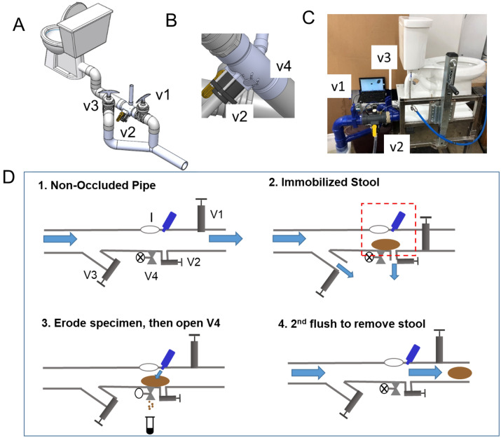
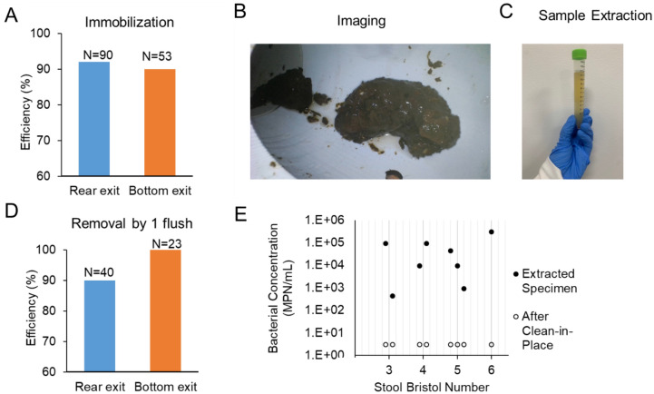
A standard flush toilet consists of a bowl that disposes of human excreta by using water to flush it through a drainpipe to another location for disposal. Critical to this design is a U-shaped section of outlet pipe that holds water—termed a P-trap or S-trap—that acts as a seal and prevents noxious gases from rising up through the toilet into the bathroom. Our design captures feces using a device integrated into the plumbing at the outlet of the toilet after the trap (Fig. 1) to preserve the odor sealing properties of the toilet. Reversible immobilization of the whole stool was achieved by solid/liquid separation and was demonstrated by flushing stool into a benchtop test bed (Fig. 1A–C). Immobilization was achieved using a combination of flow transport forces and occlusion by a gate valve V1 (Fig. 1D). When V1 was closed, inertia transported solid waste into a 3D printed custom designed component occluded by V1; water drained through valve V2 and through valve V3 located on a diversion pipe. The stool was immobilized near valve V2 and was not typically in contact with the occluding valve V1. The majority of the water drained through valve V3, ensuring the bowl was emptied with a similar speed as with a regular flush. We found that stool was immobilized in the region near valve V2 and aligned with the sampling port for 90% or more of the tests (Fig. 2A) in both types of toilets (90 flushes for the rear exit toilet and 40 for the bottom exit toilet); in the remainder of the cases the stool was typically immobilized between valve V2 and valve V1. Once stool was immobilized and the water drained, a stool image was collected through a port using a waterproof inspection camera (Depstech endoscope with 6 LED illumination) (Fig. 2B). Such images of individual stool are well suited for accurate determination of stool morphology by machine learning, as some of these investigators have previously reported52.
Figure 1.
(A) An illustration of the prototype connected to a rear exit toilet. (B) Close-up view of the bottom part of the prototype shown in (A). (C) Picture of the prototype connected to a bottom exit toilet with endoscope connected to a laptop for image collection. (D) Operating principle of the system including 4 valves, V1 through V4, and I = inspection and venting port. 1. The valve position for regular operation of the toilet, blue arrow indicates water flush direction from toilet to sewer line. 2. The stool is immobilized in the stool area (red dashed rectangle). 3. A spray-jet of buffer erodes specimen collected by gravity through V4. 4. The stool is flushed to sewer.
Figure 2.
(A) Immobilization efficiency measured with formed surrogates and human stools in rear exit and bottom exit toilets. (B) In situ image of immobilized human stool in the prototype. (C) Image of spray-eroded extracted fecal specimen. (D) Efficiency of stool removal from the immobilization region with 1 toilet flush. (E) Bacterial enumeration by most probable number (MPN) for stool specimens of different consistencies (BSFS type) for specimen extraction and after the stool was removed and clean-in-place procedure implemented.
A specimen was extracted using a high-pressure liquid stream to erode and dissolve a portion of the immobilized stool (Fig. 2C). The buffer-eroded liquid specimen was collected by gravity through a zero dead-leg valve V4 (details in section “Specimen extraction”) (Fig. 1D, step 3).
As a last step of the operation, the remainder of the stool was disposed of to the sewer through a second flush while valve V1 occlusion was removed and all other valves were closed. Removal was achieved with a single flush in 90–100% of tests (Fig. 2D).
A major concern of sampling a sticky material such as feces is to ensure minimal cross contamination between specimens. Our design included a zero dead volume sampling valve for the purpose of eliminating stagnant volume or protruding surfaces. We also made use of the availability of tap water in the toilet operation to rinse surfaces. To determine the extent of specimen-to-specimen contamination upon stool immobilization, we measured fecal coliform bacteria, which are naturally present in stool.
Fecal bacteria were quantified using a most probable number (MPN) method on samples from a 3D-printed ABS device. The MPN assay was conducted on extracted stool specimens and samples from blank flushes following the stool disposal flush. The bacterial concentration was high in the specimen samples, as expected (> 104 MPN/mL) (Fig. 2E). The sample collected after the disposal flush contained a much-reduced bacterial content, from 1 to 3 log reduction, likely associated by how “clean” the sample was removed. The clean-in-place procedure in this study consisted of a second flush. We tested specimens of different consistencies [value from BSFS 3 (sausage shaped) to BSFS 6 (mushy)] and for these tests, this flushing was adequate for the extracted volume to result in bacterial content below the limit of detection of the assay (3 MPN/mL).
Hands-free stool sampling
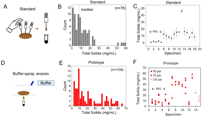
Standard sampling for fecal assays requires a person inserting a grooved stick or spatula in multiple regions of the stool and immediately diluting the specimen in buffer (Fig. 3). This process is challenging to automate within a wastewater plumbing environment because it requires a new stick for each measurement to avoid cross contamination.
Figure 3.
Specimen sampling study. Standard sampling: (A) Procedure illustration from the manufacturer. (B) Distribution of total wet solids with median value of 8 mg/mL. (C) Average and standard deviation of total solids from individual specimens, n = 3 to 5 for each specimen. BS2 = Bristol Stool Form Scale 2 indicates lumpy, hard consistency stool. Prototype sampling (D): Schematic. (E) Distribution of total wet solids collected from the prototype. (F) Total solids extracted from individual specimens using 5 s spraying time at the indicated pressure.
We propose a novel sampling approach that is readily amenable to implementation in plumbing and automation and that is informed by the specific characteristics of feces. The water content of formed feces is on average 75%53, which is high as opposed to typical paste material. For example, the soybean surrogate used in these tests was measured at 54% water content, while the average water content for watery stools in cases of diarrhea is 88%54. The high water content of stool suggested that adding a small amount of liquid was sufficient to transform the solid stool into a liquid that is easier to manipulate and less sticky.
The fecal specimen extraction in our design relied on a spray jet of buffer to erode the stool and on collection of the liquefied sample by gravity through a valved opening. The advantages of this approach are multifold: no mechanical parts inside the pipe, readily amenable to automation (of the spray and valving) and ease of cleaning. We reasoned that this approach would yield a specimen suitable for biochemical analysis since dilution in buffer is the first step of most fecal assays.
Fecal assays require a small amount of stool (~ tenths of mg) and are often tolerant of a range of values. The wet total solids content in mg/mL is a measure of the dilution of the solid amount in buffer. Molecular tests for detection of pathogens in stool recommend a mass of 50–200 mg (see SI) resulting in 20–100 mg/mL wet solids while tests for more abundant species like microbiome require a vague “small” quantity of stool. To determine the stool amount for a point-of-care (POC) assay, we measured the amount of solid collected by the stick in a commercial occult blood at-home test (Fig. 3A). Figure 3B shows the results of solid content obtained using standard sampling over 18 stools and illustrates a broad range of values, with median 8 mg/mL, average 11 mg/mL and standard deviation of 12 mg/mL. The heterogeneity of stool form (BSFS 2–6 were measured) accounts for some of this variability; however, Fig. 3C shows that replicate measurements over the same stool specimens resulted in a wide variation (CoV between 0.2 and 0.7). The performance of our proposed extraction approach (Fig. 3D) was evaluated using a custom set-up with a tunable flowrate pump and connected to a precision nozzle.
We explored the amount of solid to be extracted by spray erosion. Measurements were conducted at multiple flowrates (between 0.4 and 1.1 L/min, corresponding to pressures between 3.6 and 30 psi, according to the nozzle specifications) and different spray times (2 s, 5 s, 10 s). We collected spray-eroded specimens in vials and determined total solids by drying as described in the methods.
Figure 3E shows that spray erosion enabled us to collect a wide range of wet solids from a few mg/mL to 60 mg/mL, with the histogram showing a broad range fully covering the range of the standard sampling by using different flowrate and spray times. To ensure this approach enabled collection of adequate fecal specimen solutions from a hard stool (BSFS 2), we showed that 10 psi produces values of 2–5 mg/mL, comparable to the standard method. By increasing to 30 psi we obtained over 20 mg/mL solids. No hard lump BSFS 1 stools were available for this study, but this data and the ability to increase pressure suggests that spray erosion can be tuned to obtain specimens from the whole range of stool consistency. For softer stool, a pressure of 3.6 psi was best suited to obtain a range of 10–20 mg/mL comparable to the 8 mg/mL median concentration obtained for standard sampling (Fig. 3F).
Stool protein biomarker measurement
To demonstrate the feasibility of measuring a GI disease protein biomarker from stool specimens obtained from the prototype with spray erosion, we selected the well-established lateral flow assay (LFA) Fecal Immunochemical Test (FIT) to measure occult blood. The Fecal Immunochemical Test (FIT) for occult blood relies on detection of human hemoglobin and does not require any restrictions of food or medication intake. The FIT test features good sensitivity and excellent specificity for GI cancer55 and it is recommended worldwide and by the American Cancer Society as an annual screening for people beginning at 45 years of age who are at regular risk of GI cancer9,10,56.
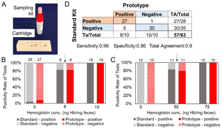
Healthy stools are negative for occult blood and can be spiked with human hemoglobin to obtain a positive readout as has been done in proficiency tests of these kits57. Numerous FDA CLIA-waved FIT lateral flow assay kits for at-home use are available with qualitative (positive/negative) output. Our study evaluated brands Pinnacle and Accutest57. Both tests feature a sampling kit and a cartridge with an immunochromatographic strip for reading (Fig. 4A). The sampling kit is a small vial, containing a few mL volume (3 mL for Pinnacle, 2.5 mL for Accutest) of buffer, with a cap that is attached to a grooved plastic stick for stool collection. After inserting the grooved stick in the stool for a prescribed number of times (6 times), the user returns the grooved stick to the vial and shakes to dissolve the stool in the buffer. The kit vial has a smaller cap to dispense a prescribed number of drops of stool solution to the cartridge. Within 5 min from solution application, the control line of the cartridge appears and, in the presence of hemoglobin (Hb), the test line color appears (Suppl. Fig. S1).
Figure 4.
Effect of standard and prototype sampling on occult blood at-home test. (A) Picture of at-home kit (Pinnacle), including a tube with buffer, a screw cap and grooved stick for sample collection, and a cartridge for reading results. (B,C) The positivity rate (percentage of positive tests) of the (B) Pinnacle brand and (C) Accutest brand for human stools spiked at varying hemoglobin (Hb) concentrations. Black arrow: nominal limit of detection of the test. Figures on the second horizontal axis are the number of replicates. (D) The agreement matrix for pair-wise comparison on the same specimens at Hb concentration above the cutoff (10 ng/mg for Pinnacle brand, 75 ng/mL for Accutest).
The Hb detection limit is set by the manufacturer and it is typically 50 ng/mL. The Hb detection limit expressed as ng Hb per mg of stool is reported as 6 ng/mg for Pinnacle and 50 ng/mg for Accutest.
We compared the nominal sensitivity of the kit by testing a range of Hb spiking concentration spanning a range above and up to the nominal threshold. Using PBS as the buffer for erosion, we obtained by spray erosion specimens with wet solid content ranging from 5 to 25 mg/mL and applied a volume equivalent to the drops (75 μL) to the cartridge (Suppl. Fig. S1). Figure 4B shows a sensitivity curve for standard sampling with 100% positive readings (n = 18) at 10 ng/mg Hb, a value above the nominal sensitivity cutoff, and 4% (n = 26) positive for unspiked specimens. The reading of the cartridge samples from the prototype was similar, with 100% positive readings above the limit of detection and 7% positive for unspiked specimens. A similar study was done on the Accutest assay using 100 μL suspension (Fig. 4C) to strengthen the case that prototype sampling achieved the same analytical detection concentration as standard sampling.
To determine sensitivity, specificity, and total agreement of the test results obtained by prototype sampling relative to the standard method, we conducted pair-wise comparison of sampling methods on the same specimens at a fixed Hb spike concentration. For the Pinnacle assay, sensitivity was 95%, specificity 83%, and a total of 38 out of 43 pairs were concordant (88%) (Fig. S2). Discordant test pairs were mostly due to prototype sampling being positive while the standard was negative. This was typically caused by a large solids concentration (60 mg/mL in two cases). For the Accutest assay (Fig. S3), 19 of 20 pairs were concordant (95%). Overall, we found 90% agreement with 96% sensitivity and 86% specificity (Fig. 4D).
Using the Pinnacle assay, we also collected proof-of-concept data suggesting no impact on assay performance by interferents such as toilet paper (covering stool during spray-erosion) and urine (stool bathed in dilute urine mimicking a toilet bowl for 60 s prior to erosion). We measured 100% agreement with truth (defined as positive/negative result for spiked/unspiked stool respectively) for n = 5 test in each condition for both toilet paper and urine.
Microbiome analysis
The objective of this experiment was to demonstrate the feasibility of microbiome analysis on specimens collected from our system. Stool specimens were obtained and sampled with a spatula for standard processing. Then, the same stool was placed in the toilet bowl, flushed and samples were collected by spray erosion. The study used 4 stools, S1, S2, S3, S4. When S4 stool was placed in the toilet bowl, 500 mL urine was also added for one minute prior to flushing to explore the possible contamination effects from urine, thereby we denote the specimen as S4u. S1 was measured in 6 biological replicates, S2, S3, S4u in triplicates for both sampling approaches. Six additional S1 replicates were centrifuged and the concentrated sludge used for analysis. A total of n = 36 samples underwent 16S rRNA microbiome analysis.
Extracted DNA concentration from the stool-sampling portal was found to be abundant (64 ng/μL as compared to 269 ng/μL for conventional sample) and more than adequate for 16S ribosomal RNA gene sequencing. All 36 samples submitted met the minimum criteria for analysis. The analysis of the relative abundance of bacterial taxonomic groups by order showed Clostridiales and Bacteroidales as the first and second most abundant, as expected in healthy subjects58 (Suppl. Fig. S4).
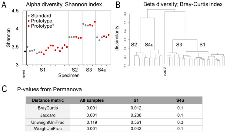
We quantified the microbial diversity within sample (α diversity) for standard sampling and our design sampling, and the difference between samples, i.e. between sampling approaches (β diversity).
Within-sample species diversity (α diversity, Shannon diversity index) did not significantly change across sampling approach (Fig. 5A). Alpha diversity analyses were performed using a linear mixed effects model with sampling approach as a fixed effect and stool as a random effect; p values were not significant, included for S4u, the specimen flushed with urine.
Figure 5.
Microbiome analysis result of n = 36 fecal specimens from stool S1, S2, S3, and s4u. (A) Within-sample microbial species diversity (Alpha diversity, Shannon index) by stool and by sampling approach. * denotes sample centrifuged prior to nucleic acid extraction. (B) Hierarchical clustering using beta diversity index showing taxonomical distance: the clustering is around the stools not around the sampling approach. (C) P-value from permanova analysis of beta diversity metric comparing sampling methods (standard vs prototype) for test groups of interest: all stool samples (controlling for stools), stool 1 and stool 4u. P < 0.05 is statistically different.
Beta diversity was calculated for each pair of specimens for multiple metrics (namely Bray–Curtis, Jaccard, UniFrac, and Weighted un-normalized UniFrac). Hierarchical clustering was performed using beta diversity metrics. A tree diagram illustration of the Bray Curtis beta diversity index in Fig. 5B shows the data clustering around specimen not sampling approach. These data illustrate that inter-individual variation of microbiome, a known feature of the microbiome of healthy individuals59 is a bigger factor than sampling approach.
A permutational multivariate analysis of variance (Permanova) of beta diversity was used to compare the two sampling approaches and test the null hypothesis.
The results were mixed depending on the metrics and the stool (Fig. 5C), with p values below p = 0.05 considered significant, indicating a difference introduced by sampling approach. We believe that the buffer type used in the spray-erosion sampling may introduce effects, since storage in different buffer types has been reported to influence fecal microbiome profiles60. Interestingly, for stool S4u, all four indices resulted in p = 0.1 or higher in the permanova analysis, supporting our hypothesis that urine contamination is a limited concern. In our design the contact time between urine and stool is limited in time as opposed to urine contaminating specimens collected with a stool collection kit.
Detection of diarrhea
The stool sampling method based on solid/liquid separation is suitable for a wide range of stool consistencies. One limitation is that it cannot be utilized for completely watery stools (i.e., BSFS 7); however, several physiological conditions result in loose or watery stools, and the occurrence of such stools is itself an important GI biomarker. Specifically, diarrhea is clinically defined as the passage of 3 loose stools in a 24-h period. In conventional practice, this diagnosis generally relies on self-reporting and thus suffers from limited accuracy that impacts treatment. Here, we propose a non-invasive method for monitoring diarrhea that relies on an analog sensor in line with toilet effluent plumbing and is compatible with the configuration described in Fig. 1.
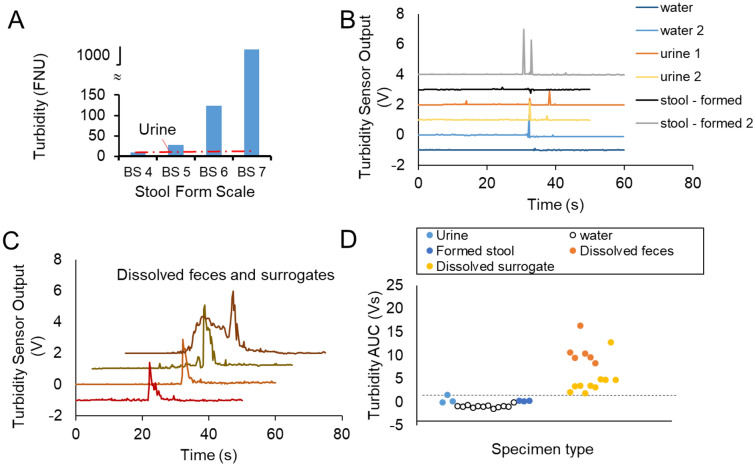
We found that nephelometric turbidity, a light-scattering-based measurement of the suspended particles in solution, is a sensitive measure of the presence of dissolved stool in a water solution. In batch static (no flow) tests we mixed water, human urine, and feces (both formed and liquid) in concentrations emulating one toilet use, as described in the Methods. Formed stools in water resulted in turbidity values ranging from 10 to 120 FNU (formazin nephelometric unit), significantly different from water baseline (T = 0.7 FNU) and from urine added to water (T = 1.1 ± 0.3 FNU, n = 5). Stool completely dissolved in water, emulating a BSFS 7 watery stool, resulted in saturation of the turbidity reading > 1000 FNU (Fig. 6A).
Figure 6.
(A) Static Turbidity meter readings from solution containing stools of different form as defined by the BSFS measured after t = 1 min of immersion. (B) Sensor output recorded over toilet flushes (at t = 30 s) for water only and urine and formed stool. (C) Sensor output recorded for dissolved specimens flushed at predetermined time corresponding to peak (curves are shifted over both vertical and horizontal axis for clarity). (D) Cluster plot of area-under-curve of sensor output by specimen type, datapoints are spaced for clarity. Dotted line represents a proposed threshold of 2 Vs separating dissolved specimen from other wastewater types.
We used a waterproof analog turbidity sensor (SEN0189) designed to monitor the haziness of water in home appliances to obtain a dynamic measurement of turbidity within wastewater plumbing. The sensor was calibrated against the Hach 2100Q turbidity meter (Suppl. Fig. S5) and found to be linear up to 30 mg/mL wet solid content, more than adequate to detect diarrhea in toilet wastewater. The effect of toilet paper on the turbidity sensor was found to be negligible. Using an amount of toilet paper informed by ASME standard 112.19.2 for Ceramic Plumbing Fixtures (in “Methods”), we performed paired measurements across different concentrations of stool surrogate (soybean paste) and/or under different conditions (stirred or settled), with and without toilet paper added. Toilet paper did not result in significantly different readings (p = 0.7334 for n = 12 paired measurements with and without toilet paper).This was not surprising since toilet paper disintegration time ranges from 10 min to hours61, and is facilitated by extensive turbulence in a sewer system, well beyond the small pipe exiting a toilet62.
We installed the turbidity probe in the benchtop setup of the rear exit toilet to evaluate its dynamic response during toilet flushing. The probe was installed in the wastewater pipe immersed in the water of the S-trap and the electronic adapter was outside of the pipe (Suppl Fig. S6). The sensor probe output was collected with the following protocol: 60 s recording, stool or urine poured in the bowl at t = 10 s, toilet flushed at t = 30 s. Figure 6B illustrates typical readout for water and formed stool: the turbulence created by a flush may result in a short noise spike during at the time of the flush, but the output is otherwise unchanged. The flushing of a turbid solution of either dissolved feces or surrogate created the signature of a peak of a few seconds wide at t = 30 s (Fig. 6C). Occasionally, the pouring of dissolved specimen in the bowl prior to flushing resulted in a turbidity sensor signal (top curve in Fig. 6C) indicating the dissolved specimen had crossed the S-trap and reached the sensor without a toilet flush. The Area-Under-Curve measured for all signals measured around the flush is plotted in Fig. 6D. Values for water, urine and formed stool are either zero or below 2 V-s; values measured for dissolved specimens are above 2 V-s indicating that the AUC metric can be used as a threshold to discriminate the presence of loose stool.
Discussion
Aversion to handling one’s own stool is arguably the most significant barrier to harnessing health data from this physiological specimen23,25,29. This study demonstrated feasibility of stool collection from toilet effluent and suitability of the specimen for biochemical analysis.
Previously published work on toilet-related stool analysis used sensors or receptacles in the toilet bowl or seat for stool imaging and did not achieve the diagnostic specificity of biomolecular assays31,34. We report a configuration that achieves solid/liquid separation of a stool specimen in a specified region of the wastewater plumbing and stool sampling by spray-erosion. This configuration is advantageous because it minimizes mechanical parts inside the sewer conduit that may foul and fail, does not induce mechanical smearing of the specimen that may cause cross-contamination, and is amenable to automated control.
This stool sampling approach occurring after the use of a conventional toilet outside the purview of the user will remove significant barriers to stool specimen collection. We envision the stool sampling system installed in the home for longitudinal monitoring of intestinal health and disease. The stool analysis may be conducted on site, by POC devices measuring a biomarker appropriate for a diagnosed chronic condition, or by laboratory analysis for health screening and research purposes.
We demonstrated analytical integrity of the sampled fecal specimen using established clinical POC kit for occult blood for screening for GI cancer. We believe the approach can be applied to additional stool biomarkers for which frequent monitoring improves clinical outcomes. For example, the approach could be used to measure fecal calprotectin which is gaining widespread use in treatment decisions for Inflammatory Bowel Disease63. Stool-based POC assays can also be used to detect inadvertent gluten exposure in patients with celiac disease on gluten-free diets14.
The sampling approach of this study yielded specimen suitable for microbiome analysis. While limited in number of samples, our data suggest that 16S rRNA microbiome analysis can be readily carried out with spray-eroded samples and that the mixing of urine with stool in wastewater does not appear to cause a measurable effect. Further studies are needed to optimize buffer selection and implementing automated sealing of liquefied specimens for shipment to a laboratory.
We note that the two assays used in this study do not depend on the exact amount of feces analyzed, as is also the case of molecular-analysis based fecal pathogen screening. Future work will examine feasibility of quantitative fecal assays, such as metabolite analysis, by analyzing the solid pellet obtained from centrifuging a spray-eroded fecal specimen.
We also demonstrated a real-time sensing approach for loose stools in wastewater. The occurrence of loose/watery stool over time is clinically important to appropriately diagnose and treat diarrhea. Despite the fact that a visual assessment of the stool can be readily implemented after toilet use, tracking of bowel movement is burdensome and prone to inaccuracy and may delay and otherwise degrade therapeutic intervention, particularly in a common and chronic condition such as Irritable Bowel Syndrome43.
This study was a feasibility demonstration and has some limitations. The test system was twice as large a toilet and would not fit within the footprint of a standard bathroom (with 12″ distance between the floor drain and the wall per building code). Ongoing engineering efforts focus on making the system more compact so that it can be installed in a bathroom with no infrastructure modification, and on automation of operation of flow valves. This study was also limited to the use of healthy human stools collected outside of the toilet system, Future work will demonstrate the approach with clinical specimens and human subjects using the system. A major factor in toilet use in the western world, toilet paper, was not systematically evaluated in this study, although preliminary data suggests that toilet paper does not impact results in POC test and turbidity measurements.
Looking at the ultimate implementation of this technology as a home monitoring system, we anticipate that the cost will be affordable because the system does not require specialty materials or high-performance actuation, and essentially relies on pipe shapes and commercially available valves to regulate water flow from the existing cistern flush. We envision a system with a cost at the midrange of current toilet prices and whose purchase may ultimately be covered by medical insurance.
In conclusion, we have presented a configuration that enables individual stool specimen collection from toilet wastewater for biochemical assays. The data here presented support the further development of the approach to achieve seamless stool biomarker data collection for a higher standard of monitoring of intestinal health and disease.
Methods
Specimens
Feces and urine samples were collected from healthy anonymous volunteers according to a protocol approved by Duke University Health System IRB 0010-5536. All the study procedures were conducted in accordance with the declaration of Helsinki and Duke University relevant regulations and procedures, and informed consent was obtained from all participants.
Feces was collected in plastic toilet hats and stored at 4 °C and before any test, it was allowed to equilibrate at room temperature for 2 h. Urine was collected in sterile 500 mL bottles and stored at 4 °C until use. As surrogate for feces, experiments were conducted with cylinders of soybean paste64, both in latex casing and uncased, as prescribed in ASME 112.19.2 Ceramic Plumbing Fixtures testing standards. Amount used in testing with surrogates is 100, 150 and 200 g or two samples of 50 g. Human feces amount used in experiments is either 50 or 100 g, unless otherwise indicated (the median wet weight of stool in high income countries is 128 g53). The volume of urine passed each time by a normal adult typically ranges from 250–400 mL, with median values reported as 233 (1400/6) mL53.
Prototype
The system was tested with commercial wash-down toilets of two types: a rear exit toilet (Saniflo 093) and a bottom exit toilet (Kohler Wellworth K-4198), with water efficient cistern (4.8 L flush). The volume of water in the toilet bowl was measured as 1.85 ± 0.17 L for the bottom exit toilet. A rear outlet P-trap connector (Signature Hardware) with rubber seal was installed at the 3″ exit of the toilets, and 3″ PVC pipes, elbows and connectors were used to connect to the custom design part. The stool immobilization and sampling pipe was drawn using SolidWorks and 3D printed in multiple versions in either PLLA and ABS material. It includes a gentle dip and sampling port opening located at the bottom to collect liquid by gravity. Valve 1 is a 3″ gate valve, V2 is 2″ gate valve; Valve 3 is a ball valve that mates to a connector included in the 3D-printed part to minimize dead volume. Valve 4 for sample extraction was a 3D printed swing-type closure with rubber stopper sized to ensure zero dead leg.
Specimen extraction
The sample collection kit of an occult blood assay (Pinnacle Biolabs) was used to measure the stool mass used in standard sampling. The stool mass on the grooved stick from the kit was measured using an analytical balance. The balance scale was also used to measure the volume of buffer in the collection kit (3 mL and 2.5 mL). The wet solid content of the suspension prepared with the standard kit was calculated by ratio.
Spray erosion was achieved with a pump (12 V PowerFlo), powered by a tunable voltage power supply. Buffer was pumped through a ¼ inch nylon tubing and an adapter to a precision nozzle (Lechlert 632.404). The flow rate was measured in the range of 0.4–1.1 Liter per minute, and spray pressure was calculated from the nozzle manufacturer chart. Fecal specimens of 35 g each were used. The liquefied specimen was collected by gravity from the bottom sampling port in a 15 mL sterile centrifuge tube.
In order to compare results of spray-erosion with the standard sampling, the wet solid content of the eroded suspension was determined. First, dry solid mass content was measured according to the conventional total solids method (oven at 105 °C for at least 4 h). Given the definition of moisture content MC (%) = (wet weight − dry weight)/ wet weight; the wet solid weight was calculated using a nominal value MC of 75% for feces. Moisture content of feces ranges between 50 and 80% with a median value of 75% across multiple studies53. All the solid content results reported in this work are wet solid weight.
MPN analysis
Bacteria were enumerated using a most probable number (MPN) method as previously described65. Samples were collected in sterile centrifuge tubes and were stored at 4 °C until plating. Serial dilutions (10−1–10−8) of each sample were made in triplicate in lysogeny broth in sterile 48-well cell culture plates. Samples were incubated at 37 °C for 48 h before being analyzed.
Occult blood assay
Two assays detecting human hemoglobin (Hb) were used: 1. the Second Generation FIT by Pinnacle Biolabs, with nominal cutoff at 50 ng/mL Hb in buffer (equivalent to 6 μg Hb /g feces); and 2. Accutest® iFOBT, by Jant Pharmacal Corporation with nominal cutoff at 50 ng/mL Hb in buffer or 50 μg Hb/g feces.
The kits were used per manufacturer instruction. The grooved stick was stabbed 6 times into the stool specimen, then the grooved stick was screwed to the buffer tube (3 mL Pinnacle, 2.5 mL Accutest), and manually shaken. A tip cover of the tube was removed to dispense (3 drops, Pinnacle measured as 75 μL, or 4 drops measured as 100 μL, Accutest) of suspension on the cartridge.
Human hemoglobin (H7379-1G, Sigma Aldrich) was used for spiking. A custom procedure was developed to spike whole stools while maintaining form. The process consists of manual folding for 50 times the whole stool mixed with spiking solution (100–500 μL of solution for 100 g of solid) using a spatula as shown in Supplementary Video S3.
Effect of urine
For this test to be reproducible fixed amounts of stool and urine were mixed in a container on the bench and then manually placed in the prototype for spray-erosion. The upper limit of urination volume of 500 mL was used. For practical reasons, the amounts were scaled down by a factor of 5. We combined 200 g/5 = 40 g feces, 500 mL/5 = 100 mL urine and 1.85/5 = 0.4 L water (representing bowl volume). After 1 min, the feces was collected with a slotted spoon and placed in the prototype for spray-erosion at 10 psi for approximately 5 s.
Effect of toilet paper on occult assay
For this test to be reproducible we combined fixed amounts of stool and toilet paper in a container and placed them in the prototype for spray-erosion. We collected the specimen with a grooved stick for the occult blood assay by standard method before adding the toilet paper. We used 40 g of stool and 6 sheets of toilet paper and combined with 400 mL of water to make the toilet paper completely moist. Using a slotted spoon, we collected the stool and toilet paper and placed it in the prototype ensuring the toilet paper was covering the stool. We conducted spray-erosion at 10 psi for approximately 5 s and collected the eroded liquid with no change of procedure.
Microbiome analysis
Stool were sampled per instructions from the Duke Genomics and Computational Biology (GCB) Microbiome Shared Resource (MSR). A metal spatula reached the core of the stool and extracted ~ 250 mg sample. Six such collections were performed from separate regions of the stool, each reaching into the core of the stool. The extracted samples were placed in a 1.2 mL cryovial.
Then the same stool was placed in the toilet bowl and flushed, and sequential spray-eroded specimens were collected in a 15 mL sterile centrifuge tube. A portion of collected liquid samples was transferred to a 1.2 mL cryovial. For specimen S1, six replicates were centrifuged at 5000 RPM for 10 min and the supernatant discarded to provide a more concentrated sludge for analysis.
After storing overnight at − 20 °C, specimens were transferred on ice to GCBMSR where they were stored at − 80 °C until processed according to standard protocols.
Total microbial DNA was extracted using protocols standardized by the Human Microbiome Roadmap Initiative. DNA was isolated using the Qiagen PowerSoil ProDNA Isolation Kit. Extracted DNA concentration was measured using Nanodrop8000 spectrometer and found as abundant as 64 ng/μL for the processed sample and 269 ng/μL for the conventional sample.
Bacterial community composition in isolated DNA samples were characterized by amplification of the V4 variable region of the 16S rRNA gene by polymerase chain reaction using the forward primer 515 and reverse primer 806 16S rRNA following the Earth Microbiome Project protocol. The primers (515F and 806R) carry unique barcodes allowing for multiplexing and co-sequencing of hundreds of samples at a time. Equimolar 16S rRNA PCR products were quantified using Promega GloMax and pooled for sequencing. Sequencing was performed on an Illumina MiSeq sequencer with 250 base-pair paired-end sequencing runs (with up to 384 samples per lane sequencing runs). Raw reads were processed into Amplicon Sequence Variant (ASV) count tables via Qiime 2.0. Data underwent quality control by denoising and dereplicating the reads and by additional filtering. Taxonomy was assigned to ASVs using the q2‐feature‐classifier classify‐sklearn naïve Bayes taxonomy classifier against the SILVA 132 database. The negative extraction control was used to filter out potential contaminant ASVs using both prevalence and post-PCR DNA quantification thresholds via the Decontam package in R.
Turbidity measurements
For static turbidity measurements, a calibrated turbidity meter Hach 2100Q (Hach, Colorado, USA) was used. A single toilet use was emulated using excreta physiological values of 130 g stool mass and 400 mL urine in 4.8 L cistern flush volume, resulting in 0.03 g/mL feces, 0.08 mL/mL urine in water vol/vol. We represented the use of toilet paper according to values of 4 balls of 6 single ply sheets required by ASME Standard 112.19.2.
For dynamic measurements, dissolved specimens were obtained from formed samples by diluting 1:1 by weight in water and mixing to obtain homogeneous suspension for replicate 100 mL test measurements.
An analog turbidity probe with cables and board (SEN0189 by DF Robot) was connected to an Arduino Uno board (DFRduino UNO v3, DF Robot). Arduino was used to develop the sensors code and Teraterm software to convert the recording into a .CSV file was used to log the turbidity sensor data using temporal resolution of 0.25 s.
Statistical analysis
All data except the microbiome data were plotted in Microsoft Excel 2016. Turbidity data were analyzed with GraphPad Prism 9.1.2. Normality of differences between paired turbidity measurements was checked by the Anderson–Darling test, with a p value of < 0.05 considered statistically significant. Since the differences in measurements failed the normality test (p = 0.0006) a non-parametric test (Wilcoxon matched-pairs signed rank test) was used to determine if the differences between paired measurements were significant (p < 0.05).
Supplementary Information
Acknowledgements
This research was funded in part by the Duke Bass Connection Program, by Duke MEDx and by philanthropic donations to the Duke Center for WaSH-AID. The funders had no role in study design, data collection and analysis, decision to publish, or preparation of the manuscript. We thank Ms. Mara Shurgot for editing the manuscript and the videos. We are grateful to Mr. Ben Snyder for helping in the recording of the video of operation of the system.
Author contributions
S.G., B.T.H. and B.R.S. conceived and planned the study. C.M.W. and K.L.S. prepared the stool samples and conducted assays, G.S.G. guided the microbiome study. G.H.M. and B.R.S. designed and assembled the engineered system and collected operational data. P.F.C. wrote the code and conducted data collection for the turbidity sensor experiments. S.G., J.R.R. and D.A.F. were involved in data interpretation. S.G., B.T.H., D.A.F. and G.S.G. wrote the manuscript. All authors have read and approved this paper.
Data availability
All data associated with this study are present in the paper or the Supplementary Materials. All information and materials can be requested from the corresponding author.
Competing interests
S.G., K.L.S., B.T.H. and B.R.S. are inventors on a patent application related to an excreta sampling toilet (PCT/US 21/18765). S.G., G.S.G. and B.R.S. are cofounders of Coprata Inc. a company that develops excreta sampling toilet technology. J.R.R. is a consultant for Coprata. D.A.F. is now employed by Eli Lilly and Company. D.A.F. was also a consultant for Exact Sciences and Guardant Health, both companies developing colorectal cancer screening tests. Other authors do not have competing interest.
Footnotes
Publisher's note
Springer Nature remains neutral with regard to jurisdictional claims in published maps and institutional affiliations.
Supplementary Information
The online version contains supplementary material available at 10.1038/s41598-022-14803-9.
References
- 1.Gambhir SS, Ge TJ, Vermesh O, Spitler R. Toward achieving precision health. Sci. Transl. Med. 2018;10(430):eaao3612. doi: 10.1126/scitranslmed.aao3612. [DOI] [PMC free article] [PubMed] [Google Scholar]
- 2.Taylor KI, et al. Outcome measures based on digital health technology sensor data: Data-and patient-centric approaches. npj Digit. Med. 2020;3(1):1–8. doi: 10.1038/s41746-020-0305-8. [DOI] [PMC free article] [PubMed] [Google Scholar]
- 3.Kernebeck S, et al. Impact of mobile health and medical applications on clinical practice in gastroenterology. World J. Gastroenterol. 2020;26(29):4182. doi: 10.3748/wjg.v26.i29.4182. [DOI] [PMC free article] [PubMed] [Google Scholar]
- 4.Mathews SC, Sakulsaengprapha V. Digital health landscape in gastroenterology and hepatology. Clin. Gastroenterol. Hepatol. 2021;19(3):421–424.e2. doi: 10.1016/j.cgh.2020.11.001. [DOI] [PubMed] [Google Scholar]
- 5.Black CJ, Ford AC. Global burden of irritable bowel syndrome: Trends, predictions and risk factors. Nat. Rev. Gastroenterol. Hepatol. 2020;17(8):473–486. doi: 10.1038/s41575-020-0286-8. [DOI] [PubMed] [Google Scholar]
- 6.Sperber AD, et al. Worldwide prevalence and burden of functional gastrointestinal disorders, results of Rome Foundation global study. Gastroenterology. 2021;160(1):99–114.e3. doi: 10.1053/j.gastro.2020.04.014. [DOI] [PubMed] [Google Scholar]
- 7.Jairath V, Feagan BG. Global burden of inflammatory bowel disease. Lancet Gastroenterol. Hepatol. 2020;5(1):2–3. doi: 10.1016/S2468-1253(19)30358-9. [DOI] [PubMed] [Google Scholar]
- 8.Arnold M, et al. Global burden of 5 major types of gastrointestinal cancer. Gastroenterology. 2020;159(1):335–349.e15. doi: 10.1053/j.gastro.2020.02.068. [DOI] [PMC free article] [PubMed] [Google Scholar]
- 9.Bibbins-Domingo K, et al. Screening for colorectal cancer: US Preventive Services Task Force recommendation statement. JAMA. 2016;315(23):2564–2575. doi: 10.1001/jama.2016.5989. [DOI] [PubMed] [Google Scholar]
- 10.Allison JE, Fraser CG, Halloran SP, Young GP. Population screening for colorectal cancer means getting FIT: The past, present, and future of colorectal cancer screening using the fecal immunochemical test for hemoglobin (FIT) Gut Liver. 2014;8(2):117. doi: 10.5009/gnl.2014.8.2.117. [DOI] [PMC free article] [PubMed] [Google Scholar]
- 11.Peng Z, et al. Advances in the diagnosis and treatment of Clostridium difficile infections. Emerg. Microbes Infect. 2018;7(1):1–13. doi: 10.1038/s41426-017-0019-4. [DOI] [PMC free article] [PubMed] [Google Scholar]
- 12.Theede K, et al. Fecal calprotectin predicts relapse and histological mucosal healing in ulcerative colitis. Inflamm. Bowel Dis. 2016;22(5):1042–1048. doi: 10.1097/MIB.0000000000000736. [DOI] [PubMed] [Google Scholar]
- 13.Colombel J-F, et al. Effect of tight control management on Crohn's disease (CALM): A multicentre, randomised, controlled phase 3 trial. Lancet. 2017;390(10114):2779–2789. doi: 10.1016/S0140-6736(17)32641-7. [DOI] [PubMed] [Google Scholar]
- 14.Comino I, et al. Fecal gluten peptides reveal limitations of serological tests and food questionnaires for monitoring gluten-free diet in celiac disease patients. Am. J. Gastroenterol. 2016;111(10):1456. doi: 10.1038/ajg.2016.439. [DOI] [PMC free article] [PubMed] [Google Scholar]
- 15.Tilg H, Adolph TE, Gerner RR, Moschen AR. The intestinal microbiota in colorectal cancer. Cancer Cell. 2018;33(6):954–964. doi: 10.1016/j.ccell.2018.03.004. [DOI] [PubMed] [Google Scholar]
- 16.Cryan, J. F. et al. The microbiota–gut–brain axis. Physiological Reviews (2019). [DOI] [PubMed]
- 17.Halfvarson J, et al. Dynamics of the human gut microbiome in inflammatory bowel disease. Nat. Microbiol. 2017;2(5):1–7. doi: 10.1038/nmicrobiol.2017.4. [DOI] [PMC free article] [PubMed] [Google Scholar]
- 18.Maruvada P, Leone V, Kaplan LM, Chang EB. The human microbiome and obesity: Moving beyond associations. Cell Host Microbe. 2017;22(5):589–599. doi: 10.1016/j.chom.2017.10.005. [DOI] [PubMed] [Google Scholar]
- 19.Cani PD. Human gut microbiome: Hopes, threats and promises. Gut. 2018;67(9):1716–1725. doi: 10.1136/gutjnl-2018-316723. [DOI] [PMC free article] [PubMed] [Google Scholar]
- 20.Khakoo NS, et al. Patient adherence to fecal calprotectin testing is low compared to other commonly ordered tests in patients with inflammatory bowel disease. Crohn’s Colitis 360. 2021;3(3):otab028. doi: 10.1093/crocol/otab028. [DOI] [PMC free article] [PubMed] [Google Scholar]
- 21.Östlund I, Werner M, Karling P. Self-monitoring with home based fecal calprotectin is associated with increased medical treatment. A randomized controlled trial on patients with inflammatory bowel disease. Scand. J. Gastroenterol. 2021;56:38–45. doi: 10.1080/00365521.2020.1854342. [DOI] [PubMed] [Google Scholar]
- 22.Puolanne A-M, Kolho K-L, Alfthan H, Färkkilä M. Is home monitoring of inflammatory bowel disease feasible? A randomized controlled study. Scand. J. Gastroenterol. 2019;54(7):849–854. doi: 10.1080/00365521.2019.1618910. [DOI] [PubMed] [Google Scholar]
- 23.Gordon NP, Green BB. Factors associated with use and non-use of the Fecal Immunochemical Test (FIT) kit for Colorectal Cancer Screening in Response to a 2012 outreach screening program: A survey study. BMC Public Health. 2015;15(1):1–12. doi: 10.1186/s12889-015-1908-x. [DOI] [PMC free article] [PubMed] [Google Scholar]
- 24.Green BB, et al. Impact of continued mailed fecal tests in the patient-centered medical home: Year 3 of the Systems of Support to Increase Colon Cancer Screening and Follow-Up randomized trial. Cancer. 2016;122(2):312–321. doi: 10.1002/cncr.29734. [DOI] [PMC free article] [PubMed] [Google Scholar]
- 25.Kalla R, et al. Patients’ perceptions of faecal calprotectin testing in inflammatory bowel disease: Results from a prospective multicentre patient-based survey. Scand. J. Gastroenterol. 2018;53(12):1437–1442. doi: 10.1080/00365521.2018.1527394. [DOI] [PubMed] [Google Scholar]
- 26.McCombie A, et al. A noninferiority randomized clinical trial of the use of the smartphone-based health applications IBDsmart and IBDoc in the care of inflammatory bowel disease patients. Inflamm. Bowel Dis. 2020;26(7):1098–1109. doi: 10.1093/ibd/izz252. [DOI] [PubMed] [Google Scholar]
- 27.Green BB, et al. Reasons for never and intermittent completion of colorectal cancer screening after receiving multiple rounds of mailed fecal tests. BMC Public Health. 2017;17(1):1–13. doi: 10.1186/s12889-017-4458-6. [DOI] [PMC free article] [PubMed] [Google Scholar]
- 28.Maréchal C, et al. Compliance with the faecal calprotectin test in patients with inflammatory bowel disease. United Eur. Gastroenterol. J. 2017;5(5):702–707. doi: 10.1177/2050640616686517. [DOI] [PMC free article] [PubMed] [Google Scholar]
- 29.Buisson A, et al. Comparative acceptability and perceived clinical utility of monitoring tools: A nationwide survey of patients with inflammatory bowel disease. Inflamm. Bowel Dis. 2017;23(8):1425–1433. doi: 10.1097/MIB.0000000000001140. [DOI] [PubMed] [Google Scholar]
- 30.Park S-M, et al. Digital biomarkers in human excreta. Nat. Rev. Gastroenterol. Hepatol. 2021;18:521–522. doi: 10.1038/s41575-021-00462-0. [DOI] [PMC free article] [PubMed] [Google Scholar]
- 31.Park S-M, et al. A mountable toilet system for personalized health monitoring via the analysis of excreta. Nat. Biomed. Eng. 2020;4:624–635. doi: 10.1038/s41551-020-0534-9. [DOI] [PMC free article] [PubMed] [Google Scholar]
- 32.Miller IJ, et al. Real-time health monitoring through urine metabolomics. npj Digit. Med. 2019;2(1):1–9. doi: 10.1038/s41746-019-0185-y. [DOI] [PMC free article] [PubMed] [Google Scholar]
- 33.Tsang W, et al. Validation of the TOTO Flowsky® uroflowmetry device. Eur. Urol. Suppl. 2017;16(3):e1959–e1960. [Google Scholar]
- 34.Zhang Z, et al. Artificial Intelligence of Toilet (AI-Toilet) for an Integrated Health Monitoring System (IHMS) using smart triboelectric pressure sensors and image sensor. Nano Energy. 2021;90:106517. [Google Scholar]
- 35.Ra M, et al. Smartphone-based point-of-care urinalysis under variable illumination. IEEE J. Transl. Eng. Health Med. 2017;6:1–11. doi: 10.1109/JTEHM.2017.2765631. [DOI] [PMC free article] [PubMed] [Google Scholar]
- 36.Kim H, Allen D. Using digital filters to obtain accurate trended urine glucose levels from toilet-deployable near-infrared spectrometers. J. Anal. Bioanal. Tech. 2016;7(5):5–8. [Google Scholar]
- 37.Ghosh P, Bhattacharjee D, Nasipuri M. Intelligent toilet system for non-invasive estimation of blood-sugar level from urine. IRBM. 2020;41(2):94–105. [Google Scholar]
- 38.Lewis S, Heaton K. Stool form scale as a useful guide to intestinal transit time. Scand. J. Gastroenterol. 1997;32(9):920–924. doi: 10.3109/00365529709011203. [DOI] [PubMed] [Google Scholar]
- 39.Ohno H, et al. Validity of an observational assessment tool for multifaceted evaluation of faecal condition. Sci. Rep. 2019;9(1):1–9. doi: 10.1038/s41598-019-40178-5. [DOI] [PMC free article] [PubMed] [Google Scholar]
- 40.Hachuel, D. et al. Augmenting gastrointestinal health: A deep learning approach to human stool recognition and characterization in macroscopic images. arXiv preprintarXiv:1903.10578 (2019).
- 41.Coletta M, Di Palma L, Tomba C, Basilisco G. Discrepancy between recalled and recorded bowel habits in irritable bowel syndrome. Aliment. Pharmacol. Ther. 2010;32(2):282–288. doi: 10.1111/j.1365-2036.2010.04322.x. [DOI] [PubMed] [Google Scholar]
- 42.Jones MP, et al. Gastrointestinal recall questionnaires compare poorly with prospective patient diaries for gastrointestinal symptoms: Data from population and primary health centre samples. Eur. J. Gastroenterol. Hepatol. 2019;31(2):163–169. doi: 10.1097/MEG.0000000000001296. [DOI] [PubMed] [Google Scholar]
- 43.Halmos E, et al. Inaccuracy of patient-reported descriptions of and satisfaction with bowel actions in irritable bowel syndrome. Neurogastroenterol. Motil. 2018;30(2):e13187. doi: 10.1111/nmo.13187. [DOI] [PubMed] [Google Scholar]
- 44.Asghar H, et al. Environmental surveillance for polioviruses in the Global Polio Eradication Initiative. J. Infect. Dis. 2014;210(suppl 1):S294–S303. doi: 10.1093/infdis/jiu384. [DOI] [PMC free article] [PubMed] [Google Scholar]
- 45.Medema G, et al. Presence of SARS-Coronavirus-2 RNA in sewage and correlation with reported COVID-19 prevalence in the early stage of the epidemic in the Netherlands. Environ. Sci. Technol. Lett. 2020;7:511–516. doi: 10.1021/acs.estlett.0c00357. [DOI] [PubMed] [Google Scholar]
- 46.Michael-Kordatou I, Karaolia P, Fatta-Kassinos D. Sewage analysis as a tool for the COVID-19 pandemic response and management: The urgent need for optimised protocols for SARS-CoV-2 detection and quantification. J. Environ. Chem. Eng. 2020;8:104306. doi: 10.1016/j.jece.2020.104306. [DOI] [PMC free article] [PubMed] [Google Scholar]
- 47.Randazzo W, et al. SARS-CoV-2 RNA in wastewater anticipated COVID-19 occurrence in a low prevalence area. Water Res. 2020;181:115942. doi: 10.1016/j.watres.2020.115942. [DOI] [PMC free article] [PubMed] [Google Scholar]
- 48.Betancourt WQ, et al. COVID-19 containment on a college campus via wastewater-based epidemiology, targeted clinical testing and an intervention. Sci. Total Environ. 2021;779:146408. doi: 10.1016/j.scitotenv.2021.146408. [DOI] [PMC free article] [PubMed] [Google Scholar]
- 49.Gibas C, et al. Implementing building-level SARS-CoV-2 wastewater surveillance on a university campus. Sci. Total Environ. 2021;782:146749. doi: 10.1016/j.scitotenv.2021.146749. [DOI] [PMC free article] [PubMed] [Google Scholar]
- 50.Scott LC, et al. Targeted wastewater surveillance of SARS-CoV-2 on a University Campus for COVID-19 outbreak detection and mitigation. Environ. Res. 2021;200:111374. doi: 10.1016/j.envres.2021.111374. [DOI] [PMC free article] [PubMed] [Google Scholar]
- 51.Welling C, et al. Predictive values of time-dense SARS-CoV-2 wastewater analysis in university campus buildings. Sci. Total Environ. 2022;835:155401. doi: 10.1016/j.scitotenv.2022.155401. [DOI] [PMC free article] [PubMed] [Google Scholar]
- 52.Zhou J, et al. Stool image analysis for digital health monitoring by smart toilets. IEEE Internet Things J. 2022 doi: 10.1109/JIOT.2022.3158886. [DOI] [Google Scholar]
- 53.Rose C, Parker A, Jefferson B, Cartmell E. The characterization of feces and urine: A review of the literature to inform advanced treatment technology. Crit. Rev. Environ. Sci. Technol. 2015;45(17):1827–1879. doi: 10.1080/10643389.2014.1000761. [DOI] [PMC free article] [PubMed] [Google Scholar]
- 54.Fischer B, et al. Faecal elastase-1: Lyophilization of stool samples prevents false low results in diarrhoea. Scand. J. Gastroenterol. 2001;36(7):771–774. doi: 10.1080/003655201300192058. [DOI] [PubMed] [Google Scholar]
- 55.Young GP, et al. Advances in fecal occult blood tests: The FIT revolution. Dig. Dis. Sci. 2015;60(3):609–622. doi: 10.1007/s10620-014-3445-3. [DOI] [PMC free article] [PubMed] [Google Scholar]
- 56.Wolf AM, et al. Colorectal cancer screening for average-risk adults: 2018 guideline update from the American Cancer Society. CA Cancer J. Clin. 2018;68(4):250–281. doi: 10.3322/caac.21457. [DOI] [PubMed] [Google Scholar]
- 57.Daly JM, Xu Y, Levy BT. Which fecal immunochemical test should I choose? J. Prim. Care Community Health. 2017;8(4):264–277. doi: 10.1177/2150131917705206. [DOI] [PMC free article] [PubMed] [Google Scholar]
- 58.King CH, et al. Baseline human gut microbiota profile in healthy people and standard reporting template. PLoS One. 2019;14(9):e0206484. doi: 10.1371/journal.pone.0206484. [DOI] [PMC free article] [PubMed] [Google Scholar]
- 59.Scepanovic P, et al. A comprehensive assessment of demographic, environmental, and host genetic associations with gut microbiome diversity in healthy individuals. Microbiome. 2019;7(1):1–15. doi: 10.1186/s40168-019-0747-x. [DOI] [PMC free article] [PubMed] [Google Scholar]
- 60.Choo JM, Leong LE, Rogers GB. Sample storage conditions significantly influence faecal microbiome profiles. Sci. Rep. 2015;5(1):1–10. doi: 10.1038/srep16350. [DOI] [PMC free article] [PubMed] [Google Scholar]
- 61.Knopp K, Tice L. Physical Properties Affecting Toilet Paper Disintegration Time. Worcester Polytechnic Institute; 2015. [Google Scholar]
- 62.Eren B, Karadagli F. Physical disintegration of toilet papers in wastewater systems: Experimental analysis and mathematical modeling. Environ. Sci. Technol. 2012;46(5):2870–2876. doi: 10.1021/es203589v. [DOI] [PubMed] [Google Scholar]
- 63.Haisma S-M, et al. Head-to-head comparison of three stool calprotectin tests for home use. PLoS One. 2019;14(4):e0214751. doi: 10.1371/journal.pone.0214751. [DOI] [PMC free article] [PubMed] [Google Scholar]
- 64.Penn R, Ward BJ, Strande L, Maurer M. Review of synthetic human faeces and faecal sludge for sanitation and wastewater research. Water Res. 2018;132:222–240. doi: 10.1016/j.watres.2017.12.063. [DOI] [PubMed] [Google Scholar]
- 65.Hawkins BT, et al. Electrochemical disinfection of repeatedly recycled blackwater in a free-standing, additive-free toilet. Water Environ. J. 2017;31(4):545–551. doi: 10.1111/wej.12277. [DOI] [PMC free article] [PubMed] [Google Scholar]
Associated Data
This section collects any data citations, data availability statements, or supplementary materials included in this article.
Supplementary Materials
Data Availability Statement
All data associated with this study are present in the paper or the Supplementary Materials. All information and materials can be requested from the corresponding author.


