Abstract
Matrix-assisted laser desorption ionization–time of flight mass spectrometry (MALDI-TOF MS) was used to investigate whole and freeze-thawed Cryptosporidium parvum oocysts. Whole oocysts revealed some mass spectral features. Reproducible patterns of spectral markers and increased sensitivity were obtained after the oocysts were lysed with a freeze-thaw procedure. Spectral-marker patterns for C. parvum were distinguishable from those obtained for Cryptosporidium muris. One spectral marker appears specific for the genus, while others appear specific at the species level. Three different C. parvum lots were investigated, and similar spectral markers were observed in each. Disinfection of the oocysts reduced and/or eliminated the patterns of spectral markers.
Cryptosporidium parvum is an obligate protozoan parasite found in surface waters. It is the etiological agent for cryptosporidiosis, a parasitic infection that causes severe gastrointestinal illness which is potentially fatal among immunocompromised individuals (4, 6, 7, 15; http://www.awwa.org/pressroom /crypto.htm). The Milwaukee, Wis., outbreak of April 1993 infected as many as 400,000 individuals, making it the largest waterborne disease outbreak in U.S. history (16). Lawsuits arising from this incident are still in litigation. The quantification of Cryptosporidium in drinking water supplies is often problematic, and much research has focused on the detection and analysis of Cryptosporidium. Furthermore, Cryptosporidium is more resistant to conventional chlorine disinfection than most other microorganisms, so research in the development of effective treatment technologies for the removal and/or disinfection of Cryptosporidium has also been a matter of high priority for the drinking water industry.
An issue often neglected in research with C. parvum is that different researchers may use oocysts from different sources. This could make comparison among studies problematic. There can be observable differences in the resistance to disinfection of C. parvum organisms from different sources and of different lots from the same source (20; J. H. Owens, R. J. Miltner, T. R. Slifko, and J. B. Rose, Proc. Water Qual. Tech. Conf. 1999). An in-house production facility has been established to support Cryptosporidium research conducted by the U.S. Environmental Protection Agency. Production conditions have been quality controlled to minimize variation among oocyst lots. The present work investigates how matrix-assisted laser desorption ionization–time of flight mass spectrometry (MALDI-TOF MS) may be used to supplement quality controls by providing a spectral marker “fingerprint” of the C. parvum oocysts.
MALDI-TOF MS has been used to produce mass spectral responses to various biomolecules derived from microbiological samples to fingerprint various microbial species (5, 8, 10, 11, 14, 22; D. D. Kryack, F. W. Schaefer, M. Ware, T. Krishmamurthy, and S. Richardson, Proc. Int. Symp. Waterborn Pathog., 1999). It has been commonly applied to bacteria and has recently been extensively reviewed (2, 22). Spectral markers, sometimes referred to as biomarkers, may be dependent on the culture conditions for the bacteria, but species and genus level distinctions are possible when the conditions have been properly controlled. Software and algorithms may be used to aid in the analysis of such markers (10, 11).
MALDI-TOF MS has also been applied to Bacillus spores (8) and fungal spores (24), but to our knowledge the successful application of MALDI-TOF MS to protozoans, such as Cryptosporidium, has not been reported in the peer-reviewed literature. One preliminary investigation reported MALDI-TOF mass spectra for Giardia cysts and referred to its application to Cryptosporidium D. D. Kryack et al., Proc. Int. Symp. Waterborn Pathog.), and another reported experimental conditions for analysis of whole C. parvum oocysts (M. A. Claydon, D. J. Evason, K. Hall, and J. Watkins, Proc. 48th Am. Soc. Mass Spectrum Conf., 2000). The key to the success of MALDI-TOF MS analysis is sample preparation. In this paper, we review the way in which a freeze-thaw procedure may be used to prepare Cryptosporidium oocyst samples for fingerprint analysis. Freeze-thawed C. parvum oocysts were compared to Cryptosporidium muris oocysts. The effects of disinfection by chlorine and ozone of C. parvum were also investigated.
MATERIALS AND METHODS
Oocyst production.
C. parvum and C. muris oocysts were propagated in house. A modified version of a protocol developed by Yang et al. (25) was used to propagate C. parvum oocysts in 6-week-old immunocompromised female C57BL/6 mice (J. Cicmanec and D. J. Reasoner, Proc. 1997 Int. Symp. Waterborne Cryptosporidium). One hundred twenty mice were administered dexamethasone phosphate (0.288 mg/liter) and tetracycline HCl (0.500 mg/liter) on alternating days via drinking water. On the eighth day of this dexamethasone-tetracycline regimen, each mouse was orally inoculated with approximately 1 million C. parvum oocysts obtained from the University of Arizona, where the Iowa strain, originally procured by Harley Moon (National Animal Disease Center, Ames, Iowa), has been propagated in Holstein bull calves. At 2 days postinfection (p.i.), the mice were placed in suspension cages over fecal collection pans. Feces were collected and washed through a series of sieves (mesh sizes, 10, 20, 60, and 100) with 0.01% (vol/vol) Tween 20. Oocysts were isolated from the fecal slurry on Sheather's sucrose and cesium chloride gradients (1) every 36 h until the number of oocysts shed/day/mouse declined to a point at which it was no longer reasonable to continue (∼3 weeks p.i.). The RN66 strain of C. muris oocysts, originally obtained from M. Iseki (Osaka University Medical School, Osaka, Japan), was propagated in female CF-1 mice (9). Each mouse was orally inoculated with approximately 2 × 10⁵ oocysts in 200 μl of phosphate-buffered saline (PBS) and housed in a suspension cage (10 mice/cage). Two weeks (p.i.), feces were collected and washed through a series of sieves (mesh sizes, 10, 20, 60, and 100) with 0.01% (vol/vol) Tween 20. Approximately 150 ml of the sieved fecal slurry was underlaid with 75 ml of 1.0 M sucrose and centrifuged at 1,200 × g for 10 min at 4°C. Oocysts at the sucrose-Tween 20 interface were isolated and washed two times. The oocyst preparation was purified further by repeating this process with 0.85 M sucrose. The oocysts were stored in PBS with penicillin (100 U/ml) and streptomycin (100 μg/ml) at 4°C for up to 30 days.
Preparation of oocyst samples for MALDI-TOF mass spectrometric analysis. (i) Oocyst washing.
For whole-oocyst analysis, oocysts suspended in PBS were placed in a 2-ml polypropylene centrifuge tube. The sample was desalted to prevent the formation of cation adducts, which tend to degrade the quality of the MALDI-TOF MS spectrum. The production of the oocysts, as detailed above, leaves the oocysts in a solution which contains a number of potential adduct-forming species. Washing also greatly reduces the quantity of residual chlorine following the disinfection studies. The following desalting procedure was used to prepare Cryptosporidium oocysts for analysis. Oocysts were centrifuged at 1,600 × g for 10 min, and the supernatant was aspirated with an automatic pipettor. A 200-μl volume of deionized water was added to the centrifuge tube, which was then capped and vortexed. The washing procedure was repeated three times. Samples were washed as many as five times; however, more than three washes did not improve the spectral quality. The washed oocysts were resuspended in 5 to 15 μl of deionized water and vortexed. The sample was then spotted on the MALDI-TOF target with a Gilson P-2 pipettor and analyzed. Samples were spotted immediately to prevent isotonic imbalances from disrupting the oocysts.
(ii) Freeze-thawing of washed oocysts.
For the samples subjected to freeze-thawing, whole oocysts were first washed as described above. The samples were then alternately frozen with liquid nitrogen and thawed in a 60°C water bath for 1-min intervals. The freeze-thaw cycle was repeated five times. The sample was then centrifuged at 16,000 × g for 15 min. The sample (0.5 μl) for MALDI analysis was carefully removed with a Gilson P-2 pipettor, with care not to disturb the pellet.
MS study.
A KOMPACT SEQ (Kratos Analytical, Ramsey, N.J.) MALDI-TOF mass spectrometer was used. Mass spectra were acquired in positive linear high-power mode, and pulsed extraction was used to improve resolution. The MALDI target was spotted with 0.5 μl of the sample solution. Before the sample dried, 0.25 μl of matrix solution (10 mg of 3,5-dimethoxy-4-hydroxy-cinnamic acid/ml in a water-acetonitrile mixture [various ratios of water-acetonitrile were investigated; mass spectral responses were similar for the various ratios, and 70:30 water-acetonitrile was chosen for convenience]) was applied on top of the sample. The resultant spot was allowed to air dry. Then, another 0.25 μl of matrix solution was applied. Mass spectra were acquired by taking 500 to 4,000 shots across the MALDI target. Results were similar for the range 500 to 4,000 shots but tended to degrade when there were less than 500 shots.
Disinfection procedure.
Chlorination of oocysts was performed by mixing 5% commercial sodium hypochlorite solution with the oocysts in a 2-ml polypropylene centrifuge tube. After the reaction period, the chlorinating solution was removed in the first step of the freeze-thaw procedure. Thus, no neutralizing agent was added. Ozonation of the oocysts was performed by mixing ozonated, deionized water with the oocyst water in a 2-ml polypropylene centrifuge tube. The ozonated water was produced by sparging deionized water with ozone gas; the ozone concentration was monitored spectrophotometrically. After the reaction period, the ozonating solution was removed in the first step of the freeze-thaw procedure.
RESULTS AND DISCUSSION
Effect of sample preparation.
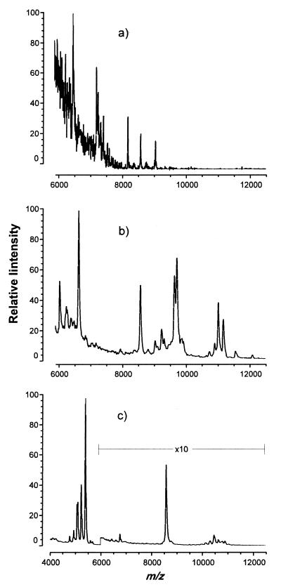
Figure 1a shows the positive-ion MALDI mass spectrum of whole C. parvum oocysts washed three times with deionized water. The mass spectrum has several discernible features, which are summarized in Table 1. Figure 1a represents ∼4 million oocysts contributing to the spot deposited on the MALDI target. Figure 1a shows the range of the mass spectrum from m/z 6,000 to 13,000. Portions of the mass spectrum in m/z 300 to 1,000 contained numerous peaks (>60), similar to another report (Claydon et al., Proc. 48th Am. Soc. Mass Spectrom. Conf.; M. A. Claydon, personal communication) of the MALDI-TOF mass spectrum of whole oocysts. A high signal/noise ratio in the region made mass assignments problematic. Therefore, the region m/z 6,000 to 13,000 was used in further studies, since these peaks were better resolved (more separated in the m/z range) and in a region of the mass spectrum with a lower signal/noise ratio.
FIG. 1.
MALDI-TOF mass spectra of C. parvum and C. muris oocysts. The portion of the mass spectra containing the spectral markers is shown. Other regions were not interesting. (a) Whole C. parvum oocysts washed in deionized water. (b) C. parvum oocysts washed and freeze-thawed. (c) C. muris oocysts washed and freeze-thawed. Note that the region between m/z 6,000 and 12,500 has been vertically expanded by a factor of 10.
TABLE 1.
Comparison of selected spectral-marker ions detected by MALDI-TOF MS for C. parvum and C. muris
| m/z | Ions detected
|
||
|---|---|---|---|
|
C. parvum
|
C. muris (freeze-thawed) | ||
| Whole | Freeze-thawed | ||
| 5,098 | × | ||
| 5,226 | × | ||
| 5,385 | × | ||
| 6,040 | × | ||
| 6,251 | × | ||
| 6,499 | × | ||
| 6,747 | × | ||
| 7,223 | × | ||
| 8,579 | × | × | × |
| 9,059 | × | ||
| 9,243 | × | ||
| 9,724 | × | ||
| 10,478 | × | ||
| 11,024 | × | ||
While the number of spectral markers may be sufficient to form a fingerprint, it would be preferable to have more spectral markers and improved sensitivity. Toward that end, samples were subjected to a freeze-thaw procedure prior to analysis. The mass spectrum shown in Fig. 1b was obtained with ∼200,000 oocysts subjected to a freeze-thaw procedure. The peaks in Fig. 1b probably represent various biomolecules liberated from the oocyst by the freeze-thaw process. To determine whether these mass spectral features were an artifact of the sample preparation procedure, we performed the same procedure on C. muris oocysts. The C. muris sample was prepared with the same freeze-thaw procedure used for C. parvum. Figure 1c shows the MALDI mass spectrum of the C. muris freeze-thaw isolate. Figure 1c differs from Fig. 1b in the peaks between m/z 6,000 and 11,000, with a common peak at m/z 8,579. Thus, the nonsimilar peaks between m/z 6,000 and 11,000 may be considered to represent a fingerprint of the freeze-thawed C. parvum. The C. muris spectrum also shows very large peaks around m/z 5,000 that do not appear for C. parvum. These peaks at m/z ∼5,000 are roughly 10 times the intensity of the other peaks for both species. While ion stability issues have precluded a direct conversion of spectral-marker intensity to molecular mass, the presence of these large peaks was intriguing and warrants further investigation.
Table 1 provides a comparison of selected spectral peaks that originate from whole C. parvum (Fig. 1a), freeze-thawed C. parvum (Fig. 1b), and freeze-thawed C. muris (Fig. 1c). One peak at m/z 8,580 appears to be common to the genus Cryptosporidium and also appears in the whole C. parvum sample. In all, there are at least six spectral markers of higher intensity (and others of lower intensity) present in each batch of C. parvum oocysts that do not appear among C. muris markers. Some spectral markers for whole C. parvum oocysts were not apparent in oocyst samples subjected to the freeze-thaw procedure. This suggests that biomolecules associated with those spectral markers may have been destroyed by the freeze-thaw procedure or separated out during the centrifugation step of the freeze-thaw procedure.
The sensitivity of this technique was estimated from the signal/noise ratio of the mass spectrum of a sample containing approximately 70,000 oocysts. To achieve a signal three times the noise, 20,000 oocysts would have to contribute to the spot in order to produce the spectral markers in Table 1. Some spectral markers were more sensitive than others. For instance, the spectral marker at m/z 11,024 was estimated to require a contribution of only 5,000 oocysts. This is probably related to the ionizability of the biomolecules and the stability of their ions. Sample handling has been implicated as being more important than the absolute sensitivity of the MALDI-TOF MS instrument in establishing a lower limit for the number of organisms that can be quantified (8). Matrix selection may also be important. The minimum number of oocysts required to produce spectral markers is not a significant factor in the present study, because a sufficient quantity of oocysts were available from the oocyst production facility. The number of oocysts required represents less than 0.5% of the total oocysts produced per batch. However, the unreasonably large volumes of water that would have to be collected and processed to obtain this number of oocysts in the environment would make an analysis of naturally occurring C. parvum impractical. Developments in sample handling for MALDI-TOF MS may help lower the detection limit for this analyte, as well as others, such as Bacillus spores (8).
Spectral markers from several batches of freeze-thawed oocysts.
In order to compare the variabilities of oocysts produced in our laboratory, several oocyst lots were collected over a 5-month period. Oocysts from each batch were subjected to MALDI-TOF MS using the freeze-thaw procedure, and their mass spectral peaks were tabulated. These peaks appear in multiple preparations, i.e., desalting, freeze-thaw, and sample spotting, of the MALDI targets from each lot. The peaks may be organized into a MALDI fingerprint, defined by (11)
 |
1 |
where for each peak i, li is the average peak location, sli is the standard deviation in peak location, hi is the average peak height, shi is the standard deviation of peak height, and pi is the fraction of replicates in which peak i appears. In an algorithm for the fingerprint analysis of bacteria (11), the peak height, hi, was not used because the variability in the peak intensities could not be dealt with objectively. Variability was experienced in the analysis of C. parvum. For instance, there was ∼75% variability in the relative intensity of the spectral marker at m/z 9,243; the cause of this variability is unknown. Since marker intensities for bacteria did not generally affect the success of the algorithm (11), they were not considered in the MALDI fingerprint (equation 1) of C. parvum.
Table 2 lists the parameters (equation 1) of the MALDI fingerprint derived from the three lots of C. parvum. In Table 2, most of the peaks appear in each lot and have pis of 1. Two of those listed have pis of 0.67; in these cases, the peak was visually present but its mass could not be confidently determined. The set of peaks between 6,796 and 7,950 appeared in only one lot, so the pi was 0.33. Also included in Table 2 is the percent error in li. Larger values of the percent error in li are associated with difficulties in mass assignment for broader and/or smaller peaks. Thus, the peaks with pis of 0.33 tend to be broader and/or smaller.
TABLE 2.
Fingerprint parameters calculated from three lots of C. parvum
| li (m/z)a | sli (m/z)a | pia | % Errorb |
|---|---|---|---|
| 6,040 | 5 | 1 | 0.08 |
| 6,251 | 2 | 1 | 0.04 |
| 6,285 | 5 | 1 | 0.08 |
| 6,398 | 6 | 1 | 0.09 |
| 6,637 | 6 | 1 | 0.09 |
| 6,796c | 14 | 0.33 | 0.20 |
| 7,220c | 15 | 0.33 | 0.20 |
| 7,567c | 11 | 0.33 | 0.14 |
| 7,739c | 8 | 0.33 | 0.10 |
| 7,950c | 12 | 0.33 | 0.15 |
| 8,579 | 10 | 1 | 0.11 |
| 8,821 | 5 | 1 | 0.06 |
| 9,036 | 6 | 1 | 0.06 |
| 9,118 | 5 | 1 | 0.05 |
| 9,178 | 4 | 0.67 | 0.04 |
| 9,243 | 9 | 1 | 0.09 |
| 9,327 | 3 | 1 | 0.04 |
| 9,645 | 2 | 1 | 0.02 |
| 9,724 | 4 | 1 | 0.04 |
| 10,755 | 3 | 1 | 0.02 |
| 10,910 | 6 | 1 | 0.05 |
| 11,024 | 7 | 1 | 0.06 |
| 11,192 | 13 | 0.67 | 0.11 |
| 12,072 | 8 | 1 | 0.06 |
Unless otherwise noted, these parameters were calculated from spectra acquired from each of three lots. The parameters are defined in the text.
% Error = 100 · sli/li.
Parameters in this row were calculated from three replicate preparations of the same lot because these peaks did not appear in the other lots.
Using the proposed fingerprint of C. parvum contained in Table 2, comparison of future lots can be made through the use of the parameters in Table 2. This fingerprint may be useful for quality control comparison with subsequent lots from in-house production, i.e., if a future lot produces a fingerprint that differs markedly from those in Table 2, action may be warranted. In considering an action level for quality control, it should be noted that in a study with bacteria (11), it was generally necessary for an arbitrary 50% of the fingerprint peaks to be present to confidently identify the organism. In Table 2, >70% of the peaks are found in all C. parvum lots. Therefore, if the calculated fingerprint parameters of an unknown C. parvum lot match an arbitrary ∼70% of the parameters in Table 2, confidence in the lot's production quality may be increased.
Escherichia coli is a potential contaminant and could produce spectral interference. A recent study of E. coli by MALDI-TOF MS (3) reported a peak resulting from E. coli at m/z 9,236, and another study (21) reported a peak resulting from E. coli at m/z 9,241. Both of these are within experimental error of m/z 9,243 (Table 2). However, E. coli produces many peaks (3, 21) not found in the C. parvum spectra. Likewise, peaks observed for C. parvum are not identified for E. coli (3, 21). As an example, an E. coli peak three times more intense than m/z 9,236 has been reported at m/z 9,537 (3), and this peak is not observed for C. parvum. The production procedure for the C. parvum oocysts contains steps to separate the oocysts from bacteria in the sample. Taking the production procedure together with the mass spectral results, it is unlikely that the mass spectra we report are influenced by E. coli contamination.
Effect of disinfection on spectral-marker patterns.
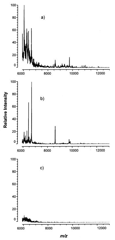
It would be interesting to relate changes in the fingerprint (Table 2) to practical C. parvum issues. While many endpoints may be studied, one relevant issue is how different lots and sources of C. parvum resist disinfection to different degrees (20; Owens et al., Proc. Water Qual. Tech. Conf.). As a first step in this direction, oocysts were subjected to both chlorine and ozone to investigate the effect of chemical disinfection on the spectral-marker patterns of C. parvum. Figure 2a shows the MALDI-TOF MS spectrum obtained from oocysts exposed to ozone (12 mg/liter for 20 min). Compared to the nondisinfected sample (Fig. 1b [1/10 the vertical scale of Fig. 2a]), there is a marked reduction in the intensity of the spectral markers, indicating that the spectral markers were destroyed along with the organism. Most were no longer detectable, with the region m/z 6,000 to 6,400 containing most of the remaining signal. For comparison, the same number of oocysts were subjected to 250 mg of chlorine/liter (a much higher chlorine concentration is required to achieve inactivation rates comparable to those achieved with ozone [12]). The mass spectrum for these oocysts (Fig. 2b) was similar to that of ozonated oocysts (Fig. 2a). When the chlorine concentration was increased to 25,000 mg/liter (far in excess of the amount needed), no spectral-marker-type features were apparent (Fig. 2c).
FIG. 2.
MALDI-TOF mass spectra of freeze-thawed C. parvum oocysts subjected to disinfection. The portion of the mass spectra containing the spectral markers is shown. Other regions were not interesting. (a) Ozone (12 mg/liter) added and reacted for 20 min. (b) Chlorine (250 mg/liter) added and reacted for 20 min. (c) Chlorine (25,000 mg/liter) added and reacted for 20 min. The vertical scale has been adjusted to 10 times the intensity of that in Fig. 1b, which does not contain disinfectant.
The presence of residual disinfectant may interfere with sample ionization and ion stability, degrading the quality of the mass spectra. Ozone, which is volatile, does not leave residual disinfectant. Therefore, the change in the mass spectrum between Fig. 1b and 2a results from chemical disinfection of the oocysts with ozone. Chlorine, added as sodium hypochlorite, is expected to chemically disinfect the oocysts, resulting in changes in the mass spectrum as well (Fig. 2b and c). However, by-products of chlorination may influence the quality of the mass spectrum. MALDI-TOF MS is generally recognized as having a high tolerance for impurities (up to 1 mol/liter, depending on the substance) compared to other mass spectrometric techniques. The chlorination by-products expected in large concentrations are chloroform, chloride ion, and residual hypochlorite ion. Chloroform is volatile and would evaporate during the drying of a spot on the MALDI-TOF MS target. The procedure of washing the oocysts three times with deionized water is designed to reduce the concentration of residual disinfectant by a factor of >1,000, from 5% to a nominal 0.005%. E. coli was successfully used as a 2% suspension in ammonium chloride solution (21), so the presence of chloride would not be expected to affect the MALDI signal. To investigate the possibility of spectral degradation, the oocysts were processed with and without 25,000 mg of sodium hypochlorite/liter. The MALDI-TOF MS target was then spotted, using horse skeleton apomyoglobin as an internal control. As another test, the target was directly spotted with the apomyoglobin with and without 25 mg of sodium hypochlorite solution/liter, which is greater than the concentration expected after the washing steps. In each case, the apomyoglobin produced a parent ion peak whose magnitude was similar within ∼50% to the other cases with no clear trends among the four cases. Given the inherent difficulties in quantification with MALDI-TOF MS (23), the agreement among these results provides confidence that the quality of the mass spectra in Fig. 2b and c is not significantly affected by residual disinfectant, i.e., the residual disinfectant does not produce complete spectral suppression. Thus, Fig. 2b and c primarily represent the destruction of the spectral markers by chlorine, not spectral degradation.
Chemical treatment of oocysts is thought to operate primarily by destroying biomolecules necessary for parasitic activity. Various antigens have been implicated in the ability of the parasite to attach itself to the host, and these antigens have been estimated in the 15-, 17-, 27-, and 47-kDa ranges (17–19). Peaks of these masses are not identifiable in the mass spectrum; however, they may form unstable ions and therefore not be detectable. Taken as a whole, Fig. 2 shows that the appearance of the spectral markers is related to the presence of disinfectants. Thus, the use of these spectral markers in a systematic study relating their presence to Cryptosporidium oocyst resistance would be warranted. A detailed cross comparison of oocysts produced by various sources was beyond the scope of this study, but such a comparison may help to explain why C. parvum oocysts obtained from different sources or lots may exhibit various degrees of resistance to disinfection, or to predict such resistance, and thereby help to answer relevant questions, i.e., whether the appearance or absence of a particular spectral marker correlates with disinfectability of a particular lot. Specifically, do oocysts from lots that have the fingerprint parameters (pi = 0.33) (Table 2) have disinfectabilities different from those of other lots?
Conclusion.
This study may have led to the first MALDI-TOF MS fingerprint of Cryptosporidium oocysts. The mass spectrum of C. parvum differs from that of C. muris, increasing confidence in the specificity of the fingerprint analysis. The analysis was relatively rapid and simple to reproduce. Oocysts from several different lots obtained at the Environmental Protection Agency produced similar mass spectra, suggesting that the technique may be used to supplement quality controls for C. parvum oocyst production. Maintaining the quality of the mass spectral fingerprint generated by the MALDI-TOF MS technique may also lead to more tightly controlled Cryptosporidium disinfection and removal experiments. The disinfection results obtained in this study suggest that further exploration of MALDI-TOF MS for C. parvum analysis is warranted, particularly an investigation of the relationship between spectral markers and the biocidal potentials of various chemical disinfectants, i.e., chlorine, ozone, and chlorine dioxide.
REFERENCES
- 1.Arrowood M J, Donaldson K. Improved purification methods for calf-derived Cryptosporidium parvum oocysts using discontinuous sucrose and cesium chloride gradients. J Eukaryot Microbiol. 1996;43:89S. doi: 10.1111/j.1550-7408.1996.tb05015.x. [DOI] [PubMed] [Google Scholar]
- 2.Bakhtiar R, Nelson R W. Electrospray ionization and matrix-assisted laser desorption ionization mass spectrometry: emerging technologies in biomedical sciences. Biochem Pharm. 2000;59:891–905. doi: 10.1016/s0006-2952(99)00317-2. [DOI] [PubMed] [Google Scholar]
- 3.Demirev P A, Ho Y-P, Ryzhov V, Fenselau C. Microorganism identification by mass spectrometry and protein database searches. Anal Chem. 1999;71:2732–2738. doi: 10.1021/ac990165u. [DOI] [PubMed] [Google Scholar]
- 4.Dubey J P, Speer C A, Fayer R, editors. Cryptosporidiosis of man and animals. Boca Raton, Fla: CRC Press; 1990. [Google Scholar]
- 5.Erhard M, von Dohren H, Jungblut P. Rapid typing and elucidation of new secondary metabolites of intact cyanobacteria using MALDI-TOF mass spectrometry. Nat Biotechnol. 1997;15:906–909. doi: 10.1038/nbt0997-906. [DOI] [PubMed] [Google Scholar]
- 6.Fayer R, editor. Cryptosporidium and cryptosporidiosis. Boca Raton, Fla: CRC Press; 1997. [Google Scholar]
- 7.Franzen C, Muller A. Cryptosporidia and microsporidia—waterborne diseases in the immunocompromised host. Diagn Microbiol Infect Dis. 1999;34:245. doi: 10.1016/s0732-8893(99)00003-6. [DOI] [PubMed] [Google Scholar]
- 8.Hathout Y, Demirev P A, Ho Y-P, Bundy J L, Ryzhov V, Sapp L, Stutler J, Jackman J, Fenselau C. Identification of Bacillus spores by matrix-assisted laser desorption ionization-mass spectrometry. Appl Environ Microbiol. 1999;65:4313–4319. doi: 10.1128/aem.65.10.4313-4319.1999. [DOI] [PMC free article] [PubMed] [Google Scholar]
- 9.Iseki M, Maekawa T, Moriya K, Uni S, Takada S. Infectivity of Cryptosporidium muris (strain RN66) in various laboratory animals. Parasitol Res. 1989;75:218–222. doi: 10.1007/BF00931279. [DOI] [PubMed] [Google Scholar]
- 10.Jarman K H, Daly D S, Petersen C E, Saenz A J, Valentine N B, Wahl K L. Extracting and visualizing matrix-assisted laser desorption/ionization time-of-flight mass spectral fingerprints. Rapid Commun Mass Spectrom. 1999;13:1586–1594. doi: 10.1002/(SICI)1097-0231(19990815)13:15<1586::AID-RCM680>3.0.CO;2-2. [DOI] [PubMed] [Google Scholar]
- 11.Jarman K H, Cebula S T, Saenz A J, Petersen C E, Valentine N B, Kingsley M T, Wahl K L. An algorithm for automated bacterial identification using matrix-assisted laser desorption/ionization mass spectrometry. Anal Chem. 2000;72:1217–1223. doi: 10.1021/ac990832j. [DOI] [PubMed] [Google Scholar]
- 12.Korich D G, Mead J R, Madore M S, Sinclair N A, Sterling C R. Effects of ozone, chlorine dioxide, chlorine, and monochloramine on Cryptosporidium parvum oocyst viability. Appl Environ Microbiol. 1990;56:1423–1428. doi: 10.1128/aem.56.5.1423-1428.1990. [DOI] [PMC free article] [PubMed] [Google Scholar]
- 13.Krishnamurthy T, Ross P L, Rajamani U. Detection of pathogenic and non-pathogenic bacteria by matrix-assisted laser desorption/ionization time-of-flight mass spectrometry. Rapid Commun Mass Spectrom. 1996;10:883–888. doi: 10.1002/(SICI)1097-0231(19960610)10:8<883::AID-RCM594>3.0.CO;2-V. [DOI] [PubMed] [Google Scholar]
- 14.Krishnamurthy T, Ross P L. Rapid identification of bacteria by direct matrix-assisted laser desorption/ionization mass spectrometric analysis of whole cells. Rapid Commun Mass Spectrom. 1996;10:1992–1996. doi: 10.1002/(SICI)1097-0231(199612)10:15<1992::AID-RCM789>3.0.CO;2-V. [DOI] [PubMed] [Google Scholar]
- 15.Lindquist H D A. Emerging pathogens of concern in drinking water. EPA 600/R-99/070. Cincinnati, Ohio: Office of Research and Development, U.S. Environmental Protection Agency; 1999. [Google Scholar]
- 16.McKenzie W R, Hoxie N J, Proctor M E, Gradus M S, Blair K A, Petersen D E, Kazmiercak J J, Addis D G, Fox K, Rose J B, Davis J P. A massive outbreak in Milwaukee of Cryptosporidium infection transmitted through the public water supply. N Engl J Med. 1994;331:161–167. doi: 10.1056/NEJM199407213310304. [DOI] [PubMed] [Google Scholar]
- 17.Moss D M, Chappel C L, Okhuysen P C, DuPont H L, Arrowood M J, Hightower A W, Lammie P J. The antibody response to 27-, 17-, and 15-kDa Cryptosporidium antigens following experimental infection in humans. J Infect Dis. 1998;178:827–833. doi: 10.1086/515377. [DOI] [PubMed] [Google Scholar]
- 18.Nesterenko M V, Woods K, Upton S J. Receptor/ligand interactions between Cryptosporidium parvum and the surface of the host cell. Biochim Biophys Acta. 1999;1454:165–173. doi: 10.1016/s0925-4439(99)00034-4. [DOI] [PubMed] [Google Scholar]
- 19.Nina J M S, McDonald V, Dyson D A, Catchpole J, Uni S, Iseki M, Chiodini P L, McAdam K P W J. Analysis of oocyst wall and sporozoite antigens from three Cryptosporidium species. Infect Immun. 1992;60:1509–1513. doi: 10.1128/iai.60.4.1509-1513.1992. [DOI] [PMC free article] [PubMed] [Google Scholar]
- 20.Rennecker J L, Marinas B J, Owens J H, Rice E W. Inactivation of Cryptosporidium parvum oocysts with ozone. Water Res. 1999;33:2481–2488. [Google Scholar]
- 21.Saenz A J, Petersen C E, Valentine N B, Gantt S L, Jarman K H, Kingsley M T, Wahl K L. Reproducibility of matrix-assisted laser desorption/ionization time-of-flight mass spectrometry for replicate bacterial culture analysis. Rapid Commun Mass Spectrom. 1999;13:1580–1585. doi: 10.1002/(SICI)1097-0231(19990815)13:15<1580::AID-RCM679>3.0.CO;2-V. [DOI] [PubMed] [Google Scholar]
- 22.van Baar B L M. Characterisation of bacteria by matrix-assisted laser desorption/ionisation and electrospray mass spectrometry. FEMS Microbiol Lett. 2000;24:193–219. doi: 10.1016/S0168-6445(99)00036-4. [DOI] [PubMed] [Google Scholar]
- 23.Walker A K, Lund C M, Kinsel G R, Nelson K D. Quantitative determination of the peptide retention of polymeric substances using matrix-assisted laser desorption/ionization mass spectrometry. J Am Soc Mass Spectrom. 2000;11:62–68. doi: 10.1016/S1044-0305(99)00117-8. [DOI] [PubMed] [Google Scholar]
- 24.Welham K J, Domin M A, Johnson K, Jones L, Ashton D S. Characterization of fungal spores by laser desorption/ionization time-of-flight mass spectrometry. Rapid Commun Mass Spectrom. 2000;14:307–310. doi: 10.1002/(SICI)1097-0231(20000315)14:5<307::AID-RCM823>3.0.CO;2-3. [DOI] [PubMed] [Google Scholar]
- 25.Yang S, Healey M C, Du C. Infectivity of preserved Cryptosporidium parvum oocysts for immunosuppressed adult mice. FEMS Immunol Med Microbiol. 1996;13:141–145. doi: 10.1016/0928-8244(95)00096-8. [DOI] [PubMed] [Google Scholar]


