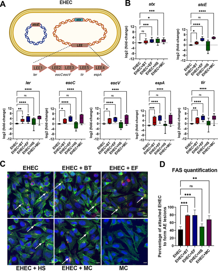
FIG 1.
Bacteroides thetaiotaomicron and Enterococcus faecalis, but not commensal Escherichia coli, enhance EHEC virulence. (A) A schematic of the EHEC major virulence genes: locus of enterocyte effacement (LEE), stx (encoding for Shiga toxin), and stcE (encoding for StcE mucinase). (B) EHEC was anaerobically grown alone or in coculture with commensal strains, and transcription of virulence genes was measured by targeted qRT-PCR. (C) FAS assay to detect actin pedestal formation on HeLa cells infected with mCherry-expressing EHEC and commensal strains for 6 h. Actin and DNA were stained with FITC-phalloidin (green) and DAPI (blue), respectively. White arrows indicate sites of actin polymerization (pedestals). Original magnification: 63×. Scale bar: 10 μm. (D) Quantification of FAS showing the percentage of attached EHEC cells to form actin pedestals. The number of attached EHEC and actin pedestals were enumerated in multiple fields (n = 4). Error bars represent the means ± SD from at least three independent experiments. MC, mixed culture (B. theta, E. faecalis, and commensal E. coli). Statistical significance was determined by one-way ANOVA followed by a post hoc Tukey test. *, P < 0.05; **, P < 0.01; ***, P < 0.001; ****, P < 0.0001; ns, not significant.

