Abstract
Background and Aims
Hepatitis B surface antigen (HBsAg) clearance is significantly more common in children with chronic hepatitis B (CHB) than in adults; however, the possible influencing factors related to HBsAg loss have yet to be found. This study aimed to explore the efficacy of long-term interferon (IFN)α therapy in treating children with CHB and analyzed the factors influencing functional cure after treatment.
Methods
A total of 236 children aged 1–6 years and diagnosed with CHB via liver biopsy were included in the study, all receiving IFNα treatment (IFNα-2b monotherapy, IFNα-2b followed by lamivudine [LAM] or IFNα-2b combined with LAM) and followed up for 144 weeks. A comprehensive analysis was conducted on clinical data, including biochemical items, serum markers of hepatitis B virus (HBV) and immunological indexes, and logistic regression analysis was used to screen the influencing factors related to HBsAg loss.
Results
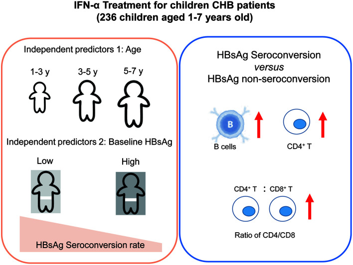
The cumulative loss rates of HBsAg were 79.5%, 62.1% and 42.1% at 144 weeks after the start of treatment in the 1–3 years-old group, 3–5 years-old group and 5–7 years-old group, respectively (p<0.05). IFNα-2b combined with LAM treatment displayed the highest HBsAg loss rates compared with monotherapy and sequential treatment (p=0.011). Younger baseline age and lower HBsAg levels were independent factors for the prediction of HBsAg loss (p<0.05). The baseline PreS1 and hepatitis B core antibody levels in the HBsAg loss group were lower than those in the HBsAg non-loss group. In addition, the PreS1 level was positively corelated with the level of HBsAg, HBV DNA and liver inflammation.
Conclusions
Long-term treatment with IFNα was effective in achieving HBsAg loss in CHB children aged 1–6 years-old. Age less than 3 years-old and lower HBsAg levels are independent predictors of functional cure in children with CHB.
Keywords: Chronic hepatitis B; Children; interferon, IFN; Therapeutic efficacy; Lymphocytes; Predictors
Graphical abstract
Introduction
Hepatitis B virus (HBV) infection has been a major threat to global public health. It is estimated that approximately 50,000–100,000 children are infected per year, with 90% of the cases evolving into chronic infection.1 According to European data, less than 2% children under 3 years-old (y) develop natural hepatitis B e antigen (HBeAg) seroconversion annually, compared with 4–5% in children over 3 y. The annual natural clearance rate of hepatitis B surface antigen (HBsAg) is only 0.6–1%.2 Fortunately, compared with adults, antiviral treatment tends to be more effective in achieving HBeAg and HBsAg seroconversion in HBV-infected children.3,4 The treatment of children with chronic hepatitis B (CHB) aims to maximize the long-term inhibition of HBV replication and reduce the risk of cirrhosis and liver cancer, and for some target patients, “functional cure” should be pursued as far as possible.5–7
Interferon (IFN)α is the first choice for the antiviral treatment of CHB in children. Studies have shown that age is an important predictor of IFNα efficacy, and starting antiviral treatment before 5 y can result in a higher HBsAg loss rate.8–10 The host immune response plays a key role in the determination of functional cure. Our previous data in adults showed that the reversal of excessive activation and functional impairment of B cells was related to HBsAg seroconversion;11 however, the correlation between immunological indicators and disease outcome in children with CHB remains unclear. This study investigated the effect of IFNα on CHB children and identified the relationship between immune factors and HBsAg loss.
Methods
Patients
This retrospective study included 236 children aged 1–6 y. All children were admitted from 2012 to 2018 and diagnosed with CHB according to the American Association for the Study of Liver Diseases (AASLD) 2018 Hepatitis B Guidance.6 The treatments consisted of IFNα-2b (3 MU/m2 to 5 MU/m2, QOD) monotherapy (n=60), IFN sequential lamivudine (LAM) (3 mg/kg/day) (n=73, for which serum HBV DNA declined to <2 log10 after 12 weeks) and IFN combined LAM (3 mg/kg/day) (n=103). After 12 weeks, IFN monotherapy was initiated for the children whose serum HBV DNA declined to <2 log10, and LAM (3 mg/kg/day) was subsequently added (n=73). These children were evaluated every 1 to 3 months. A total of 160 children were treated and followed up for at least 144 weeks, with 76 children dropping out. Clinical and immunologic data were collected and analyzed retrospectively. The following cases were excluded: 1) those who were coinfected with hepatitis A, C, D, or E; 2) those with other known metabolic diseases or immune disorders; and 3) those who had received immunosuppressive or other immunoregulatory agents (including thymosin, etc.) or systemic cytotoxic drugs in the last 6 months. This study was approved by the Medical Ethics Committee of the Fifth Medical Center of the General Hospital of PLA.
Efficacy assessments
The lower limit of detection of HBV DNA was 40 IU/mL (by real-time quantitative polymerase chain reaction; Roche COBAS AmpliPrep). HBeAg seroconversion was defined as the loss of HBeAg and appearance of anti-HBe. HBsAg loss was defined as serum HBsAg <0.05 IU/mL (HBsAg quantification by Roche COBAS HBsAgII-Q, with 0.05 IU/mL as the lower limit of detection). Lymphocyte subsets were detected by flow cytometry (Becton, Dickinson and Company, BD Biosciences), and liver function was assessed at each visit. The primary outcome was the proportion of patients with HBsAg seroconversion 144 weeks after the start of the treatment. The secondary outcomes included switches in virologic and serologic biomarkers at other time points.
Statistical analysis
Statistical analysis was conducted using SPSS Statistics 20.0 (IBM Corp.). The statistical data of the normally distributed continuous variables are expressed as the mean±standard deviation, and the comparison between the two groups was performed by t test. The statistics of nonnormally distributed measurement data are expressed by median (interquartile range), and the Mann-Whitney U test was used for comparisons between the two groups. One-way ANOVA was used for the multigroup comparison. The count data are expressed as percentages, and the chi-squared test or Fisher’s exact probability method was used for comparisons between the two groups. Kaplan-Meier analysis was used to compare the cumulative HBsAg loss rate. Multifactorial analysis was performed by binary logistic regression analysis. All the tests in the study were two-sided, and a p value of 0.05 or less was considered to indicate statistical significance in all tests.
Results
Clinical characteristics of 236 children with HBV infection
A total of 236 children were enrolled in this study, including 131 boys and 105 girls, with the median age being 2.79±1.51 y. According to the age at the first visit, the children were subdivided into the ≥1 to <3 y group (boys/girls: 58/54), the ≥3 to <5 y group (boys/girls: 52/38), and the ≥5 to ≤6 y group (boys/girls: 21/13). The mean duration of the follow-up period was 129.4±51.8 weeks (range: 12–278 weeks) for all of the pediatric patients. Among these children, 86.9% were HBeAg+. HBV genotyping was available for 226 patients, and genotypes B and C accounted for 22.1% (50) and 73% (165) respectively among the patients. Liver biopsy was performed in 232 (98.3%) children. In addition, 46.6% of the children showed mild inflammation (G<2), and 68.1% of them showed mild fibrosis (S<2). The treatments consisted of IFN monotherapy (n=60), IFN sequential LAM (n=73) and IFN combined LAM (n=103).
Efficacy analysis of antiviral treatment
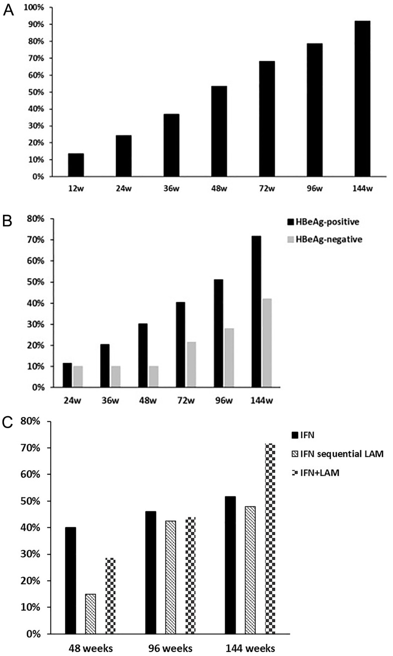
Among the 205 HBeAg+ children (boys/girls: 125/80), 141 children completed the 144-week follow-up. The HBeAg seroconversion rate demonstrated gradual increase, being 13.6%, 24.2%, 36.8%, 53.5%, 68.2%, 78.6%, and 91.9% at each follow-up time point during the 144 weeks of antiviral treatment for the HBeAg+ CHB children (Fig. 1A). The HBsAg loss rates among the HBeAg+ children were 11.4%, 20.5%, 30.1%, 40.4%, 51.1%, and 71.8% at each follow-up time point during the 144 weeks of treatment. Of 31 HBeAg- children (boys/girls: 17/14), 19 completed the 144-week follow-up. The HBsAg loss rates among the HBeAg- children were 10.0%, 10.0%, 10.0%, 21.4%, 28.0%, and 42.1% at each follow-up time point. We also found that the HBsAg loss rate of the two groups significantly varied at each time point (p<0.05), indicating that children who were HBeAg+ at baseline may achieve a higher HBsAg loss rate 144 weeks after antiviral therapy (Fig. 1B). However, because of limitation by the small sample size of the HBeAg- group, the low HBsAg loss rate in the group needs further investigation in future.
Fig. 1. HBsAg loss rate in CHB children undergoing 144-week antiviral treatment.
(A) The proportion of HBeAg seroconversion in HBeAg+ CHB children. (B) The proportion of HBsAg loss in HBeAg+ children (black) vs. HBeAg- children (gray). (C) HBsAg loss rates among different treatment regimens after 144 weeks of treatment. HBsAg, Hepatitis B surface Antigen; CHB, chronic hepatitis B; HBeAg, Hepatitis B e Antigen.
According to the treatment regimen, the children were divided into three groups, namely the IFN monotherapy group, sequential treatment group, and combination treatment group. At the 48th week, the HBsAg loss rates of the three groups were 40.0% (24/60), 15.0% (11/73) and 28.6% (28/98) (χ2=10.465, p=0.005) (Fig. 1C). The HBsAg loss rates of the three groups at the 96th week were 46.0% (28/60), 42.5% (31/73) and 44.0% (37/84) (χ2=0.238, p=0.888). At the 144th week, the HBsAg loss rates were 71.6% (48/67) in the combination treatment group, 51.7% (31/60) in the monotherapy group and 47.9% (35/73) in the sequential treatment group (χ2=8.999, p=0.011).
Cumulative proportion of HBsAg loss
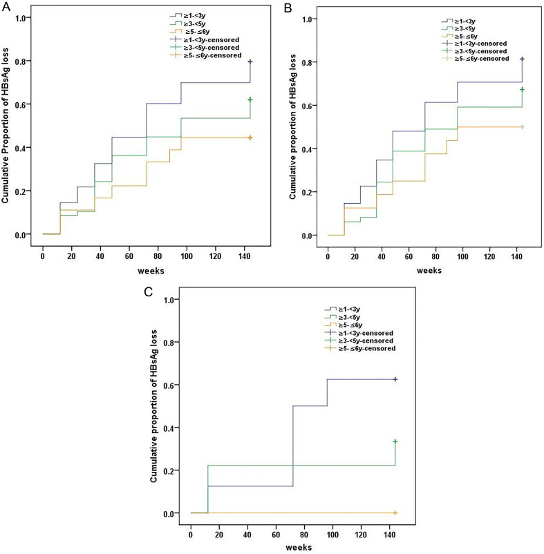
The 160 children who completed follow-up were divided into three age-based groups, as follows: 1–3 y (≥1 to <3 y), 3–5 y (≥3 to <5 y), and 5–7 y (≥5 to <7 y). The Kaplan-Meier analysis showed that the cumulative proportions of HBsAg loss during the 144-week follow-up were 58.9%, 40.0% and 23.5% in the 1–3 y, 3–5 y and 5–7 y groups, respectively (Fig. 2A). The respective mean of the amount of time used to achieve HBsAg loss in the three groups were 77.1 weeks, 93.7 weeks and 104.2 weeks, compared with the 86.2 weeks mean for all patients. The cumulative HBsAg loss curves for the three groups were different, and the pooled overall strata showed that there was a significant difference in the cumulative HBsAg loss rate among the three groups (log rank p=0.001). The pairwise over strata comparisons showed that there were significant differences in the HBsAg loss rate between the 1–3 y group and the 3–5 y group (p=0.01) and between the 1–3 y group and the 5–6 y group (p=0.002). There was no significant difference in the HBsAg loss rate between the 3–5 y group and the 5–6 y group (p=0.129).
Fig. 2. Cumulative proportion of HBsAg loss in CHB children with different ages during the 144-week follow-up period.
(A) The cumulative proportions of HBsAg loss in 160 children. (B)The cumulative proportions of HBsAg loss in HBeAg+ children. (C) The cumulative proportions of HBsAg loss in HBeAg- children. The data were calculated by Kaplan-Meier test. HBsAg, Hepatitis B surface Antigen; CHB, chronic hepatitis B; HBeAg, Hepatitis B e Antigen.
We also identified a change in the cumulative HBsAg loss rate in HBeAg+ CHB children (Fig. 2B). The cumulative proportions of HBsAg loss during the 144-week follow-up were 81.3%, 67.3% and 44.1% respectively in the three age groups (≥1 to <3 y vs. ≥5 to ≤6 y, p=0.030). The mean of the amount of time used to achieve HBsAg loss in the three groups were 75.2 weeks, 89.9 weeks and 99.2 weeks, respectively. For HBeAg- CHB children, the change of the cumulative HBsAg loss rate was also assessed (Fig. 2C). The cumulative proportions of HBsAg loss during the 144-week follow-up were 62.5% and 33.3% in the 1–3 y and 3–5 y groups, respectively (p>0.05). None of the HBsAg- children aged 5–6 y achieved HBsAg loss.
Predictors of HBsAg loss in CHB children
Of the 160 children who completed the 144-week follow-up, only 110 children achieved the goal of HBsAg loss. Sex, genotyping, HBV DNA, HBsAg and alanine aminotransferase level did not vary between the HBsAg loss group and the HBsAg non-loss group (p>0.05), but there was a significant difference in age between the two groups (p<0.05). The B cell count and CD4/CD8 ratio in the HBsAg loss group were both higher than those in the HBsAg non-loss group (p=0.07 and p=0.059, respectively). Age, sex, HBV DNA, HBeAg, HBsAg, alanine aminotransferase, aspartate aminotransferase, B cell count and CD4/CD8 ratio were taken as independent variables, and prognosis was taken as the dependent variable. Binary multivariate logistic regression analysis showed that age at the start of treatment and HBsAg level were influencing factors for the prognosis of CHB children after 144 weeks of treatment (Table 1). We examined the HBsAg loss in children with CHB between the ages of 1 and 6 y, and revealed the role played by age and the level of HBsAg in this process by showing that children of lower age and with lower HBsAg levels are more likely to obtain HBsAg loss.
Table 1. Predictors of HBsAg seroconversion in CHB children.
| HBsAg loss (n=110) | HBsAg non-loss (n=50) | Univariate | OR (95% CI) | Multivariate | |
|---|---|---|---|---|---|
| Age in years at treatment | 2 (1, 6) | 3 (2, 6) | 0.002 | 1.553 (1.019–2.369) | 0.041 |
| Sex, male (%) | 53 (48.2) | 32 (64) | 0.065 | 1.264 (0.418–3.820) | 0.678 |
| Treatment regimens | 31/34/45 | 8/22/20 | 0.158 | ||
| HBV genotype, C (%) | 73 (66.4) | 33 (66) | 0.229 | ||
| HBeAg+, % | 102 (92.7) | 39 (78) | 0.011 | 4.982 (0.726–32.569) | 0.094 |
| Log (HBV DNA), IU/mL | 7.8 (7.11, 8.2) | 8.17 (6.59, 8.57) | 0.764 | 0.533 (0.256–1.111) | 0.093 |
| Log (HBsAg), IU/mL | 4.25 (3.56, 4.55) | 4.27 (3.57, 4.7) | 0.472 | 6.329 (1.315–30.471) | 0.021 |
| ALT, U/L | 85.5 (53, 128) | 125 (60.75, 174.75) | 0.065 | 1.001 (0.993–1.008) | 0.864 |
| AST, U/L | 76 (59.25, 112) | 106 (62.75, 201.25) | 0.014 | 1.005 (0.996–1.014) | 0.318 |
| Lymphocytes, cell/µL | 4,140 (3,515, 5,550) | 2,285 (1,864, 3,506.25) | 0.303 | ||
| T cell, cell/µL | 2,660 (2,267, 3,623.5) | 3,785 (2,820, 5,187.5) | 0.497 | ||
| CD4+ T cell, cell/µL | 1,581 (1,175, 1,985.5) | 1,273.5 (929.75, 1,852) | 0.121 | ||
| CD8+ T cell, cell/µL | 934 (765, 1,275) | 872 (635.25, 1,193.25) | 0.959 | ||
| B cell, cell/µL | 1,079 (752.5, 1,368.5) | 764 (607, 1,137.75) | 0.070 | 0.999 (0.995–1.004) | 0.832 |
| NK cell, cell/µL | 236 (151.5, 443.5) | 207 (153, 521.25) | 0.593 | ||
| CD4/CD8 | 1.59 (1.295, 2.01) | 1.52 (1.02, 1.78) | 0.059 | 0.528 (0.064–4.331) | 0.552 |
ALT, alanine aminotransferase; AST, aspartate aminotransferase; CI, confidence interval; NK, natural killer; OR, odds ratio; HBsAg, Hepatitis B surface Antigen; HBeAg, Hepatitis B e Antigen.
Serological profiles
The dynamic changes in lymphocyte subsets at different time points are shown in Table 2. After 12 weeks of treatment, ALT and HBV DNA levels dropped significantly from the baseline level, while the lymphocyte counts revealed a decline in both the HBeAg+ and HBeAg- groups. In the group of patients with HBsAg loss during follow-up, the absolute lymphocyte count was significantly lower compared with the group that is without. In addition, the CD4+/CD8+ T cell ratio was significantly lower than that before the treatment (p<0.05). At the 24th week, 36th week and the 48th week, the CD8+ T cell count, B cell count, natural killer cell count and CD4+/CD8+ T cell ratio in the HBsAg loss group were significantly higher than those in the HBsAg non-loss group (p<0.05).
Table 2. Dynamic of HBV DNA, HBsAg, ALT, AST and lymphocyte subsets of HBeAg+ patients at various visiting time-points.
| HBsAg non-loss group | |||||
|---|---|---|---|---|---|
| Week | 0 (n=32) | 12 (n=30) | 24 (n=17) | 36 (n=21) | 48 (n=20) |
| Log (DNA), IU/mL | 7.8±1.5 | 5.1±2.5 | 2.8±1.7 | 1.9±0.6 | 2.1±0.7 |
| Log (HBsAg), IU/mL | 4.3±0.7 | 3.8±0.9 | 3.8±1.0 | 3.4±0.8 | 3.3±1.1 |
| ALT, U/L | 158.8±170.2 | 39.1±40.9 | 28.3±15.6 | 23.0±13.3 | 23.8±13.6 |
| AST, U/L | 144.1±140.2 | 96.3±108.6 | 62.5±32.9 | 52.0±18.7 | 55.6±18.3 |
| Lymphocytes, cell/µL | 4,300±2,106 | 4,237±1,816 | 4,141±1,875 | 3,455±1,789 | 3,406±1,564 |
| T cell, cell/µL | 2,841±1,550 | 2,785±1,239 | 2,688±1,212 | 2,278±1,257 | 2,113±961 |
| CD4+T cell, cell/µL | 1,458±712 | 1,290±594 | 1,230±593 | 991±467 | 1,003±447 |
| CD8+T cell, cell/µL | 1,086±783 | 1,158±583 | 1,107±551 | 1,068±748 | 896±526 |
| B cells, cell/µL | 930±498 | 971±510 | 934±552 | 805±415 | 915±515 |
| NK cells, cell/µL | 377±3,226 | 330±260 | 398±220 | 267±166 | 291±216 |
| CD4/CD8% | 1.48±0.5 | 1.23±0.4 | 1.26±0.4 | 1.09±0.4* | 1.25±0.4 |
| HBsAg loss group | |||||
|---|---|---|---|---|---|
| Week | 0 (n=69) | 12 (n=71) | 24 (n=43) | 36 (n=50) | 48 (n=50) |
| Log (DNA), IU/mL | 7.60±1.2 | 4.381±2.4 | 2.96±1.8 | 1.88±0.5 | 1.63±0.2 |
| Log (HBsAg), IU/mL | 4.03±1.0 | 2.54±1.9 | 1.81±2.3 | 0.49±2.1 | −0.51±1.3 |
| ALT, U/L | 114.7±126.7 | 30.06±27.5 | 32.59±32.1 | 21.14±14.6 | 27.42±34.0 |
| AST, U/L | 105.5±102.3 | 84.3±72.9 | 69.2±32.9 | 59.7±26.3 | 58.4±34.3 |
| Lymphocytes, cell/µL | 4,670±1,822 | 4,181.55±1,765 | 3,750±1,404* | 4,282±1,862 | 4,057±1,669 |
| T cells, cell/µL | 3,014±1,180 | 2,562±1,036 | 2,278±810* | 2,494±985 | 2,406±972* |
| CD4+T cells, cell/µL | 1,674±765* | 1,348±630 | 1,168±454 | 1,270±592 | 1,203±579 |
| CD8+T cells, cell/µL | 1,059±447 | 971±461 | 895±369 | 980±465 | 956±394 |
| B cells, cell/µL | 1,175±652 | 1,197±670 | 1,039±539 | 1,216±693 | 1,039±511 |
| NK cells, cell/µL | 341±255 | 314±204* | 318±181* | 429±356 | 478±400 |
| CD4/CD8% | 1.68±0.6* | 1.47±0.5 | 1.38±0.4 | 1.34±0.4 | 1.27±0.4 |
*p<0.05 as compared to those in HBsAg non-loss group at the same time-points after antiviral treatment. HBsAg, Hepatitis B surface Antigen; HBeAg, Hepatitis B e Antigen; ALT, alanine aminotransferase; AST, aspartate aminotransferase; NK, natural killer.
PreS1 and hepatitis B core antibody (HBcAb) gradually decreased with prolonged antiviral treatment time
We monitored the changes in PreS1 and HBcAb concentrations in children aged 1–6 y and found that PreS1 and HBcAb gradually decreased as the antiviral treatment was prolonged. Importantly, the baseline PreS1 levels were significantly higher in the HBsAg loss group (n=15, 3.86±1.55 log IU/mL) than in the HBsAg non-loss group (n=20, 3.31±1.60 log IU/mL), while the baseline HBcAb levels were significantly lower in the HBsAg loss group (2.59±0.97 log IU/mL) compared with the HBsAg non-loss group (3.18±0.41 log IU/mL, p<0.05). Analysis revealed a positive correlation between serum PreS1 and liver inflammatory score (Kendall’s tau-b=0.343, p=0.018), but no significant correlations between HBcAb and liver inflammation and fibrosis scores were observed. Instead of HBcAb, serum PreS1 was found to be positively correlated with HBV DNA and HBsAg in plasma (r=0.365, p=0.034; r=0.403, p=0.02).
Discussion
With mother-to-child vertical transmission being the cause of the majority of CHB cases in adults in East and Southeast Asia, the region outnumbers Europe and America in terms of CHB pediatric patients.12,13 Prompt antiviral treatment during immune activation or reactivation is important, for it can reduce the risk of cirrhosis or liver cancer and can achieve functional cure in some children.14 Since HBsAg clearance is less common in the adult cohort, it is difficult to observe the influencing factors related to it. Given the greater prevalence of HBsAg clearance among CHB children, we conducted a comprehensive analysis on the clinical data of liver biopsies collected from 236 CHB children and explored the possible influencing factors related to HBsAg loss in this study.
Previous studies have shown that 96 weeks of pegylated (Peg)-IFNα-2a treatment may result in higher HBeAg seroconversion rates and higher HBsAg clearance rates compared with 48-week Peg-IFNα-2a treatment in HBeAg+ CHB children,4,15,16 suggesting that combination or sequential therapy with IFN and nucleotide analogues may be adopted to achieve better efficacy.9,17,18 Our data showed higher HBsAg loss rates in the combination treatment group compared with the IFN monotherapy group and sequential treatment group after 144 weeks of therapy. These data also suggested the potential of longer treatment to achieve higher HBsAg loss rates in CHB children.
The factors influencing HBsAg loss remain unknown. A study from China showed that antiviral therapy imitated in children before 5 y could allow for achievement of a higher HBsAg clearance rate.19 Another study reported that much higher HBsAg clearance rate could be achieved in children who received antiviral treatment when they were younger than 3 y.20 Our data showed that the HBsAg loss rate after antiviral treatment in the 1–3 y group was significantly higher than that in the 3–5 y and 5–6 y groups. In addition, the children in the 1–3 y group achieved HBsAg loss more quickly than those in the other two groups. Multivariate logistic regression analysis showed that age and HBsAg level were influencing factors for HBsAg loss in CHB patients.
Previous studies have shown that host immune responses play an active role in HBV clearance and liver pathogenesis when it comes to chronic hepatitis, especially HBV-specific T and B cells, which are important for virus clearance and disease transformation.21,22 This study describes the dynamic changes in some virological, biochemical and immunological parameters associated with HBsAg loss during IFN therapy in CHB children. We found that the immune cell proportion is an important factor associated with HBsAg loss. For example, the baseline B cell count and CD4/CD8 ratio were higher in the HBsAg loss group than in the HBsAg non-loss group. These data indicated that monitoring peripheral lymphocyte subsets was helpful in predicting prognosis and antiviral treatment efficacy in CHB children. Notably, the lower HBsAg loss rate was observed in HBeAg- group as compared to HBeAg+ group, which was possibly due to the small sample size of the HBeAg- group. Future studies should comprehensively assess the biochemical markers, serum markers and immunological factors associated with HBsAg loss in a larger cohort of children CHB carriers in multiple center trials.
Our data also suggested that baseline virological factors are closely correlated with HBsAg loss. Positive correlation is also identified between the levels of HBsAg and HBV DNA in children with CHB and their serum PreS1 levels, which were significantly higher in the HBsAg loss group than in the HBsAg non-loss group. Indeed, previous studies have also demonstrated similar association between PreS1 levels and HBeAg and HBV DNA levels in adult CHB patients.23 These data suggested that PreS1 could be used to predict the antiviral effect in children with CHB. Furthermore, recent studies have shown that baseline HBcAb levels can not only be used to predict the efficacy of antiviral therapy but are also correlated with the degree of liver inflammation and liver fibrosis in CHB patients.24 A few clinical studies have examined the relationship between baseline HBcAb levels and antiviral efficacy in CHB children. Our results showed that the serum baseline HBcAb levels were significantly lower in children who achieved HBsAg loss than in children who did not achieve HBsAg loss. Notably, no correlation was observed between the serum HBcAb levels and the liver inflammation and fibrosis scores in CHB children, which is different from adult cases. Clinical trials with larger cohorts are warranted to confirm the difference between children and adult patients with CHB.
In conclusion, CHB children may achieve functional cure in the clinic more easily with a relatively higher cumulative proportion of HBsAg loss than adult CHB patients after long-term antiviral treatment. Age and HBsAg level were significantly associated with the achievement of HBsAg loss in children with CHB. Future studies should investigate the functional roles of several key immunological and virological factors, such as B cells, CD4/CD8, PreS1 and HBcAb, at baseline, which may be good predictors of antiviral therapy efficacy.
Abbreviations
- ALT
alanine aminotransferase
- AST
aspartate aminotransferase
- CHB
chronic hepatitis B
- CI
confidence interval
- HBV
hepatitis B virus
- HBcAb
hepatitis B core antibody
- HBeAg
hepatitis B e antigen
- HBsAg
hepatitis B surface antigen
- IFN
Interferon
- LAM
lamivudine
- NK
natural killer
- OR
odds ratio
Data sharing statement
All data are available upon request.
References
- 1.Ott JJ, Stevens GA, Groeger J, Wiersma ST. Global epidemiology of hepatitis B virus infection: new estimates of age-specific HBsAg seroprevalence and endemicity. Vaccine. 2012;30:2212–2219. doi: 10.1016/j.vaccine.2011.12.116. [DOI] [PubMed] [Google Scholar]
- 2.Fattovich G, Bortolotti F, Donato F. Natural history of chronic hepatitis B: special emphasis on disease progression and prognostic factors. J Hepatol. 2008;48:335–352. doi: 10.1016/j.jhep.2007.11.011. [DOI] [PubMed] [Google Scholar]
- 3.Zhu S, Dong Y, Wang L, Liu W, Zhao P. Early initiation of antiviral therapy contributes to a rapid and significant loss of serum HBsAg in infantile-onset hepatitis B. J Hepatol. 2019;71(5):871–875. doi: 10.1016/j.jhep.2019.06.009. [DOI] [PubMed] [Google Scholar]
- 4.Wirth S, Zhang H, Hardikar W, Schwarz KB, Sokal E, Yang W, et al. Efficacy and Safety of Peginterferon Alfa-2a (40KD) in Children With Chronic Hepatitis B: The PEG-B-ACTIVE Study. Hepatology. 2018;68(5):1681–1694. doi: 10.1002/hep.30050. [DOI] [PubMed] [Google Scholar]
- 5.European Association for the Study of the Liver Electronic address eee, European Association for the Study of the L. EASL 2017 Clinical Practice Guidelines on the management of hepatitis B virus infection. J Hepatol. 2017;67:370–398. doi: 10.1016/j.jhep.2017.03.021. [DOI] [PubMed] [Google Scholar]
- 6.Terrault NA, Lok ASF, McMahon BJ, Chang KM, Hwang JP, Jonas MM, et al. Update on prevention, diagnosis, and treatment of chronic hepatitis B: AASLD 2018 hepatitis B guidance. Hepatology. 2018;67:1560–1599. doi: 10.1002/hep.29800. [DOI] [PMC free article] [PubMed] [Google Scholar]
- 7.Chinese Society of Infectious Diseases CMA The guidelines of prevention and treatment for chronic hepatitis B (2019 version) Zhonghua Gan Zang Bing Za Zhi. 2019;27(12):938–961. doi: 10.3760/cma.j.issn.1007-3418.2019.12.007. [DOI] [PubMed] [Google Scholar]
- 8.Jonas MM, Lok AS, McMahon BJ, Brown RS, Wong JB, Ahmed AT, et al. Antiviral therapy in management of chronic hepatitis B viral infection in children: A systematic review and meta-analysis. Hepatology. 2016;63:307–318. doi: 10.1002/hep.28278. [DOI] [PubMed] [Google Scholar]
- 9.Kobak GE, MacKenzie T, Sokol RJ, Narkewicz MR. Interferon treatment for chronic hepatitis B: enhanced response in children 5 years old or younger. J Pediatr. 2004;145:340–345. doi: 10.1016/j.jpeds.2004.05.046. [DOI] [PubMed] [Google Scholar]
- 10.Wu JF, Chiu YC, Chang KC, Chen HL, Ni YH, Hsu HY, et al. Predictors of hepatitis B e antigen-negative hepatitis in chronic hepatitis B virus-infected patients from childhood to adulthood. Hepatology. 2016;63(1):74–82. doi: 10.1002/hep.28222. [DOI] [PubMed] [Google Scholar]
- 11.Xu X, Shang Q, Chen X, Nie W, Zou Z, Huang A, et al. Reversal of B-cell hyperactivation and functional impairment is associated with HBsAg seroconversion in chronic hepatitis B patients. Cellular & molecular immunology. 2015;12:309–316. doi: 10.1038/cmi.2015.25. [DOI] [PMC free article] [PubMed] [Google Scholar]
- 12.Lok AS, McMahon BJ. Chronic hepatitis B. Hepatology. 2007;45:507–539. doi: 10.1002/hep.21513. [DOI] [PubMed] [Google Scholar]
- 13.Choe HJ, Choe BH. What physicians should know about the management of chronic hepatitis B in children: East side story. World J Gastroenterol. 2014;20:3582–3589. doi: 10.3748/wjg.v20.i13.3582. [DOI] [PMC free article] [PubMed] [Google Scholar]
- 14.Marcellin P, Asselah T. Long-term therapy for chronic hepatitis B: hepatitis B virus DNA suppression leading to cirrhosis reversal. J Gastroenterol Hepatol. 2013;28:912–923. doi: 10.1111/jgh.12213. [DOI] [PubMed] [Google Scholar]
- 15.European Association for the Study of the Liver EASL 2017 Clinical Practice Guidelines on the management of hepatitis B virus infection. J Hepatol. 2017;67(2):370–398. doi: 10.1016/j.jhep.2017.03.021. [DOI] [PubMed] [Google Scholar]
- 16.Liu Y, Li H, Yan X, Wei J. Long-term efficacy and safety of peginterferon in the treatment of children with HBeAg-positive chronic hepatitis B. J Viral Hepat. 2019;26(Suppl 1):69–76. doi: 10.1111/jvh.13154. [DOI] [PubMed] [Google Scholar]
- 17.Mieli-Vergani G, Bansal S, Daniel JF, Kansu A, Kelly D, Martin C, et al. Peginterferon Alfa-2a (40KD) Plus Lamivudine or Entecavir in Children with Immune-tolerant Chronic Hepatitis B. J Pediatr Gastroenterol Nutr. 2021;73(2):156–160. doi: 10.1097/MPG.0000000000003118. [DOI] [PubMed] [Google Scholar]
- 18.Dong Y, Li M, Zhu S, Gao X, Zhao P. De novo combination antiviral therapy in e antigen-negative chronic hepatitis B virus-infected paediatric patients with advanced fibrosis. J Viral Hepat. 2020;27(12):1338–1343. doi: 10.1111/jvh.13372. [DOI] [PubMed] [Google Scholar]
- 19.Zhu S, Dong Y, Xu Z, Wang L, Chen D, Gan Y, et al. A retrospective study on HBsAg clearance rate after antiviral therapy in children with HBeAg-positive chronic hepatitis B aged 1-7 years. Chin J Hepatol. 2016;24:738–743. doi: 10.3760/cma.j.issn.1007-3418.2016.10.005. [DOI] [PubMed] [Google Scholar]
- 20.Zhu S, Dong Y. A retropective study on the liver pathological characteristics and the effect of antiviral treatment for 1 to 7 years old chidren with hetitis B e antigen negative chronic hepatitis B. Chin J Pediatr. 2016;54:587–591. doi: 10.3760/cma.j.issn.0578-1310.2016.08.006. [DOI] [PubMed] [Google Scholar]
- 21.Li HJ, Zhai NC, Song HX, Yang Y, Cui A, Li TY, et al. The Role of Immune Cells in Chronic HBV Infection. J Clin Transl Hepatol. 2015;3(4):277–283. doi: 10.14218/JCTH.2015.00026. [DOI] [PMC free article] [PubMed] [Google Scholar]
- 22.Khanam A, Chua JV, Kottilil S. Immunopathology of Chronic Hepatitis B Infection: Role of Innate and Adaptive Immune Response in Disease Progression. Int J Mol Sci. 2021;22(11):5497. doi: 10.3390/ijms22115497. [DOI] [PMC free article] [PubMed] [Google Scholar]
- 23.Cavallone D, Ricco G, Oliveri F, Colombatto P, Moriconi F, Coco B, et al. Do the circulating Pre-S/S quasispecies influence hepatitis B virus surface antigen levels in the HBeAg negative phase of HBV infection? Aliment Pharmacol Ther. 2020;51(12):1406–1416. doi: 10.1111/apt.15753. [DOI] [PubMed] [Google Scholar]
- 24.Li MR, Lu JH, Ye LH, Sun XL, Zheng YH, Liu ZQ, et al. Quantitative hepatitis B core antibody level is associated with inflammatory activity in treatment-naive chronic hepatitis B patients. Medicine (Baltimore) 2016;95:e4422. doi: 10.1097/MD.0000000000004422. [DOI] [PMC free article] [PubMed] [Google Scholar]


