Abstract
Practical sessions facilitate teaching, critical thinking, and coping skills, especially among medical students and professionals. Currently, in ophthalmology, virtual and augmented reality are employed for surgical training by using three-dimensional (3D) eyeball models. These 3D models when printed can be used not only for surgical training but also in teaching ophthalmic residents and fellows for concept learning through tactile 3D puzzle assembly. 3D printing is perfectly suited for the creation of complex bespoke items in a cost-effective manner, making it ideal for rapid prototyping. Puzzle making, when combined with 3D printing can evolve into a different level of learning in the field of ophthalmology. Though various 3D eyeball models are currently available, complex structures such as the cerebral venous system and the circle of Willis have never been 3D printed and presented as 3D puzzles for assembling and learning. According to our knowledge, this concept of ophthalmic pedagogy has never been reported. In this manuscript, we discuss in detail the 3D models created by us (patent pending), for printing into multiple puzzle pieces for effective tactile learning by cognitive assembling.
Keywords: 3D Models, 3D Printing, 3D Puzzle, Tactile Learning
Three-dimensional (3D) printing, or additive manufacturing, is the process of creating 3D solid objects from a digital file designed using computer-aided design (CAD) or digital 3D computer graphics software.[1] Toys and puzzles are used to improve intellectual knowledge along with playfulness in young children. Similarly, this gamification can be used for skillful and tactile learning for ophthalmologists and optometrists. These tools create a memory in the brain and make teaching easy by developing their cognizance, especially when it is a 3D model with actual measurement and color.[2] In addition, holding a model in hand will definitely improve the actual learning experience.
Innovation
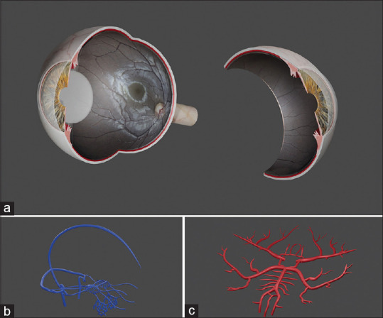
3D printed models created by us (patent pending) can be extensively used in the field of ophthalmology, especially for teaching.[3,4,5] We have created a 3D eyeball model [Fig. 1] with detailed microscopic structures.[6] We have also created models associated with ophthalmology, such as the cerebral venous system and circle of Willis in 3D computer graphics software, and then separated the model just like the pieces of a puzzle [Figs. 2–4 and Video Clip 1].
Figure 1.

Image showing the models created in the 3D computer graphics software. (a) Eyeball model with TrueColor high-resolution confocal fundus image. (b) Cerebral venous system. (c) Circle of Willis
Figure 2.

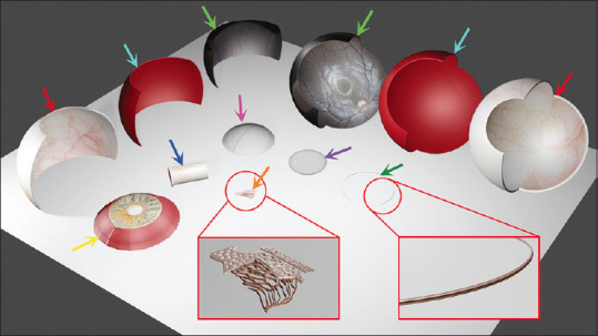
Image showing the eyeball model split into different parts: sclera (red arrows), choroid (light blue arrows), retina (light green arrows), iris and ciliary body (yellow arrow), optic nerve (dark blue arrow), cornea (rose arrow), lens (purple arrow), trabecular meshwork (orange arrow), and anterior chamber angle (green arrow) for 3D puzzle printing
Figure 4.

Image showing the circle of Willis split into different parts: posterior communicating artery (green arrow) puzzle, posterior cerebral artery, superior cerebellar artery, anterior inferior cerebellar artery, vertebral artery, basilar artery, and pontine artery (yellow arrow) puzzle complex, anterior communicating artery, anterior cerebral artery (blue arrow) puzzle complex, the internal carotid artery, middle cerebral artery and ophthalmic artery (dark blue arrows) puzzle complex for 3D puzzle printing
Figure 3.

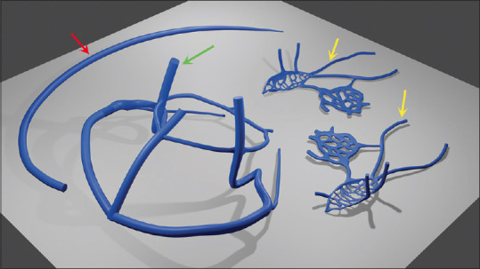
Image showing the separate parts of the cerebral venous system split into the superior sagittal sinus (red arrow) puzzle, superior and inferior petrosal sinus, internal jugular vein, and transverse sinus (green arrow) puzzle complex, superior and inferior ophthalmic vein, cavernous sinus, and pterygoid venous plexus (yellow arrows) puzzle complex for 3D puzzle printing
Printing the Models
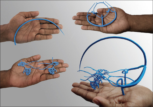
These miniature models can be printed with polylactic acid (PLA) material separately using the 3D printing technology, and then they can be joined to create the final model [Fig. 5] by the neophytes, making learning more interactive. The cerebral venous and arterial system 3D models that we have created, are far deeper yet simpler, than the other 2D and 3D models currently available. It is feasible to print and we have made it in the proportion of an adult human brain. However, the same can be made in a bigger proportion if needed. Eye models with complex microscopic structures such as the angles of the anterior chamber and the usage of TrueColor confocal multimodal fundus images for retina makes these models unique. When used as a puzzle, these 3D models become more productive and effective.[7] The 3D eye ball model cannot be 3D printed with multiple colors as such. First, each part was printed in a single-color material with adequate supports. Then, we stuck the 2D color print paper or plastic onto the model. For the retina, thermoplastic polyurethane (TPU) material was used for 3D printing. For unicoloured structures such as the circle of Willis, red spray paint [Video Clip 2] can be used after 3D printing the model, or the model itself can be printed in red.
Figure 5.

Image showing the 3D printed puzzle pieces of the cerebral venous system and the final model of the cerebral venous system after the puzzle pieces were joined together
Economics
The economics involved in 3D printing models is as follows:
PLA (1kg) - Rs. 1,000
TPU (1kg) - Rs. 3,000
Circle of Willis Model (400g) - Rs. 500
Cerebral Venous System Model (400g) - Rs. 500
Eyeball Model (1 kg) - Rs. 2,000
Duration of printing
The duration of printing the 3D models is as follows:
The circle of Willis - 10 hours
The cerebral venous system - 5 hours
The eyeball - 48 hours
Basic or beginner informative knowledge about 3D printing is required to get the models 3D printed. The 3D models created by us are currently available for free download from the website (meh.org.in), under the RESEARCH & DEVELOPMENT Tab.
Discussion
3D printing has already started to evolve over the past few years and can be used in various applications in the field of ophthalmology.[3] Now it is time to use it extensively in the field of teaching in ophthalmology as there are no real human eye anatomical models that can be used for learning and teaching because of its complex microscopic structures. There are many virtual anatomical dissection ophthalmic tools, which are very complex. In contrast, we have proposed printing real-time 3D models in the actual size of the human eyeball, or with a much larger size, printed for the microscopic structures such as the trabecular meshwork for better tactile understanding. These models, when created as 3D puzzles, improve the art of teaching and learning, especially among neophytes for yielding fruitful results. It can also be applied in other fields beyond ophthalmology.
Conclusion
3D printing, ophthalmology and ophthalmology related anatomical structures as puzzles; and assembling these puzzles can pave the way for a new-age of cognitive tactile learning in the field of ophthalmology. According to our knowledge, this concept of ophthalmic pedagogy has never been reported in the literature.
Declaration of patient consent
The authors certify that they have obtained all appropriate patient consent forms. In the form the patient(s) has/have given his/her/their consent for his/her/their images and other clinical information to be reported in the journal. The patients understand that their names and initials will not be published and due efforts will be made to conceal their identity, but anonymity cannot be guaranteed.
Financial support and sponsorship
Nil.
Conflicts of interest
There are no conflicts of interest.
Videos available on: www.ijo.in
References
- 1.3D printing. Wikipedia. 2021. [Last accessed on 2021 Oct 05]. Available from: https://en.wikipedia.org/w/index.php?title=3D_printing&oldid=1045923488 .
- 2.Sehnonimo. 3D Jigsaw Puzzles and Their Benefits. HubPages. [Last accessed on 2021 Oct 05]. Available from: https://discover.hubpages.com/games-hobbies/3D-Puzzle .
- 3.Akkara JD, Kuriakose A. The magic of three-dimensional printing in ophthalmology. Kerala J Ophthalmol. 2018;30:209–15. Available from: http://www.kjophthal.com/text.asp?2018/30/3/209/247598 . [Google Scholar]
- 4.Adams JW, Paxton L, Dawes K, Burlak K, Quayle M, McMenamin PG. 3D printed reproductions of orbital dissections: A novel mode of visualising anatomy for trainees in ophthalmology or optometry. Br J Ophthalmol. 2015;99:1162–7. doi: 10.1136/bjophthalmol-2014-306189. [DOI] [PubMed] [Google Scholar]
- 5.Dave TV, Tiple S, Vempati S, Palo M, Ali MJ, Kaliki S, et al. Low-cost three-dimensional printed orbital template-assisted patient-specific implants for the correction of spherical orbital implant migration. Indian J Ophthalmol. 2018;66:1600–7. doi: 10.4103/ijo.IJO_472_18. [DOI] [PMC free article] [PubMed] [Google Scholar]
- 6.Ramesh PV, Aji K, Joshua T, Ramesh SV, Ray P, Raj PM, et al. Immersive photoreal new-age innovative gameful pedagogy for e-ophthalmology with 3D augmented reality. Indian Journal of Ophthalmology. 2022;70:275–80. doi: 10.4103/ijo.IJO_2133_21. [DOI] [PMC free article] [PubMed] [Google Scholar]
- 7.Harring ton Z. 7 Spectacular Benefits of Puzzles (Hint: They’re Not What You’d Think!) |Melissa &Doug Blog. [Last accessed on 2021 Oct 05]. Available from: https://www.melissaanddoug. com/on/demandware.store/Sites-melissaanddoug-Site/en_US/Blog-PostView?postId=7-spectacular-benefits-of-puzzles-hint-theyre-not-what-youd-think .
Associated Data
This section collects any data citations, data availability statements, or supplementary materials included in this article.


