Abstract
Background
Transmissible viral proventriculitis (TVP) causes significant economic loss to the poultry industry. However, the exact causative agents are obscure. Here we examine the virome of proventriculus from specified pathogen free (SPF) chickens that reproduced by infection of proventricular homogenate from broiler chicken with TVP using long read sequencing of the Pacific Biosciences RSII platform. The normal SPF chickens were used as control.
Results
Our investigation reveals a virome of proventriculitis, including three Gyrovirus genera of the Aneloviridae: Gyrovirus homsa1 (GyH1) (also known as Gyrovirus 3, GyV3) (n = 2662), chicken anemia virus (CAV) (n = 482) and Gyrovirus galga1 (GyG1) (also known as avian Gyrovirus 2, AGV2) (n = 11); a plethora of novel CRESS viral genomes (n = 26) and a novel genomovirus. The 27 novel viruses were divided into three clusters. Phylogenetic analysis showed that the GyH1 strain was more closely related to the strains from chicken (MG366592) than mammalian (human and cat), the GyG1 strain was closely related to the strains from cat in China (MK089245) and from chicken in Brazil (HM590588), and the CAV strain was more closely related to the strains from Germany (AJ297684) and United Kingdom (U66304) than that previously found in China.
Conclusion
In this study, we revealed that Gyrovirus virome showed high abundance in chickens with TVP, suggesting their potential role in TVP, especially GyH1. This study is expected to contribute to the knowledge of the etiology of TVP.
Supplementary Information
The online version contains supplementary material available at 10.1186/s12917-022-03339-9.
Keywords: Transmissible viral proventriculitis, Virome, Gyrovirus, Novel CRESS virus, Pacific Biosciences RSII platform
Background
Transmissible viral proventriculitis (TVP) is an infectious disease reported in all types of chickens and has significant impact on the poultry industry. The typical pathological lesions of TVP are necrosis of oxynticopeptic cells, lymphocytic infiltration and hyperplastic ductal epithelium which replaces the glandular epithelium [1]. Due to the lesions in the proventriculus, affected birds suffer from maldigestion, poor feed conversion and stunted growth [2].
The first case of TVP was reported by Kouwenhoven in 1978 in Netherlands [3]. They reported a case of proventriculitis in commercial chicken broilers and proved that TVP was induced by an infectious factor. Since then, TVP cases have been identified and reported worldwide. However, the etiology of TVP has not been explicitly defined so far. Several studies have supported the association between chicken proventricular necrosis virus (CPNV) and TVP [4]. CPNV was detected in commercial broiler chickens as an adenovirus-like virus (R11/3 strain) for the first time in the USA [5]. Consequently, the R11/3 strain virus was identified as Birnavirus, and named CPNV [6, 7]. Since then, CPNV was reported in the USA, Spain, France, UK, Poland and Brazil [8–13]. Studies on TVP etiopathology also imply the other viruses involvement, including infectious bursal disease viruses (IBDV) of the Birnaviridae family, infectious bronchitis viruses (IB) of the Coronaviridae family, reoviruses (REO), picornaviruses, fowl adenoviruses (FAdV), adeno-like viruses, Gyrovirus homsa1 (GyH1, also known as Gyrovirus 3, GyV3), Cyclovirus (CyCV) of the Anelloviridae [14, 15] and chicken Circovirus (CCV) of the Circoviridae [16].
To identify the definite viruses of TVP, many researchers employed metagenomic analysis, such as next generation sequencing (NGS) technology [17]. However, NGS has several serious limitations such as short read lengths (approximately 400 bp) and amplification biases [18]. Moreover, some viruses have high CG content regions, which is generally challenging to amplify and thus poorly resolved by short-read sequencing. These factors restrict our ability to understand the real landscape of the real viral genomes in TVP.
Recently, a novel long reads third-generation sequencing (TGS) technology, single molecular real-time (SMRT) sequencing using the PacBio RSII, has been developed [19, 20]. The advantages of TGS is the long-read length that allows for greater certainty in read overlap and assembly, thus providing better resolution of repetitive regions and structural variants [21]. The average sequencing read length from the current PacBio RSII instrument is about 12 kbp. The technology has been applied to various research such as determination of the full-length genome sequences of bacteria, viruses, haplotype genes, variant transcriptome or human genes [22, 23].
In this study, we collected TVP cases from broiler flocks, which showed significant stunted growth and were examined by histopathology, and were PCR negative for infectious bursa disease virus (IBDV), avian leukosis virus subgroup J (ALV-J), reticuloendotheliosis virus (REV), Marek’s disease virus (MDV), infectious bronchitis virus (IBV), reovirus (ReoV) and fowl adenovirus 4 (FAdV-4). The supernatant of proventricular homogenate was inoculated into one-day-old specified pathogen free (SPF) chicks to observe the process of TVP. At 21dpi, the infected chickens showed typical TVP, and the supernatant of proventriculi homogenate was pooled, extracted DNA/RNA, and performed long-read sequencing by PacBio RSII platform, and the same day old SPF chickens were used as control.
Materials and methods
Sample preparation
Chickens are diagnosed with TVP based on the clinic symptoms, gross and histological lesions of the proventriculus, such as, stunted growth, slightly enlarged proventriculus, necrosis of oxynticopeptic cells, lymphocytic infiltration and hyperplastic ductal epithelium which replaces the glandular epithelium. Thirty broiler chickens with TVP between 10 and 35 days of age were collected from five Chinese farms. Chickens were only present with lymphocytic proventriculitis, and PCR was negative for CPNV, IBDV, ALV-J, REV, MDV, IBV, ReoV and FAdV-4.
To reproduce TVP in SPF chickens, the supernatant of ten proventriculi homogenates from broiler chickens with TVP were pooled and used to reproduce TVP in SPF chickens. Briefly, twenty one-day-old SPF chicks were intraperitoneally inoculated with 1 mL supernatant of proventriculi homogenates from broiler chickens with TVP, and ten one-day-old SPF chicks were intraperitoneally inoculated with PBS as control. They reared in SPF chicken isolators to prevent exogenous bacteria/viruses. Three chickens of the infected group and two chickens of the control group were euthanized and necropsied at 7, 14, 21, 28 and 35 dpi. After sacrifice, a complete necropsy was performed and histology lesions were observed. Fifteen proventriculi were collected from chickens with TVP or ten control chickens for sequencing sample preparation. Briefly, 3 g proventricular tissues was sheared from SPF chickens with TVP and homogenized in 12 mL phosphate buffered saline (PBS), and then centrifugated at 2400 × g, 4 °C for 30 min. The supernatant was transferred to a fresh tube and centrifugated at 5,000 × g, 4 °C for 15 min. The supernatant was removed and filtered through sterile 0.22 µm syringe filters (Sartorius). The filtered supernatant was then ultracentrifuged (Eppendorf) for 5 h at 113,000 × g, 4 °C. The supernatant was removed and the pellet was resuspended in 1 mL sterile PBS buffer, pH 7.2.
Total DNA and RNA extraction
To remove the exogenous nucleic acids, RNase A and DNase 1 (100 U) (Thermo Scientific) were added according to the manufacturer's guidelines. The suspension was incubated in a water bath at 37 °C for 30 min followed by inactivation by adding ethylenediaminetetraacetic acid (EDTA) (Thermo Scientifific), and incubated at 65 °C for 10 min. Samples were divided into two fractions for DNA and RNA extraction and placed on ice.
Total RNA was extracted from samples as prepared above using the Ribopure RNA extraction kit (Qiagen) according to the manufacturer’s guidelines, and then RNA integrity was detected by agarose gel electrophoresis for 28 s and 18 s brightness ratio. The purity of RNA was confirmed using Nano Drop (Thermo Fisher Scientific, MA, USA) that revealed an 260/280 ratio from 1.9 to 2.2. RNA was reverse transcribed into cDNA by the RNA Reverse Transcription kit (Qiagen). The process was 42℃ 15 min, reverse transcription reaction; 95℃ 3 min, enzyme inactivation. Total DNA was extracted using the Viral DNA Mini Kit (Qiagen) according to the manufacturer’s guidelines. To produce enough genomic material for sequencing, the cDNA and DNA were mixed at 1:1 together for whole genome amplification (WGA). WGA reactions was carried out using the Repli-g Cell WGA and WTA Kit (Qiagen) according to the manufacturer’s guidelines using random primers.
Sequencing and data analysis
The library for single-molecule real-time (SMRT) sequencing was constructed with an insert size of 20 kb using the SMRT bell TM Template kit (version 1.0). Briefly, the process were that fragment and concentrate DNA, repair DNA damage and ends, prepare blunt ligation reaction, purify SMRTbell Templates with 0.45X AMPure PB Beads, size-selection using the BluePippin System, repair DNA damage after size-selection. At last, the library quality was assessed on the Qubit® 2.0 Fluorometer (Thermo Scientific) and detected the insert fragment size by Agilent 2100(Agilent Technologies). Sequencing reactions were performed by the PacBio Sequel sequencer (BGI-Shenzhen, China) with Sequel Sequencing Kit 2.1.
All the raw data were trimmed by SOAPnuke v.1.5.2. The trimmed reads were mapped to the host genome using SOAP2 software to identify and remove host originated reads. The quality of generated sequences was evaluated using FastQC tool [24]. Both reads and generated contigs were subjected to blastx with GenBank database (https://www.ncbi.nlm.nih.gov, last accessed on 10 August 2020). Only those with an e-value lower than 10−5 were considered [25]. The contigs were analyzed and annotated using Geneious v. 8.1.3 software. The contings were assembled by the SMRT Link software (v5.0.1), the arrow algorithm was used to correct and count the variant sites in the preliminary assembly results.
Genome organization and sequence analysis was performed using the SnapGene viewer software. Multiple sequence alignment was performed using the Clustal W program, and the comparison of sequence identity was performed using MegAlign software (DNAStar). Phylogenetic analysis of genes was performed using MEGA 7.0 using the neighbor-joining method with 1,000 bootstrap replicates.
Results and discussion
To confirm the TVP in broiler chickens and avoid the interference of unnecessary factors, we reproduced TVP in SPF chicks. The clinical manifestations, gross and histology lesions of infected chicks were the same as previous report (Supplementary Figure) [26]. Fifteen proventriculi from infected chickens and ten proventriculi from control chickens were pooled and sampled, respectively.
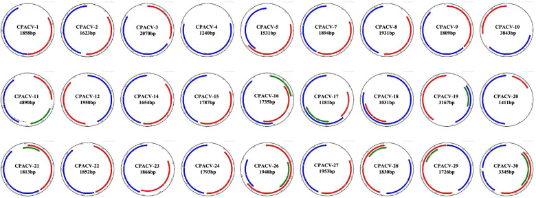
Using the PacBio RSII platform, we identified 3193 haplotype complete genomes of single-stranded DNA (ssDNA) viruses from 15 proventriculi samples of SPF chickens with TVP. Of these, 2662 genomes (83.37%) were Gyrovirus homsa1(GyH1) (also known as Gyrovirus 3, GyV3), 482 genomes (15.10%) were chicken anemia virus (CAV), and 11 genomes (0.34%) were Gyrovirus galga1 (GyG1) (also known as avian Gyrovirus 2, AGV2). 26 genomes were unique unclassified novel CRESS DNA viruses and a novel Genomovirus (Fig. 1). The three viruses of GyH1, CAV and GyG1 belong to Gyrovirus genus of Anelloviridae. They were present together in the form of haplotype virome in proventriculitis for the first time, indicating the high correlation between the virome and proventriculitis. The detail information of the 27 novel viruses was shown in Table 1. Among them, 59.26% (16/27) viruses were associated with environment, 3.7% (1/27) viruses (genomovirus, MW237851) were associated with plants, and the rest of them (10/27) were associated with animal tissues or feces. Three of the novel CRESS DNA viruses (MT975440, MW237843, MW237845) have a similar genome organization, i.e., their CP is encoded on the virion sense and their Rep on the complementary sense. The rest of novel CRESS DNA viruses genome organization showed clockwise sense.
Fig. 1.
Genome organization of 26 novel CRESS virus and one Genomovirus. CAPCV is the temporary name for these viruses. The direction, relative size, and reading frame of each Open Reading Frame (ORF) are indicated by the location, length, and color, respectively, of the arrows located internally to the virus genomic. Red, blue and green represent replication-associated protein (Rep), represents capsid protein (Cap) and VP3, respectively. All ORF are predicted using the SnapGene viewer software
Table 1.
Identified 26 novel CRESS virus and a Genomovirus in proventriculus of broiler chickens with TVP
| Number | Viral Classification | Complete genome | Temporary name | GenBank accession no | Homology virus | Accession no of homology virus | Identity | Source | GC% |
|---|---|---|---|---|---|---|---|---|---|
| 1 | CRESS virus | 1858 | CPACV 1 | MT951397 | putative replicase | AXH75828.1 | 39% | animal metagenome | 45.86 |
| 2 | CRESS virus | 1623 | CPACV 2 | MT975438 | Lake Sarah-associated circular virus-22 | YP_009237603.1 | 45% | Lake water | 46.77 |
| 3 | CRESS virus | 2070 | CPACV 3 | MT975439 | Lake Sarah-associated circular virus-12 | YP_009237573.1 | 38% | Lake water | 38.84 |
| 4 | CRESS virus | 1240 | CPACV 4 | MT975440 | capsid protein | AWW06122.1 | 38% | mouse tissue | 38.39 |
| 5 | CRESS virus | 1531 | CPACV 5 | MT975441 | Lake Sarah-associated circular virus-22 | YP_009237602.1 | 46% | Lake water | 42.65 |
| 6 | CRESS virus | 1894 | CPACV 7 | MW001861 | Lake Sarah-associated circular virus-25 | YP_009237609.1 | 38% | Lake water | 44.03 |
| 7 | CRESS virus | 1931 | CAPCV 8 | MW001862 | putative Rep | AUM61779.1 | 31% | wastewater | 42.31 |
| 8 | CRESS virus | 1809 | CAPCV 9 | MW001863 | putative replicase | AXH74921.1 | 34% | animal metagenome | 42.29 |
| 9 | CRESS virus | 3843 | CPACV 10 | MW001864 | capsid protein | AQU11778.1 | 46% | Sphagnum-dominated peatlands | 30.39 |
| 10 | CRESS virus | 4890 | CPACV 11 | MW001865 | capsid protein | AXH76335.1 | 40% | macaque stool | 35.26 |
| 11 | CRESS virus | 1950 | CAPCV 12 | MW001866 | capsid protein | AUM61702.1 | 26% | animal metagenome | 32.72 |
| 12 | CRESS virus | 1654 | CAPCV 14 | MW237841 | capsid protein | AYP28960.1 | 58% | animal metagenome | 47.1 |
| 13 | CRESS virus | 1787 | CAPCV 15 | MW237842 | capsid protein | AYP28960.1 | 58% | animal metagenome | 43.93 |
| 14 | CRESS virus | 1735 | CAPCV 16 | MW237843 | Rep | AYP28961.1 | 70% | crucian tissue | 51.76 |
| 15 | CRESS virus | 1181 | CAPCV 17 | MW237850 | capsid protein | AUM61760.1 | 32% | wastewater | 41.32 |
| 16 | CRESS virus | 1031 | CAPCV 18 | MW237844 | Rep | AYP28914.1 | 30% | animal metagenome | 40.54 |
| 17 | Genomovirus | 3167 | CAPCV 19 | MW237851 | Rep | QCS35885.1 | 32% | Capybara | 41.36 |
| 18 | CRESS virus | 1411 | CPACV 20 | MW237845 | capsid protein | AXH74652.1 | 35% | animal metagenome | 44.65 |
| 19 | CRESS virus | 1813 | CPACV 21 | MW237852 | Lake Sarah-associated circular virus-25 | YP_009237609.1 | 38% | Lake water | 43.19 |
| 20 | CRESS virus | 1852 | CPACV 22 | MW237846 | Lake Sarah-associated circular virus-12 | ALE29613.1 | 29% | Lake water | 40.17 |
| 21 | CRESS virus | 1866 | CPACV 23 | MW237847 | Lake Sarah-associated circular virus-24 | YP_009237609.1 | 38% | Lake water | 42.82 |
| 22 | CRESS virus | 1793 | CPACV 24 | MW237848 | Rep | AUM61918.1 | 62% | environmental samples | 43.56 |
| 23 | CRESS virus | 1948 | CAPCV 26 | MW237853 | Lake Sarah-associated circular virus-23 | YP_009237604.1 | 62% | Lake water | 48.41 |
| 24 | CRESS virus | 1953 | CPACV 27 | MW237849 | capsid protein | AUM61778.1 | 58% | environmental samples | 40.91 |
| 25 | CRESS virus | 1830 | CPACV 28 | MW237854 | capsid protein | AUM61778.1 | 58% | environmental samples | 44.81 |
| 26 | CRESS virus | 1726 | CAPCV 29 | MW237855 | Rep | AUM61966.1 | 45% | wastewater | 47.8 |
| 27 | CRESS virus | 3345 | CPACV 30 | MW237856 | Lake Sarah-associated circular virus-14 | AIF34806.1 | 34% | Lake water | 50.16 |
Identity: The homology with sequence in NCBI database
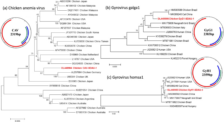
Phylogenetic analysis showed that the CAV strain (OL448984) was more closely related to the strains from Germany (AJ297684) and the United Kingdom (U66304) than viruses previously found in China (Fig. 2a) [27, 28]. This newly emerging CAV strains in China may originated from European countries, and the global circulation of commercial broilers has accelerated the spread of CAV. The GyG1 (OL448986) was closely related to the strains from cats in China (MK089245) and from chicken in Brazil (HM590588) (Fig. 2b) [29]. GyG1 found in cat feces may be derived from chickens consumed during the diet, chicken may be the original host of GyG1. The GyH1 (OL448985) was more closely related to the strains from chicken (MG366592) than mammalian (human and cat) (Fig. 2c) [14]. This suggested that GyH1 had a certain host specificity and it seemed very likely that GyH1 may, in fact, be a virus of avian origin. Overall, the three species GyVs were identified in proventriculus of chickens with TVP, indicating gyroviruses are highly resistant to gastric acid, which gives them the ability to persist and spread in the environment [30].
Fig. 2.
Genome organization and phylogenetic analysis of three gyroviruses. a Chicken anemia virus (CAV); b Gyrovirus galga1 (GyG1); c Gyrovirus homsa1 (GyH1). The reference sequences of other gyrovirus strains were obtained from GenBank database. Bootstrap values are presented at key nodes, used to evaluate the credibility of the branch. The bar indicates genetic distance. The virus strains identified in the study are shown with red
The diversity of proventriculitis lesions indicated that multiple viruses play a role in TVP. Several other viruses, including adenovirus [1], picornavirus [17] and CPNV [10], have been reported to be associated with TVP, although none of them could be definitively proved as cause of TVP. In this study, we revealed a new virome associated with TVP. This provides further insight into the true causes of TVP and future effective prevention and control of the disease.
Conclusions
In this study, we reproduced the TVP in SPF chickens, and using long read sequencing in Pacbio-RSII sequencing platform to investigate the real etiology of TVP. The results revealed that Gyrovirus virome showed high abundance in chickens with TVP, suggesting their potential role in TVP, especially GyH1. This study is expected to contribute to the knowledge of the etiology of TVP.
Supplementary Information
Additional file 1. Supplementary figure.
Acknowledgements
The authors are grateful to all veterinary practitioners and farm owners for help to collect the samples.
Authors’ contributions
ZQC designed the experiment and wrote the manuscript. TXY and GL did all experiments. DFZ and LPH analyzed the data. XJH, RQL and GHW discussed and reviewed the final manuscript. The author(s) read and approved the final manuscript.
Funding
The study was supported by grants from the Shandong Modern Agricultural Technology & Industry System (SDAIT-11–04).
Availability of data and materials
The datasets generated and/or analysed during the current study are available in the National Center for Biotechnology Information (NCBI) repository, under these GenBank accession numbers MT951397, MT975438, MT975439, MT975440, MT975441, MW001861, MW001862, MW001863, MW001864, MW001865, MW001866, MW237841, MW237842, MW237843, MW237850, MW237844, MW237851, MW237845, MW237852, MW237846, MW237847, MW237848, MW237853, MW237849, MW237854, MW237855, MW237856, OL448984, OL448985 and OL448986.
Declarations
Ethics approval and consent to participate
The animal care and use protocol was approved by the Shandong Agricultural University Animal Care and Use Committee. All the experimental animals of this study were cared for and maintained throughout the experiments strictly following the ethics and biosecurity guidelines approved by the Institutional Animal Care and Use Committee of Shandong Agricultural University. The study was carried out in compliance with the ARRIVE guidelines.
Consent for publication
Not applicable.
Competing interests
There are no competing interests in the study.
Footnotes
Publisher’s Note
Springer Nature remains neutral with regard to jurisdictional claims in published maps and institutional affiliations.
Tianxing Yan and Gen Li contributed equally to this work.
References
- 1.Goodwin MA, Hafner S, Bounous DI, Latimer KS, Player EC, Niagro FD, Campagnoli RP, Brown J. Viral proventriculitis in chickens. Avian Pathol. 1996;25(2):369–379. doi: 10.1080/03079459608419147. [DOI] [PubMed] [Google Scholar]
- 2.Dormitorio TV, Giambrone JJ, Hoerr FJ. Transmissible proventriculitis in broilers. Avian Pathol. 2007;36(2):87–91. doi: 10.1080/03079450601142588. [DOI] [PubMed] [Google Scholar]
- 3.Kouwenhoven B, Davelaar FG, Van Walsum J. Infectious proventriculitis causing runting in broilers. Avian Pathol. 1978;7(1):183–187. doi: 10.1080/03079457808418269. [DOI] [PubMed] [Google Scholar]
- 4.Grau-Roma L, Schock A, Nofrarías M, Ali Wali N, de Fraga AP, Garcia-Rueda C, de Brot S, Majó N. Retrospective study on transmissible viral proventriculitis and chicken proventricular necrosis virus (CPNV) in the UK. Avian Pathol. 2020;49(1):99–105. doi: 10.1080/03079457.2019.1677856. [DOI] [PubMed] [Google Scholar]
- 5.Guy JS, Smith LG, Evans ME, Barnes HJ. Experimental reproduction of transmissible viral proventriculitis by infection of chickens with a novel adenovirus-like virus (isolate R11/3) Avian Dis. 2007;51(1):58–65. doi: 10.1637/0005-2086(2007)051[0058:EROTVP]2.0.CO;2. [DOI] [PubMed] [Google Scholar]
- 6.Guy JS, West AM, Fuller FJ. Physical and genomic characteristics identify chicken proventricular necrosis virus (R11/3 virus) as a novel birnavirus. Avian Dis. 2011;55(1):2–7. doi: 10.1637/9504-081610-Reg.1. [DOI] [PubMed] [Google Scholar]
- 7.Guy JS, West MA, Fuller FJ, Marusak RA, Shivaprasad HL, Davis JL, Fletcher OJ. Detection of chicken proventricular necrosis virus (R11/3 virus) in experimental and naturally occurring cases of transmissible viral proventriculitis with the use of a reverse transcriptase-PCR procedure. Avian Dis. 2011;55(1):70–75. doi: 10.1637/9586-102110-Reg.1. [DOI] [PubMed] [Google Scholar]
- 8.Marusak RA, West MA, Davis JF, Fletcher OJ, Guy JS. Transmissible viral proventriculitis identified in broiler breeder and layer hens. Avian Dis. 2012;56(4):757–759. doi: 10.1637/10216-042412-Case.1. [DOI] [PubMed] [Google Scholar]
- 9.Noiva R, Guy JS, Hauck R, Shivaprasad HL. Runting Stunting Syndrome Associated with Transmissible Viral Proventriculitis in Broiler Chickens. Avian Dis. 2015;59(3):384–387. doi: 10.1637/11061-031115-Case.1. [DOI] [PubMed] [Google Scholar]
- 10.Grau-Roma L, Reid K, de Brot S, Jennison R, Barrow P, Sánchez R, Nofrarías M, Clark M, Majó N. Detection of transmissible viral proventriculitis and chicken proventricular necrosis virus in the UK. Avian Pathol. 2017;46(1):68–75. doi: 10.1080/03079457.2016.1207751. [DOI] [PubMed] [Google Scholar]
- 11.Śmiałek M, Gesek M, Dziewulska D, Niczyporuk JS, Koncicki A. Transmissible Viral Proventriculitis Caused by Chicken ProVentricular Necrosis Virus Displaying Serological Cross-Reactivity with IBDV. Animals (Basel) 2020;11(1):8. doi: 10.3390/ani11010008. [DOI] [PMC free article] [PubMed] [Google Scholar]
- 12.Hauck R, Stoute S, Senties-Cue CG, Guy JS, Shivaprasad HL. A Retrospective Study of Transmissible Viral Proventriculitis in Broiler Chickens in California: 2000–18. Avian Dis. 2020;64(4):525–531. doi: 10.1637/aviandiseases-D20-00057. [DOI] [PubMed] [Google Scholar]
- 13.Leão PA, Amaral CI, Santos WHM, Moreira MVL, de Oliveira LB, Costa EA, Resende M, Wenceslau R, Ecco R. Retrospective and prospective studies of transmissible viral proventriculitis in broiler chickens in Brazil. J Vet Diagn Invest. 2021;33(3):605–610. doi: 10.1177/10406387211004106. [DOI] [PMC free article] [PubMed] [Google Scholar]
- 14.Li G, Yuan S, He M, Zhao M, Hao X, Song M, Zhang L, Qiao C, Huang L, Zhang L, et al. Emergence of gyrovirus 3 in commercial broiler chickens with transmissible viral proventriculitis. Transbound Emerg Dis. 2018;65(5):1170–1174. doi: 10.1111/tbed.12927. [DOI] [PubMed] [Google Scholar]
- 15.Yan T, Li G, Zhou D, Yang X, Hu L, Cheng Z. Novel Cyclovirus Identified in Broiler Chickens With Transmissible Viral Proventriculitis in China. Front Vet Sci. 2020;7:569098. doi: 10.3389/fvets.2020.569098. [DOI] [PMC free article] [PubMed] [Google Scholar]
- 16.Li G, Yuan S, Yan T, Shan H, Cheng Z. Identification and characterization of chicken circovirus from commercial broiler chickens in China. Transbound Emerg Dis. 2020;67(1):6–10. doi: 10.1111/tbed.13331. [DOI] [PubMed] [Google Scholar]
- 17.Kim HR, Yoon SJ, Lee HS, Kwon YK. Identification of a picornavirus from chickens with transmissible viral proventriculitis using metagenomic analysis. Arch Virol. 2015;160(3):701–709. doi: 10.1007/s00705-014-2325-7. [DOI] [PubMed] [Google Scholar]
- 18.Gu W, Miller S, Chiu CY. Clinical Metagenomic Next-Generation Sequencing for Pathogen Detection. Annu Rev Pathol. 2019;14:319–338. doi: 10.1146/annurev-pathmechdis-012418-012751. [DOI] [PMC free article] [PubMed] [Google Scholar]
- 19.Goodwin S, McPherson JD, McCombie WR. Coming of age: ten years of next-generation sequencing technologies. Nat Rev Genet. 2016;17(6):333–351. doi: 10.1038/nrg.2016.49. [DOI] [PMC free article] [PubMed] [Google Scholar]
- 20.Eid J, Fehr A, Gray J, Luong K, Lyle J, Otto G, Peluso P, Rank D, Baybayan P, Bettman B, et al. Real-time DNA sequencing from single polymerase molecules. Science. 2009;323(5910):133–138. doi: 10.1126/science.1162986. [DOI] [PubMed] [Google Scholar]
- 21.Kumar KR, Cowley MJ, Davis RL. Next-Generation Sequencing and Emerging Technologies. Semin Thromb Hemost. 2019;45(7):661–673. doi: 10.1055/s-0039-1688446. [DOI] [PubMed] [Google Scholar]
- 22.Chaisson MJ, Huddleston J, Dennis MY, Sudmant PH, Malig M, Hormozdiari F, Antonacci F, Surti U, Sandstrom R, Boitano M, et al. Resolving the complexity of the human genome using single-molecule sequencing. Nature. 2015;517(7536):608–611. doi: 10.1038/nature13907. [DOI] [PMC free article] [PubMed] [Google Scholar]
- 23.Schirmer M, Sloan WT, Quince C. Benchmarking of viral haplotype reconstruction programmes: an overview of the capacities and limitations of currently available programmes. Brief Bioinform. 2014;15(3):431–442. doi: 10.1093/bib/bbs081. [DOI] [PubMed] [Google Scholar]
- 24.de Sena Brandine G, Smith AD. Falco: high-speed FastQC emulation for quality control of sequencing data. F1000Res. 1874;2019:8. doi: 10.12688/f1000research.21142.1. [DOI] [PMC free article] [PubMed] [Google Scholar]
- 25.Caesar L, Cibulski SP, Canal CW, Blochtein B, Sattler A, Haag KL. The virome of an endangered stingless bee suffering from annual mortality in southern Brazil. J Gen Virol. 2019;100(7):1153–1164. doi: 10.1099/jgv.0.001273. [DOI] [PubMed] [Google Scholar]
- 26.Li G, Zhou D, Zhao M, Liu Q, Hao X, Yan T, Yuan S, Zhang S, Cheng Z. Kinetic analysis of pathogenicity and tissue tropism of gyrovirus 3 in experimentally infected chickens. Vet Res. 2021;52(1):120. doi: 10.1186/s13567-021-00990-2. [DOI] [PMC free article] [PubMed] [Google Scholar]
- 27.Todd D, Scott AN, Ball NW, Borghmans BJ, Adair BM. Molecular basis of the attenuation exhibited by molecularly cloned highly passaged chicken anemia virus isolates. J Virol. 2002;76(16):8472–8474. doi: 10.1128/JVI.76.16.8472-8474.2002. [DOI] [PMC free article] [PubMed] [Google Scholar]
- 28.Meehan BM, Todd D, Creelan JL, Connor TJ, McNulty MS. Investigation of the attenuation exhibited by a molecularly cloned chicken anemia virus isolate by utilizing a chimeric virus approach. J Virol. 1997;71(11):8362–8367. doi: 10.1128/jvi.71.11.8362-8367.1997. [DOI] [PMC free article] [PubMed] [Google Scholar]
- 29.Rijsewijk FA, Dos Santos HF, Teixeira TF, Cibulski SP, Varela AP, Dezen D, Franco AC, Roehe PM. Discovery of a genome of a distant relative of chicken anemia virus reveals a new member of the genus Gyrovirus. Arch Virol. 2011;156(6):1097–1100. doi: 10.1007/s00705-011-0971-6. [DOI] [PubMed] [Google Scholar]
- 30.Welch J, Bienek C, Gomperts E, Simmonds P. Resistance of porcine circovirus and chicken anemia virus to virus inactivation procedures used for blood products. Transfusion. 2006;46(11):1951–1958. doi: 10.1111/j.1537-2995.2006.01003.x. [DOI] [PubMed] [Google Scholar]
Associated Data
This section collects any data citations, data availability statements, or supplementary materials included in this article.
Supplementary Materials
Additional file 1. Supplementary figure.
Data Availability Statement
The datasets generated and/or analysed during the current study are available in the National Center for Biotechnology Information (NCBI) repository, under these GenBank accession numbers MT951397, MT975438, MT975439, MT975440, MT975441, MW001861, MW001862, MW001863, MW001864, MW001865, MW001866, MW237841, MW237842, MW237843, MW237850, MW237844, MW237851, MW237845, MW237852, MW237846, MW237847, MW237848, MW237853, MW237849, MW237854, MW237855, MW237856, OL448984, OL448985 and OL448986.


