ABSTRACT
By fusing catalytically dead Cas9 (dCas9) to active domains of histone deacetylase (Sir2a) or acetyltransferase (GCN5), this CRISPR interference/activation (CRISPRi/a) system allows gene regulation at the transcriptional level without causing permanent changes in the parasite genome. However, the constitutive expression of dCas9 poses a challenge for studying essential genes, which may lead to adaptive changes in the parasite, masking the true phenotypes. Here, we developed a leak-free inducible CRISPRi/a system by integrating the DiCre/loxP regulon to allow the expression of dCas9-GCN5/-Sir2a upon transient induction with rapamycin, which allows convenient transcriptional regulation of a gene of interest by introducing a guide RNA targeting its transcription start region. Using eight genes that are either silent or expressed from low to high levels during asexual erythrocytic development, we evaluated the robustness and versatility of this system in the asexual parasites. For most genes analyzed, this inducible CRISPRi/a system led to 1.5- to 3-fold up-or downregulation of the target genes at the mRNA level. Alteration in the expression of PfK13 and PfMYST resulted in altered sensitivities to artemisinin. For autophagy-related protein 18, an essential gene related to artemisinin resistance, a >2-fold up- or downregulation was obtained by inducible CRISPRi/a, leading to growth retardation. For the master regulator of gametocytogenesis, PfAP2-G, a >10-fold increase of the PfAP2-G transcripts was obtained by CRISPRa, resulting in >4-fold higher gametocytemia in the induced parasites. Additionally, inducible CRISPRi/a could also regulate gene expression in gametocytes. This inducible epigenetic regulation system offers a fast way of studying gene functions in Plasmodium falciparum.
IMPORTANCE Understanding the fundamental biology of malaria parasites through functional genetic/genomic studies is critical for identifying novel targets for antimalarial development. Conditional knockout/knockdown systems are required to study essential genes in the haploid blood stages of the parasite. In this study, we developed an inducible CRISPRi/a system via the integration of DiCre/loxP. We evaluated the robustness and versatility of this system by activating or repressing eight selected genes and achieved up- and downregulation of the targeted genes located in both the euchromatin and heterochromatin regions. This system offers the malaria research community another tool for functional genetic studies.
KEYWORDS: human malaria, Plasmodium falciparum, dCas9, gene regulation, DiCre
INTRODUCTION
Despite tremendous efforts in controlling malaria, malaria still accounted for an estimated 229 million cases and 409,000 deaths globally in 2019, according to the World Malaria Report 2020. The emergence and spread of Plasmodium falciparum resistance to artemisinin-combination therapies have created an obstacle to malaria control and elimination. Therefore, a better understanding of the fundamental biology of malaria parasites via functional genomic studies is critical for identifying novel targets for antimalarial development and elucidating the drug resistance mechanism. However, genetic manipulation in P. falciparum relies exclusively on homologous recombination because Plasmodium lacks RNA interference (RNAi) and the canonical nonhomologous end-joining (NHEJ) repair mechanism (1, 2). Thus, essential genes for blood-stage development cannot be knocked out conventionally since blood-stage parasites are haploid. To overcome this limitation, several conditional genetic modification (knockdown or knockout) systems have been developed to regulate genes of interest at the DNA, transcript, or protein level. For example, the inducible Cre recombinase (DiCre), which recognizes the loxP sites, catalyzes the excision or inversion of the flanked DNA fragment upon treatment with rapamycin (Rap) (3–5). The TetR-DOZI and glmS ribozyme systems regulate gene expression at the posttranscription level (6–8). The TetR-DOZI system regulates the translation of the target mRNA by binding TetR-DOZI to the TetR aptamers inserted in the 3′ or 5′ untranslated region (UTR) of the target mRNA, blocking its translation and promoting mRNA degradation. The addition of anhydrotetracycline (aTc) removes the TetR-DOZI protein from the aptamers, allowing protein translation (8, 9). The glmS ribozyme is inserted in the 3′ UTR of a target gene, and it cleaves the target mRNA upon induction with glucosamine, leading to a reduction in mRNA expression (6). To regulate protein expression at the protein level, an engineered version of the human FKBP12 is fused to the protein of interest, targeting it to degradation, while the fusion protein is stabilized by the cell-permeable synthetic molecule Shield 1 (10–12).
The discovery of the CRISPR/Cas9 system has revolutionized genome editing in a large number of organisms (13, 14). Its applications to malaria parasites have dramatically expanded the toolkits for functional studies, allowing genes to be tagged, disrupted, or deleted and point mutations to be generated (15–17). With this system, the Cas9 endonuclease binds to a single guide RNA (sgRNA), which contains the Cas9 binding domain and 20 customizable nucleotides complementary to a target DNA site. The protospacer adjacent motif (PAM), a sequence immediately downstream from the target region, is required for recruiting Cas9 to the target site to cause a double-strand break (DSB). Depending on the organisms, this break is repaired by homology-directed repair (HDR) using donor DNA or the error-prone NHEJ, causing gene mutation, insertion, or deletion with or without genetic scarring (13–18). In addition to gene editing, this system has been systematically repurposed using the catalytically dead Cas9 (dCas9) for epigenetic editing referred to as CRISPR interference/activation (CRISPRi/a) by tethering an activator, silencer, or modifier of histone/DNA to the dCas9 (19–25). With this modification, dCas9, guided by sgRNA, binds to the promoter/enhancer region with the same efficiency as the active Cas9 but cannot generate a DSB, resulting in the blockade of the transcriptional process or alteration of epigenetic activities of the neighboring target genes. CRISPRi has been successfully adapted in P. falciparum and Plasmodium yoelii to knock down gene expression by simply guiding dCas9 alone to the region upstream of the gene of interest (26, 27). Xiao et al. designed a CRISPRi/a system by fusing dCas9 with either the histone acetyltransferase (HAT) domain of PfGCN5 or the histone deacetylase (HDAC) domain of PfSir2a, a class III histone deacetylase, to epigenetically modify the promoter chromatin and achieve upregulation or downregulation of the target gene in P. falciparum (28). However, with constitutive expression of the dCas9, this system may present a challenge for studying certain essential genes, whose altered expression resulting from the dCas9 system may be too deleterious to allow parasite growth. In addition, constant gene activation or silencing may cause adaptation of the parasites and mask the true phenotype.
To solve the potential problem associated with the constant expression of dCas9, we attempted to incorporate the inducibility of dCas9 expression into this system. We established two inducible dCas9 expression modules using the TetR-DOZI and the DiCre/loxP systems and selected the latter for leak-free inducible expression of dCas9-GCN5 and dCas9-Sir2a. We implemented the inducible expression of dCas9-GCN5 and dCas9-Sir2a to achieve targeted up- and downregulation of genes located in both the euchromatin and heterochromatin regions.
RESULTS
Generation of the inducible CRIPRi/a systems.
CRISPR-guided dCas9 fused with the HAT or HDAC creates a valuable tool to regulate target gene expression in P. falciparum (28). To introduce inducibility into this system, we attempted the TetR-DOZI-based gene knockdown and the DiCre-based conditional deletion. To conditionally activate the expression of dCas9-GCN5 or -Sir2a using the TetR-DOZI regulon, we insert the 10 aptamer repeats in the 3′ UTR of the dCas9-GCN5/-Sir2a expression cassette (Fig. S1A). This would lead to translation inhibition of the dCas9-fusion protein in the absence of aTc and induced expression of the fusion protein when aTc is added in culture (Fig. S1A). Immunofluorescence assay (IFA) did not detect fluorescent signals when the parasites were cultured without aTc. Thirty hours after adding 0.5 μM aTc to the synchronized ring-stage parasites, robust fluorescent signals were detected in the nuclei of the parasites, indicating dCas9-fusion protein expression (Fig. S1B). Western blotting further confirmed the induced expression of the fusion proteins after adding aTc for 30 h (Fig. S1C). However, we also detected low-level dCas9-fusion protein expression in the absence of aTc (Fig. S1C), suggesting leaky expression of the fusion protein, which agrees with published studies (8, 29, 30).
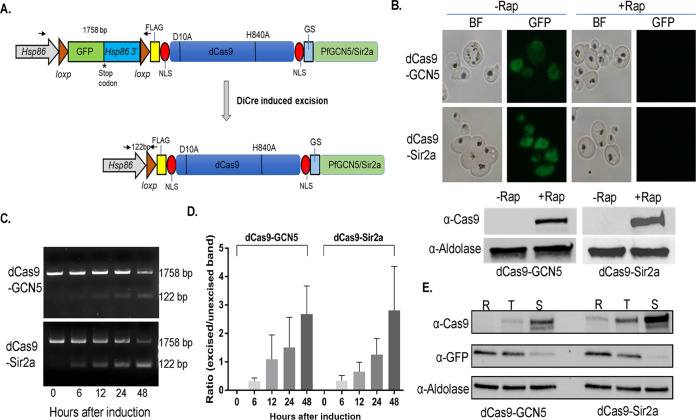
To build a DiCre-based induction system, we inserted a green fluorescent protein (GFP) expression cassette between the hsp86 promoter and the dCas9-GCN5 or dCas9-Sir2a open reading frame. The inclusion of a stop codon at the end of GFP and the hsp86 3′ UTR sequence allowed GFP expression but prevented the expression of the downstream dCas9-GCN5 or -Sir2a. Two loxP sites were inserted before the GFP start codon and the end of hsp86 3′ UTR to allow inducible excision of the loxP-flanked fragment via activation of the DiCre recombinase by rapamycin (Rap), resulting in the inducible expression of dCas9-GCN5 and dCas9-Sir2a (Fig. 1A). The transgenic parasite lines are named loxPed GFP-dCas9-GCN5 and -Sir2a. These constructs were transfected into the P. falciparum clone constitutively expressing DiCre (5) to obtain the transgenic parasite lines. As expected, GFP was ubiquitously expressed in the parasites (Fig. 1B, upper panel), whereas no dCas9-GCN5 or Cas9-Sir2A was detected by Western blotting using the anti-Cas9 antibodies before Rap induction (Fig. 1B, lower panel).
FIG 1.
Generation of inducible dCas9-GCN5/Sir2a systems using DiCre/loxP. (A) Schematic diagram illustrates the DiCre/loxP-inducible CRISPR/dCas9 system. GFP with stop codon and Hsp86 3′UTR flanked by loxP sequence was inserted before dCas9-GCN5/Sir2a. Rap is added to activate DiCre to remove sections between loxP sites, leading to the expression of dCas9-GCN5/Sir2a. FLAG, FLAG tag; D10A and H840A, two mutations in Cas9; NLS, nuclear localization signal; GS, glycine-serine protein domain linker. Two small black arrows in opposite directions show the locations of primers for PCR in panel C. The numbers between the arrows indicate the expected sizes of PCR products. (B) Live images and Western blots show the GFP and dCas9-fusion protein expression before (−Rap) and 48 h after (+Rap) DiCre was activated by the addition of Rap. (C) The efficiency of DiCre excision of loxPed GFP cassette was measured by PCR with specific primers shown in panel A. The gradual decrease and increase of unexcised and excised bands at the sizes 1,758 bp and 122 bp are shown in agarose gels, respectively. BF, bright field. (D) Three biological replicates were performed for the experiment in panel C to depict a bar graph showing the ratio of excised to unexcised bands over time elapsed after Rap treatment; pixel intensities of DNA bands were measured using ImageJ. (E) Western blots show the induction of dCas9-GCN5 and dCas9-Sir2a protein expression and decrease of GFP expression at ring (R), trophozoite (T), and schizont (S) stages after Rap treatment. Rap was added at 0 to 6 h postinvasion (hpi) for 2 h.
To monitor the DiCre recombinase excision dynamics and efficiency induced by Rap, parasites at the early ring stage were treated with 100 nM Rap for 2 h, and parasite genomic DNA was isolated at 0, 6, 12, 24, and 48 h. Excision of the gfp-hsp86 3′ UTR fragment was identified by PCR amplification of the loxP locus before excision (1,785 bp) and after excision (122 bp). PCR analysis showed that the loxP excision level and excised/unexcised ratio increased over time (Fig. 1C and D). Concomitantly, Western blotting confirmed the declining expression of GFP and gradually increased expression of the dCas9 fusion protein, with the dCas9 fusion protein level reaching the highest at the schizont stage (Fig. 1E). Live-cell fluorescence microscopy and Western blotting also detected the disappearance of the GFP fluorescent signals and the appearance of dCas9 fusion proteins 48 h after Rap treatment (Fig. 1B). Similar DiCre recombinase excision dynamics starting at the trophozoite and schizont stage were detected, indicating that excision is not stage-specific (Fig. S2). Additionally, there was no noticeable change in the parasite growth and developmental progression before and after Rap induction compared to those of the wild-type parasites. These results indicated that the expression of dCas9-GCN5/Sir2a in the DiCre system was leak free and could be robustly induced in the same intraerythrocytic developmental cycle (IDC).
Regulation of gene expression using the DiCre/loxP-inducible dCas9 system.
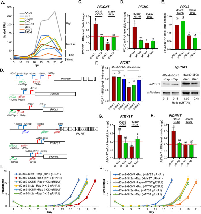
We first tested the DiCre system as a leak-free inducible dCas9-GCN5/Sir2a system for the proof-of-principle regulation of eight genes that are expressed at high (PfGCN5), medium (clathrin heavy chain: PfCHC, PfK13, PfATG18, and PfCRT), and low (PfMYST and PfDNMT) or silent (PfAP2-G) during the IDC (31) (Fig. 2A, Table S1). Of the eight genes selected, all except PfDNMT or PfAP2-G are essential for the IDC. For each gene, 2 to 3 sgRNAs were selected around the transcriptional start sites (TSSs) (Fig. 2B, 3A, 4A, Table S2) (32, 33). The constructs expressing these sgRNAs were transfected individually into either the loxPed GFP-dCas9-Sir2a or the loxPed GFP-dCas9-GCN5 parasite line. Resistant parasites after selection harboring different sgRNAs were treated with Rap at 0 to 6 h postinvasion (hpi) for 2 h. Total RNA was harvested at 40 to 46 hpi to measure the target gene mRNA level by reverse transcriptase-quantitative PCR (RT-qPCR).
FIG 2.
Repression and activation of selected genes by inducible dCas9 systems. (A) Transcriptional expression levels of eight selected genes during IDC based on the transcriptomic profile by RNA-seq. (B) Schematic diagrams show the location of TSSs and gRNAs in six selected genes. (C to E) The transcriptional changes of PfGCN5, PfCHC, and PfK13 in loxPed GFP-dCas9-GCN5 or Sir2a parasite lines after Rap treatment, respectively. (F) Left panel: RT-qPCR analysis of PfCRT transcript using 3 different sgRNAs (gRNA1 to 3) with dCas9-GCN5 and dCas9-Sir2a systems. Right panel: Western blot of PfCRT protein expression levels after activation and repression of PFCRT by using an anti-PfCRT antibody with anti-aldolase as a loading control. (G and H) The transcription levels of PfMYST and PfDNMT in the loxPed GFP-dCas9-GCN5 or Sir2a parasite lines after Rap treatment. Three replicates of RT-qPCR were performed for each gRNA and the paired t tests were conducted (ns, not significant; *, P < 0.05; **, P < 0.01; ***, P < 0.001). (I and J) Three replicates of recovery assays show the parasite growth after DHA treatment of parasite lines targeting PfK13 (I) and PfMYST (J) by dCas9-GCN5/Sir2a with respective gRNA1, compared to the parasite lines with gRNA but without Rap induction and the parasite lines without gRNA but with Rap induction.
FIG 3.
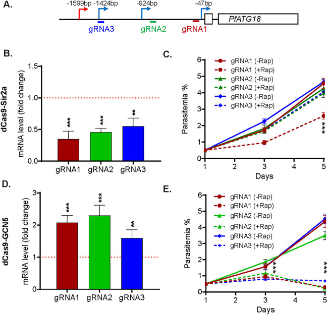
Repression and activation of PfATG18 by inducible dCas9 system. (A) Schematic diagram shows the location of TSSs and gRNAs in the 5′ UTR of PfATG18. (B and D) The changes of PfATG18 transcriptions were analyzed by RT-qPCR in dCas9-Sir2a (B) and dCas9-GCN5 (D) parasite lines with gRNA1-3 after Rap treatment compared to the transcriptions without Rap treatment, respectively. Paired t tests were conducted (**, P < 0.01; ***, P < 0.001) from three replicates. (C and E) Parasite growth phenotype after repression and activation of PfATG18 using gRNA1-3 in dCas9-Sir2a (C) and dCas9-GCN5 (E) systems was investigated by measuring the parasitemia from three replicates, respectively. Compared to the growth rate in the parasites without Rap treatment (−Rap), parasites harboring gRNA1 in dCas9-Sir2a system grew significantly more slowly at day 5 posttreatment (+Rap) (***, P < 0.001, two-way ANOVA), whereas parasites harboring any gRNA (1/2/3) in dCas9-GCN5 system grew significantly more slowly at days 3 and 5 posttreatment (+Rap) (***, P < 0.001, two-way ANOVA).
FIG 4.
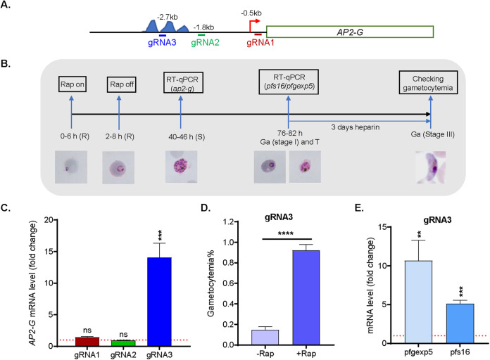
Activation of AP2-G by the inducible dCas9-GCN5 system. (A) Schematic diagram shows the location of TSS and gRNAs in the 5′ UTR of AP2-G. The areas containing AP2-G/G5 binding motifs are labeled with three blue peaks. (B) A diagram illustrates the experimental workflow for the analysis of activation of AP2-G by the inducible system dCas9-GCN5. R, ring; T, trophozoite; S, schizont; Ga: gametocyte. (C) The changes of AP2-G transcription were analyzed by RT-qPCR in the parasites harboring inducible dCas9-GCN5 system and one of three gRNAs (gRNA1 to 3) compared to the transcription without Rap induction. Paired t tests were conducted (ns, not significant; ***, P < 0.001) from three replicates. (D) Three replicates of gametocytemia were counted 5 days after parasites harboring inducible dCas9-GCN5 system and gRNA3 were treated by Rap (+Rap) compared to the same parasites without Rap treatment (−Rap) (****, P < 0.0001, paired t test). (E) The two early gametocytes makers, pfgexp5 and pfs16, were analyzed by RT-qPCR in the parasites harboring the dCas9-GCN5 system and gRNA3 at 76 to 82 hpi when the gametocytogenesis started (stage 1) and asexual parasites were at the trophozoite stage. The paired t tests were conducted (**, P < 0.01, ***, P < 0.001) from three replicates.
Since the CRISPRi/a system developed here would inhibit or activate gene expression by modifying the chromatin environment near the TSS of the target genes, we were interested in using this system to downregulate highly expressed genes, regulate intermediately expressed genes in both directions, and activate lowly expressed or silent genes. For all genes tested, we could change the levels of target genes expression after Rap-induced dCas9-GCN5 or dCas9-Sir2 expression, but the extent of the change varied greatly among genes and was influenced substantially by the locations of the sgRNAs relative to the major TSS (Fig. 2C to H).
For the PfGCN5 gene, the two sgRNAs selected were both embedded in the multiple TSSs that span ~1.2 kb in the 5′ UTR of the gene, with gRNA1 located ~400 bp from the major TSS and gRNA2 much farther (~1 kb) from the major TSS (Fig. 2B). As expected, gRNA1 resulted in ~2-fold downregulation of PfGCN5 in the loxPed GFP-dCas9-Sir2a parasite, whereas gRNA2 did not cause any changes in PfGCN5 expression (Fig. 2C). Interestingly, despite that PfGCN5 is a relatively highly expressed gene, gRNA1 in the loxPed GFP-dCas9-GCN5 parasite still increased PfGCN5 expression by ~2-fold (Fig. 2C). Although PfGCN5 is an essential gene for the IDC, these changes in PfGCN5 mRNA levels did not result in significant changes in asexual growth under the standard culture conditions. Similarly, gRNA1 and gRNA2 for PfCHC were located at 0.5 and 1 kb, respectively, upstream of the major TSS (Fig. 2B). Whereas gRNA1 led to an ~3-fold increase in PfCHC expression in the loxPed GFP-dCas9-GCN5 parasite line, gRNA2 did not have any noticeable effect on PfCHC expression (Fig. 2D).
Two drug resistance genes, PfK13 and PfCRT, were selected to evaluate both upregulation and downregulation of expression. The two gRNAs for PfK13 were both relatively far from the main TSS (~400 bp and ~700 bp, respectively) (Fig. 2B). In the loxPed GFP-dCas9-GCN5 parasite line, both gRNAs led to 1.4- to 1.7-fold upregulation of PfK13, whereas their downregulation effects in the loxPed GFP-dCas9-Sir2a parasite line were very modest (20 to 30% reduction) (Fig. 2E). For PfCRT, none of the three gRNAs had any evident effects on PfCRT expression in the loxPed GFP-dCas9-GCN5 parasite line, whereas gRNA1 and gRNA2, both located downstream of the major TSS, resulted in ~40% and ~30% reductions, respectively, in PfCRT mRNA level in the loxPed GFP-dCas9-Sir2a parasite line (Fig. 2F). We further compared the PfCRT protein levels in these parasite lines expressing gRNA1. We found that PfCRT protein was decreased by >50% in the loxped GFP-dCas9-Sir2a parasite line after Rap induction (Fig. 2F), consistent with the change in PfCRT mRNA level.
We were also interested in using the dCas9-GCN5 system to upregulate lowly expressed genes. For the two epigenetic regulators selected for evaluation, the two gRNAs for PfMYST were more than 400 bp upstream of the major TSS, while gRNA1 for PfDNMT was 180 bp from the major TSS (Fig. 2B). Regardless of the locations of the gRNAs, no noticeable or modest downregulation of PfMYST or PfDNMT was observed in the loxPed GFP-dCas9-Sir2a line. In the loxPed GFP-dCas9-GCN5 parasites, the PfMYST gRNA1 led to ~1.5-fold upregulation of PfMYST expression, while PfDNMT gRNA1 increased PfDNMT expression by almost 2-fold (Fig. 2G and H).
We monitored the growth phenotypes of the parasites only after we induced changes in the mRNA levels of the tested genes. Except for PfATG18 and PfAP2-G, no changes in the growth curve and the developmental progression of the parasite lines were observed when the target genes were regulated using the dCas9-GCN5 and the dCas9-Sir2a approaches.
Altered sensitivities to artemisinin after regulation of PfK13 and PfMYST.
PfK13 mutation is known as the marker of artemisinin resistance (34, 35), while PfMYST was found to be involved in DNA repair (36). Recently, enhanced DNA repair was also identified to be associated with artemisinin resistance (37). To reveal whether alteration of expression in PfK13 and PfMYST could change the sensitivities to artemisinin, we treated loxPed GFP-dCas9-GCN5/Sir2a parasite lines harboring gRNA1 targeting PfK13 and PfMYST with 1 μM dihydroartemisinin (DHA) at the early ring stage for 24 h and measured the time of parasite reappearance (recovery assay). Compared to the parasite lines with gRNA but without Rap induction and the parasite lines without gRNA but with Rap induction, downregulation of PfK13 (dCas9-Sir2A-K13gRNA1, +Rap) and upregulation of PfMYST (dCas9-GCN5-MYSTgRNA1, +Rap) resulted in earlier recovery, whereas upregulation of PfK13 (dCas9-GCN5-K13gRNA1, +Rap) and downregulation of PfMYST (dCas9-Sir2A-MYSTgRNA1, +Rap) led to later recovery (Fig. 2I and J). These results are consistent with the finding that the decreased K13 abundance reduced the artemisinin resistance (38–40) and suggest that PfMYST is involved in artemisinin resistance probably by the protection of DNA damage caused by artemisinin (41).
Growth inhibition resulting from bidirectional regulation of PfATG18 expression.
PfATG18 is essential for the IDC (42–44), and a mutation in PfATG18 was associated with decreased sensitivities to DHA, artemether, and piperaquine in the samples from the China-Myanmar border (45). The major TSS of PfATG18 is 1.6 kb from the ATG, and the three gRNAs selected are all located downstream from the major TSS. Interestingly, regardless of the locations of the gRNAs, all led to an ~2-fold reduction in PfATG18 expression in the loxPed GFP-dCas9-Sir2a parasite lines. Specifically, the PfATG18 mRNA levels were decreased to ~30%, 50%, and 60% of those in untreated parasites for gRNA 1, 2, and 3, respectively (Fig. 3B). Only the parasite line with gRNA1 yielding ~70% transcript reduction showed growth retardation, with the day 5 parasitemia reaching 2.6% compared to 4.7% in the untreated parasites (Fig. 3B and C; analysis of variance [ANOVA], P < 0.001). In contrast, the other two parasite lines did not show noticeable growth defects, suggesting that the degree of PfATG18 mRNA reduction at 50% or lower did not result in a growth phenotype change under the standard culture conditions (Fig. 3C). It is noteworthy that PfATG18 knockdown (KD) using the destabilization domain also resulted in a slower growth phenotype (42).
Early studies found that episomal expression of PfATG18 did not cause any noticeable changes in growth phenotype (44, 46). Since it is not known whether episomal expression resulted in PfATG18 overexpression, we wanted to see whether conditional upregulation of PfATG18 in the loxPed GFP-dCas9-GCN5 parasite line would lead to growth phenotype changes. After Rap treatment at the ring stage (0 to 6 hpi), the PfATG18 mRNA measured at 40 to 44 hpi was elevated by ~2-, 2.3-, and 1.6-fold in parasites expressing gRNA1, 2, and 3, respectively, compared to that of the untreated parasites (Fig. 3D). Surprisingly, all parasite lines showed severe growth retardation on day 3 and day 5, with more profound effects starting from the second IDC (Fig. 3E, ANOVA, P < 0.001).
Activation of HP1-controlled silent gene PfAP2-G via dCas9-GCN5.
In P. falciparum, heterochromatin protein 1 (HP1) binds H3K9me3 to control ~425 genes localized mainly in the subtelomere regions (47–50). These include silent genes (e.g., PfAP2-G) and genes that are expressed in a mutually exclusive manner (e.g., the var gene family with ~60 members) during the IDC. The ApiAP2 (AP2) transcription factor PfAP2-G is a master regulator of gametocytogenesis (51–54), and its activation led to the conversion of asexual-stage parasites to sexual development (55, 56). We selected PfAP2-G to evaluate whether the inducible dCas9-GCN5 system could activate the HP1-controlled silent gene during the IDC. Three gRNAs were selected. gRNA 1 is located in the proximity of the weak TSS (−0.5 kb) for the lowly expressed PfAP2-G (17, 32, 33, 57) (Fig. 4A). Since PfAP2-G and PfAP2-G5 were found to regulate PfAP2-G expression by binding their respective motifs located between −2.0 kb and −3.5 kb (52, 58), gRNAs 2 and 3 were designed to localize downstream and within (−1.8 kb and −2.7 kb) these motifs, respectively (Fig. 4A). In the loxPed GFP-dCas9-GCN5 parasite line, the mRNA level of PfAP2-G was measured at 40 to 46 hpi after the dCas9-GCN5 expression was induced at the ring stage (0 to 6 hpi) (Fig. 4B and C). RT-qPCR analysis of PfAP2-G did not detect any increased expression of PfAP2-G in parasites with either gRNA1 or 2 over the uninduced controls (Fig. 4C), despite that the gRNA1 is localized close to the TSS of the gene. In contrast, PfAP2-G expression was increased by ~14-fold in the parasite line harboring gRNA3 (Fig. 4C).
To determine whether elevated PfAP2-G expression was correlated with increased gametocytogenesis, parasite cultures were treated with heparin for 3 days starting from the second cycle after Rap treatment (76 to 82 hpi) to eliminate asexual-stage parasites (Fig. 4B). The loxPed GFP-dCas9-GCN5 line harboring gRNA3 had an increase in gametocytemia more than 4-fold that in the untreated parasites (Fig. 4D, unpaired two-tailed Student’s test, P < 0.0001). As expected from the mRNA analysis, the other two gRNAs did not lead to an increase in gametocytemia (data not shown). There are many known early gametocyte markers, including Pfs16 and Pfgexp5 (52, 59, 60). Consistently, RT-qPCR analysis of parasites in the second cycle after Rap treatment (76 to 82 hpi) (Fig. 4B) showed concomitant upregulation of Pfgexp5 and Pfs16 by more than 10- and 5-fold, respectively, in the parasite line with gRNA3 compared to that in the untreated parasites (Fig. 4E). These results demonstrated that the silent gene PfAP2-G could be conditionally activated in the inducible dCas9-GCN5 system, leading to elevated sexual conversion.
The inducible CRIPRi/a systems regulate gene expression in gametocytes.
The DiCre/loxP system has been successfully used to excise loxPed genes in gametocytes (61). This triggered us to determine whether CRIPRi/a systems can regulate gene expression in the gametocyte stage. Consistently, Rap treatment at day 5 (stage III) gametocytes induced excision of loxPed GFP cassette (Fig. S3A). PfATG18 gRNA1 was chosen because it substantially altered the PfATG18 expression in the asexual stage (Fig. 3). Forty-eight hours after Rap induction starting at stage III gametocytes, PfATG18 mRNA was elevated by ~3.3-fold and decreased to ~30% in dCas9-GCN5 and -Sir2a parasite lines, respectively, compared to that in the Rap-induced parasite line without gRNA1 or noninduced gametocyte with gRNA1 (Fig. S3B). Only upregulation of PfATG18 expression resulted in a significant decrease of gametocytemia as measured 3 days after induction (Fig. S3C). Taken together, the inducible CRIPRi/a system functions in sexual-stage parasites.
DISCUSSION
Our recent study has shown that epigenetic editing by dCas9 fused to the HAT domain of PfGCN5 or the deacetylase domain of PfSir2a allows for activating or silencing the gene of interest without leaving a genetic scar (28). This CRISPR/dCas9 system relies on the constantly coexpressed dCas9 and sgRNA, leading to strong activation/repression of the target genes. This constitutive expression could limit certain applications if the level of regulation of the target gene achieved with this dCas9 system is detrimental to the IDC. Therefore, we aimed to introduce inducibility to this dCas9-GCN5/Sir2a system. From experimentation with the TetR-DOZI and DiCre/loxP conditional modules to control the timing of CRISPR/dCas9 expression, we selected the DiCre/loxP module since there was no background expression of dCas9-GCN5 and -Sir2a before the addition of Rap, whereas TetR-DOZI conditional system showed a leaky expression (Fig. S1), and induction of dCas9 fusion proteins by addition of aTc did not result in more sex conversion (gametocyte) when PfAP2-G gRNAs were used (data not shown), probably because the leaky expression caused less magnification of induction than that of DiCre/loxP system. We then tested the suitability of this inducible system for regulating genes with expression levels ranging from highly expressed to silent genes during the IDC.
Our data further confirmed that the degree of gene activation or repression achieved with the inducible dCas9 systems depended primarily on the location of gRNA relative to the TSSs of the target genes. In most cases, the sgRNAs located closest to the major TSSs produced the most potent activation or repression effect (Fig. 2). Additionally, from the results of PfCRT and PfATG18, gRNAs located downstream of major TSSs lead to the repression of target genes, probably due to the blockade of the transcriptional process by the gRNA/dCas9 complex. Since most P. falciparum genes use multiple TSSs, selecting several sgRNAs around the TSSs will identify the one with the strongest effects on the target gene. For PfAP2-G, only the sgRNA3 localized to the PfAP2-G/PfAP2-G5 binding motifs triggered substantial activation of PfAP2-G expression (Fig. 4), suggesting the influence of the local chromatin status of the sgRNA-target site. Thus, additional factors such as transcription factor binding sites, the location of active or silent histone marks, and more accurate TSSs will need to be considered to optimize sgRNA selection. However, searching for suitable sgRNAs for Cas9 requiring the PAM sequence NGG in the extremely AT-rich genome of P. falciparum is challenging. More recently, a new type of Cas, Cas12a (also called Cpf1), has emerged as an alternative nuclease to Cas9 (62, 63). One advantage of Cas12a is that the preferred PAM sequence is T-rich (TTTV, V can be C, G, or A), which could alleviate the difficulties of designing sgRNAs in P. falciparum.
The inducible dCas9 system reported here offers in situ upregulation of genes to study the effect of gene overexpression. One approach to studying gene function is the overexpression of the full-length or truncated gene, often from an episome. However, continuous overexpression might cause the parasite to adapt to the change such as the attenuation of the endogenous target gene, which may mask the true phenotype resulting from overexpression of the target gene. Continuous episomal overexpression of a target gene sometimes is toxic or detrimental to the parasite. For example, ectopic overexpression of PfSR1 was toxic and caused the parasites to constrain PfSR1 overexpression for survival (64). In Toxoplasma gondii, transient but not stable overexpression of MYST-A was achieved (65). Here, we showed that the inducible dCa9-GCN5 system allowed upregulation of almost all genes tested, even including highly expressed genes such as PfGCN5 and PfCHC. Interestingly, overexpression of PfATG18 by ~2-folds in the dCas9-GCN5 parasites resulted in substantial growth defects. In contrast, no growth defect was reported when PfATG18 was episomally expressed (44, 46), suggesting that the parasites might have adapted to the constant overexpression by attenuating endogenous gene expression. Although we did not test the potential off-target effects of the sgRNAs, it is unlikely that all three strictly selected sgRNAs for PfATG18 had additional targets in the P. falciparum genome. Similar growth defects were also found when PfATG18 transcription was increased by more than 3-fold in gametocytes, suggesting that PfATG18 is critical for the parasite survival given that this gene was found to be involved in the food vacuole dynamics, autophagy-like pathway, and apicoplast biogenesis in the parasites (42, 44, 46).
We have shown that the activation of the DiCre recombinase by Rap was rapid, resulting in high-level induction of the dCas9-fusion proteins in the same IDC (Fig. 1). With this, observation of phenotypic changes may begin in the first IDC or, at most, the second IDC, which was illustrated for the PfATG18 gene (Fig. 3). Although under the standard culture conditions no growth phenotype changes were observed for six of the eight genes tested, the phenotypic changes under other conditions (e.g., stress, drug exposure) probably will elucidate the tested genes’ functions. Indeed, alteration of the expression of PfK13 and PfMYST resulted in changes in sensitivities to artemisinin (Fig. 2). Although dCas9-GCN5 activated PfAP2-G and displayed a significantly higher sexual conversion (~1%), this conversion rate is relatively limited compared to the extremely high rate (~90%) when PfAP2-G was driven by a strong constitutive promoter (calmodulin) (55).
Collectively, we showed the values of the DiCre/loxP-based, inducible dCas9-GCN5/Sir2a systems for functional gene studies in P. falciparum. To increase the robustness of the gene regulation, multiple sgRNAs should be tried to select the one with the strongest effect and no off-targets. In addition, replacing the dCas9 with dCas12 in this system may alleviate the challenges in choosing the most appropriate sgRNAs.
MATERIALS AND METHODS
Molecular cloning.
To generate TetR-based inducibility into the dCas9-GCN5 and -Sir2a systems, the 10× aptamer was amplified from the pMG75 ATPase4 plasmid (8, 9) using the primers aptamer F/R (Table S3) and inserted into 3′ UTR of the dCas9-GCN5/Sir2a expression cassette at PacI, resulting in pdCas9-GCN5/Sir2a-10× aptamer. We generated a TetR-DOZI-expressing plasmid, pTetR-DOZI, from pMG75 ATPase4 by deleting the ATPase4 homologous region and the 10× aptamer-containing 3′ UTR.
To integrate the DiCre/loxP conditional module into the dCas9-GCN5 or dCas9-Sir2a expression cassette (28), we amplified gfp flanked by two loxP sequences using primers LoxP-GFP-F and LoxP-GFP-R (Table S3). The PCR product was inserted into XhoI-digested vector pUF1-dCas9-GCN5 or -Sir2a using the in-fusion HD cloning kit (Clontech). The hsp86 3′ UTR was amplified using primers Hsp86-F and Hsp86-R (Table S3) and cloned at the XmaI site in pUF1-dCas9-GCN5 or -Sir2a, immediately downstream of gfp. Two or three sgRNAs were designed for each tested gene based on predicted TSS sites according to the sgRNA selection criteria (Table S2 and S3) (17). Individual sgRNAs were cloned into the plasmid pL6-CS-WR (28).
Plasmodium culture and transfection.
The P. falciparum 3D7 clone was used for generating the TetR-based expression system. A P. falciparum clone engineered to express DiCre constitutively (5) was kindly provided by Michael Blackman. Parasites were maintained in human red blood cells (RBCs) and cultured at 37°C in a gas mixture of 5% CO2, 3% O2, and 92% N2 as described previously (36). Parasite transfection was done using the RBC loading method (66). For the TetR-based inducible dCas9-GCN5 or -Sir2a system, pTetR-DOZI and pdCas9-GCN5/Sir2a-10× aptamer plasmids were transfected into 3D7, followed by drug selection with WR99210 at 2.5 nM and blasticidin-S-HCl (BSD) at 2.5 μg/mL. The DiCre/loxP-based plasmids were introduced into the DiCre-expressing parasite line, and resistant parasites were selected with BSD.
Rapamycin-induced excision and dCas9-fusion protein expression in the DiCre/loxP system.
Highly synchronized rings were obtained by incubating purifying schizonts via 65/35% Percoll gradient with fresh RBCs for 4 h. Rap at 100 nM was added to the synchronized rings for 2 h, and parasites were harvested 6, 12, 24, and 48 h later. The same experiments were also performed starting from the trophozoite and schizont stages. For excision analysis in gametocytes, the synchronized gametocytes were induced by previously described methods (67). On day 2 after gametocyte induction, heparin was added to the culture for 3 days to eliminate asexual-stage parasites. Rap at 100 nM was added to day 5 (stage III) gametocytes for 2 h, and the gametocytes were harvested 6, 12, 24, and 48 h later. Parasite genomic DNA was isolated using the proteinase K digestion and phenol-chloroform extraction method. PCR was performed with the same amount of genomic DNA using primers Ex-F and Ex-R (Table S3). PCR products were separated by agarose gel and stained with ethidium bromide. Pixel intensities of DNA bands were measured using ImageJ. Three biological replicates were performed for each experiment.
To measure the expression of the dCas9 fusion proteins and GFP in the parasites before and after DiCre excision events, rings were treated with Rap for 2 h and then were harvested 6, 26, and 40 h later to represent ring, trophozoite, and schizont stages, respectively. Western blotting was done with mouse anti-GFP (1:3,000, ThermoFisher), anti-Cas9 (1:1,000, Sigma), and rabbit anti-PfAldolase (1:3,000, kindly provided by Tobias Spielmann) antibodies as primary antibodies, followed by horseradish peroxidase (HRP)-conjugated goat anti-rabbit IgG and goat anti-mouse IgG (Sigma) as secondary antibodies. The GFP fluorescence signals in live parasites were captured using a Nikon Eclipse E600 epifluorescence microscope.
Induced dCas9-fusion protein expression in the TetR-DOZI system.
Western blotting was used to measure dCas9 expression in the TetR-DOZI-based inducible dCas9-GCN5 or -Sir2a system. Ring-stage parasites expressing both TetR-DOZI and the dCas9-fusion proteins were equally divided into two cultures and were treated with aTc at 0.5 μM or ethanol (vehicle control) for 30 h. Then, parasites were harvested for Western blotting with rabbit anti-FLAG antibodies (1:1,000, Abcam) and rabbit anti-PfAldolase as the primary antibodies and HRP-conjugated goat anti-rabbit IgG as secondary antibodies. The wild-type 3D7 was used as a negative control.
IFA was performed to detect the expression of dCas9 according to an established method (68, 69). Briefly, parasitized RBCs were fixed with 4% paraformaldehyde and 0.0075% glutaraldehyde, followed by quenching with 50 mM glycine. Fixed cells were then treated with 0.5% Triton X-100 and blocked in 3% bovine serum albumin (BSA) for 1 h. The anti-FLAG antibodies and fluorescein isothiocyanate (FITC)-conjugated goat anti-rabbit IgG (Sigma) were used as primary and secondary antibodies. Images were captured using a Nikon Eclipse E600 epifluorescence microscope.
RNA extraction and RT-qPCR.
Parasites were isolated after treating the parasitized RBCs with 0.1% (vol/vol) saponin, and total RNAs were extracted using the Quick-RNA MiniPrep kit (Zymo Research). Reverse transcriptase and real-time PCR were performed by using SuperScript III RT (Invitrogen) and Faststart Universal SYBR green master mix (Roche). The relative expression of each gene in the Rap- or dimethyl sulfoxide (DMSO)-treated parasites was normalized to seryl-tRNA synthetase (PF3D7_0717700) and calculated using the 2−ΔΔCT method (70). All the qPCR primer sequences are listed in Table S3. Three biological replicates were performed for each experiment.
Growth phenotypic analysis.
The growth phenotype of Pfatg18 gRNA1-3 parasite lines was compared in triplicate. Cultures were tightly synchronized as described above. Cultures starting at 0.5% ring were treated with Rap or DMSO, and parasitemia was monitored daily using Giemsa staining. Phenotypic analysis of Pfap2-g was performed following the experimental workflow in Fig. 3. Briefly, the highly synchronized ring-stage parasites (0 to 6 hpi) were immediately treated with Rap or DMSO for 2 h. The transcription of Pfap2-g and early gametocyte markers (pfgexp5/pfs16) was measured at 40 to 46 hpi and 76 to 82 hpi using RT-qPCR (Fig. 3B, Table S3), respectively. Heparin was added afterward for 3 days to eliminate asexual-stage parasites (71). The gametocytemia was calculated after heparin treatment using Giemsa-stained smears. To study the gametocyte growth phenotype after targeting PfATG18, the synchronized gametocytes were induced by the methods described previously (67). On day 2 after gametocyte induction, heparin was added to the culture for 3 days to eliminate asexual-stage parasites. Rap at 100 nM was added to day 5 gametocytes for 2 h and the gametocytemia was measured on the next day by Giemsa staining for 4 days.
Recovery assay.
This assay was performed based on established methods with some modifications (72–75). Briefly, the highly synchronized early ring stage (1 to 3 h) parasites at 2% parasitemia were treated with 1 μM DHA for 24 h. Subsequently, the parasites were cultured in a drug-free medium and parasite growth was examined daily using Giemsa staining. The amount of time needed for each parasite culture to reach 10% parasitemia was recorded.
Statistical analysis.
For growth phenotype experiments, three independent biological replicates were performed. The results are presented as mean ± standard deviation (SD) and analyzed by one- or two-way ANOVA or paired t test. For gene expression (RT-qPCR) upon induction by Rap and DSMO, ΔCTs from +Rap and −Rap from three biological replicates were analyzed by paired t test.
Supplementary Material
ACKNOWLEDGMENTS
This study was funded by the National Institute of Allergy and Infectious Diseases, National Institutes of Health (1R21AI149202 to J.M.), and the startup fund from Morsani College of Medicine to J.M. We want to thank Michael Blackman for providing a P. falciparum clone engineered to express DiCre constitutively and Tobias Spielmann for providing the anti-PfAldolase antibodies.
J.M. and L.C. conceptualized the study. B.X. and L.J. provided resources. X. Liang, R.B., and F.A.S. developed the methodology and performed the investigations. X. Li, J.Q., B.X., and H.M. supported the investigation. J.M., L.C., and L.J. reviewed and edited the paper, and J.M. supervised the study.
We declare no conflicts of interest.
Footnotes
Supplemental material is available online only.
Contributor Information
Jun Miao, Email: jmiao1@usf.edu.
Björn F. C. Kafsack, Weill Cornell Medicine
Heather Painter, U.S. Food & Drug Administration.
REFERENCES
- 1.Baum J, Papenfuss AT, Mair GR, Janse CJ, Vlachou D, Waters AP, Cowman AF, Crabb BS, de Koning-Ward TF. 2009. Molecular genetics and comparative genomics reveal RNAi is not functional in malaria parasites. Nucleic Acids Res 37:3788–3798. doi: 10.1093/nar/gkp239. [DOI] [PMC free article] [PubMed] [Google Scholar]
- 2.Kirkman LA, Lawrence EA, Deitsch KW. 2014. Malaria parasites utilize both homologous recombination and alternative end joining pathways to maintain genome integrity. Nucleic Acids Res 42:370–379. doi: 10.1093/nar/gkt881. [DOI] [PMC free article] [PubMed] [Google Scholar]
- 3.Knuepfer E, Napiorkowska M, van Ooij C, Holder AA. 2017. Generating conditional gene knockouts in Plasmodium - a toolkit to produce stable DiCre recombinase-expressing parasite lines using CRISPR/Cas9. Sci Rep 7 doi: 10.1038/s41598-017-03984-3. [DOI] [PMC free article] [PubMed] [Google Scholar]
- 4.Jones ML, Das S, Belda H, Collins CR, Blackman MJ, Treeck M. 2016. A versatile strategy for rapid conditional genome engineering using loxP sites in a small synthetic intron in Plasmodium falciparum. Sci Rep 6. doi: 10.1038/srep21800. [DOI] [PMC free article] [PubMed] [Google Scholar]
- 5.Collins CR, Das S, Wong EH, Andenmatten N, Stallmach R, Hackett F, Herman J-P, Müller S, Meissner M, Blackman MJ. 2013. Robust inducible Cre recombinase activity in the human malaria parasite Plasmodium falciparum enables efficient gene deletion within a single asexual erythrocytic growth cycle. Mol Microbiol 88:687–701. doi: 10.1111/mmi.12206. [DOI] [PMC free article] [PubMed] [Google Scholar]
- 6.Prommana P, Uthaipibull C, Wongsombat C, Kamchonwongpaisan S, Yuthavong Y, Knuepfer E, Holder AA, Shaw PJ. 2013. Inducible knockdown of plasmodium gene expression using the glmS ribozyme. PLoS One 8:e73783. doi: 10.1371/journal.pone.0073783. [DOI] [PMC free article] [PubMed] [Google Scholar]
- 7.Rajaram K, Liu HB, Prigge ST. 2020. Redesigned TetR-aptamer system to control gene expression in Plasmodium falciparum. mSphere 5. doi: 10.1128/mSphere.00457-20. [DOI] [PMC free article] [PubMed] [Google Scholar]
- 8.Ganesan SM, Falla A, Goldfless SJ, Nasamu AS, Niles JC. 2016. Synthetic RNA-protein modules integrated with native translation mechanisms to control gene expression in malaria parasites. Nat Commun 7:10727. doi: 10.1038/ncomms10727. [DOI] [PMC free article] [PubMed] [Google Scholar]
- 9.Goldfless SJ, Wagner JC, Niles JC. 2014. Versatile control of Plasmodium falciparum gene expression with an inducible protein-RNA interaction. Nat Commun 5:5329. doi: 10.1038/ncomms6329. [DOI] [PMC free article] [PubMed] [Google Scholar]
- 10.Banaszynski LA, Chen L-C, Maynard-Smith LA, Ooi AGL, Wandless TJ. 2006. A rapid, reversible, and tunable method to regulate protein function in living cells using synthetic small molecules. Cell 126:995–1004. doi: 10.1016/j.cell.2006.07.025. [DOI] [PMC free article] [PubMed] [Google Scholar]
- 11.Armstrong CM, Goldberg DE. 2007. An FKBP destabilization domain modulates protein levels in Plasmodium falciparum. Nat Methods 4:1007–1009. doi: 10.1038/nmeth1132. [DOI] [PubMed] [Google Scholar]
- 12.Russo I, Oksman A, Vaupel B, Goldberg DE. 2009. A calpain unique to alveolates is essential in Plasmodium falciparum and its knockdown reveals an involvement in pre-S-phase development. Proc Natl Acad Sci USA 106:1554–1559. doi: 10.1073/pnas.0806926106. [DOI] [PMC free article] [PubMed] [Google Scholar]
- 13.Adli M. 2018. The CRISPR tool kit for genome editing and beyond. Nat Commun 9. doi: 10.1038/s41467-018-04252-2. [DOI] [PMC free article] [PubMed] [Google Scholar]
- 14.Wang HF, Russa ML, Qi LS. 2016. CRISPR/Cas9 in genome editing and beyond. Annu Rev Biochem 85:227–264. doi: 10.1146/annurev-biochem-060815-014607. [DOI] [PubMed] [Google Scholar]
- 15.Ghorbal M, Gorman M, Macpherson CR, Martins RM, Scherf A, Lopez-Rubio J-J. 2014. Genome editing in the human malaria parasite Plasmodium falciparum using the CRISPR-Cas9 system. Nat Biotechnol 32:819–821. doi: 10.1038/nbt.2925. [DOI] [PubMed] [Google Scholar]
- 16.Wagner JC, Platt RJ, Goldfless SJ, Zhang F, Niles JC. 2014. Efficient CRISPR-Cas9-mediated genome editing in Plasmodium falciparum. Nat Methods 11:915–918. doi: 10.1038/nmeth.3063. [DOI] [PMC free article] [PubMed] [Google Scholar]
- 17.Ribeiro JM, Garriga M, Potchen N, Crater AK, Gupta A, Ito D, Desai SA. 2018. Guide RNA selection for CRISPR-Cas9 transfections in Plasmodium falciparum. Int J Parasitol 48:825–832. doi: 10.1016/j.ijpara.2018.03.009. [DOI] [PMC free article] [PubMed] [Google Scholar]
- 18.Bryant JM, Baumgarten S, Glover L, Hutchinson S, Rachidi N. 2019. CRISPR in parasitology: not exactly cut and dried!. Trends Parasitol 35:409–422. doi: 10.1016/j.pt.2019.03.004. [DOI] [PubMed] [Google Scholar]
- 19.Qi LS, Larson MH, Gilbert LA, Doudna JA, Weissman JS, Arkin AP, Lim WA. 2013. Repurposing CRISPR as an RNA-guided platform for sequence-specific control of gene expression. Cell 152:1173–1183. doi: 10.1016/j.cell.2013.02.022. [DOI] [PMC free article] [PubMed] [Google Scholar]
- 20.Gilbert LA, Larson MH, Morsut L, Liu Z, Brar GA, Torres SE, Stern-Ginossar N, Brandman O, Whitehead EH, Doudna JA, Lim WA, Weissman JS, Qi LS. 2013. CRISPR-mediated modular RNA-guided regulation of transcription in eukaryotes. Cell 154:442–451. doi: 10.1016/j.cell.2013.06.044. [DOI] [PMC free article] [PubMed] [Google Scholar]
- 21.Sander JD, Joung JK. 2014. CRISPR-Cas systems for editing, regulating and targeting genomes. Nat Biotechnol 32:347–355. doi: 10.1038/nbt.2842. [DOI] [PMC free article] [PubMed] [Google Scholar]
- 22.Brocken DJW, Tark-Dame M, Dame RT. 2018. dCas9: a versatile tool for epigenome editing. Curr Issues Mol Biol 26:15–32. doi: 10.21775/cimb.026.015. [DOI] [PubMed] [Google Scholar]
- 23.Anton T, Karg E, Bultmann S. 2018. Applications of the CRISPR/Cas system beyond gene editing. Biol Methods Protoc 3:bpy002. doi: 10.1093/biomethods/bpy002. [DOI] [PMC free article] [PubMed] [Google Scholar]
- 24.Mitsunobu H, Teramoto J, Nishida K, Kondo A. 2017. Beyond Native Cas9: manipulating genomic information and function. Trends Biotechnol 35:983–996. doi: 10.1016/j.tibtech.2017.06.004. [DOI] [PubMed] [Google Scholar]
- 25.Dominguez AA, Lim WA, Qi LS. 2016. Beyond editing: repurposing CRISPR-Cas9 for precision genome regulation and interrogation. Nat Rev Mol Cell Biol 17:5–15. doi: 10.1038/nrm.2015.2. [DOI] [PMC free article] [PubMed] [Google Scholar]
- 26.Walker MP, Lindner SE. 2019. Ribozyme-mediated, multiplex CRISPR gene editing and CRISPR interference (CRISPRi) in rodent-infectious Plasmodium yoelii. J Biol Chem 294:9555–9566. doi: 10.1074/jbc.RA118.007121. [DOI] [PMC free article] [PubMed] [Google Scholar]
- 27.Baumgarten S, Bryant JM, Sinha A, Reyser T, Preiser PR, Dedon PC, Scherf A. 2019. Transcriptome-wide dynamics of extensive m(6)A mRNA methylation during Plasmodium falciparum blood-stage development. Nat Microbiol 4:2246–2259. doi: 10.1038/s41564-019-0521-7. [DOI] [PMC free article] [PubMed] [Google Scholar]
- 28.Xiao B, Yin S, Hu Y, Sun M, Wei J, Huang Z, Wen Y, Dai X, Chen H, Mu J, Cui L, Jiang L. 2019. Epigenetic editing by CRISPR/dCas9 in Plasmodium falciparum. Proc Natl Acad Sci USA 116:255–260. doi: 10.1073/pnas.1813542116. [DOI] [PMC free article] [PubMed] [Google Scholar]
- 29.Polino AJ, Nasamu AS, Niles JC, Goldberg DE. 2020. Assessment of biological role and insight into druggability of the Plasmodium falciparum protease plasmepsin V. ACS Infect Dis 6:738–746. doi: 10.1021/acsinfecdis.9b00460. [DOI] [PMC free article] [PubMed] [Google Scholar]
- 30.Tang Y, Meister TR, Walczak M, Pulkoski-Gross MJ, Hari SB, Sauer RT, Amberg-Johnson K, Yeh E. 2019. A mutagenesis screen for essential plastid biogenesis genes in human malaria parasites. PLoS Biol 17:e3000136. doi: 10.1371/journal.pbio.3000136. [DOI] [PMC free article] [PubMed] [Google Scholar]
- 31.Bartfai R, Hoeijmakers WAM, Salcedo-Amaya AM, Smits AH, Janssen-Megens E, Kaan A, Treeck M, Gilberger TW, Françoijs KJ, Stunnenberg HG. 2010. H2A.Z demarcates intergenic regions of the plasmodium falciparum epigenome that are dynamically marked by H3K9ac and H3K4me3. PLoS Pathog 6:e1001223. doi: 10.1371/journal.ppat.1001223. [DOI] [PMC free article] [PubMed] [Google Scholar]
- 32.Adjalley SH, Chabbert CD, Klaus B, Pelechano V, Steinmetz LM. 2016. Landscape and dynamics of transcription initiation in the malaria parasite Plasmodium falciparum. Cell Rep 14:2463–2475. doi: 10.1016/j.celrep.2016.02.025. [DOI] [PMC free article] [PubMed] [Google Scholar]
- 33.Chappell L, Ross P, Orchard L, Russell TJ, Otto TD, Berriman M, Rayner JC, Llinás M. 2020. Refining the transcriptome of the human malaria parasite Plasmodium falciparum using amplification-free RNA-seq. BMC Genomics 21:395. doi: 10.1186/s12864-020-06787-5. [DOI] [PMC free article] [PubMed] [Google Scholar]
- 34.Mok S, Stokes BH, Gnädig NF, Ross LS, Yeo T, Amaratunga C, Allman E, Solyakov L, Bottrill AR, Tripathi J, Fairhurst RM, Llinás M, Bozdech Z, Tobin AB, Fidock DA. 2021. Artemisinin-resistant K13 mutations rewire Plasmodium falciparum's intra-erythrocytic metabolic program to enhance survival. Nat Commun 12:530. doi: 10.1038/s41467-020-20805-w. [DOI] [PMC free article] [PubMed] [Google Scholar]
- 35.Siddiqui FA, Liang X, Cui L. 2021. Plasmodium falciparum resistance to ACTs: emergence, mechanisms, and outlook. Int J Parasitol Drugs Drug Resist 16:102–118. doi: 10.1016/j.ijpddr.2021.05.007. [DOI] [PMC free article] [PubMed] [Google Scholar]
- 36.Miao J, Fan Q, Cui L, Li X, Wang H, Ning G, Reese JC, Cui L. 2010. The MYST family histone acetyltransferase regulates gene expression and cell cycle in malaria parasite Plasmodium falciparum. Mol Microbiol 78:883–902. doi: 10.1111/j.1365-2958.2010.07371.x. [DOI] [PMC free article] [PubMed] [Google Scholar]
- 37.Xiong A, Prakash P, Gao X, Chew M, Tay IJJ, Woodrow CJ, Engelward BP, Han J, Preiser PR. 2020. K13-mediated reduced susceptibility to artemisinin in Plasmodium falciparum is overlaid on a trait of enhanced DNA damage repair. Cell Rep 32:107996. doi: 10.1016/j.celrep.2020.107996. [DOI] [PMC free article] [PubMed] [Google Scholar]
- 38.Yang T, Yeoh LM, Tutor MV, Dixon MW, McMillan PJ, Xie SC, Bridgford JL, Gillett DL, Duffy MF, Ralph SA, McConville MJ, Tilley L, Cobbold SA. 2019. Decreased K13 abundance reduces hemoglobin catabolism and proteotoxic stress, underpinning artemisinin resistance. Cell Rep 29:2917–2928.e5. doi: 10.1016/j.celrep.2019.10.095. [DOI] [PubMed] [Google Scholar]
- 39.Siddiqui G, Srivastava A, Russell AS, Creek DJ. 2017. Multi-omics based identification of specific biochemical changes associated with PfKelch13-mutant artemisinin-resistant Plasmodium falciparum. J Infect Dis 215:1435–1444. doi: 10.1093/infdis/jix156. [DOI] [PubMed] [Google Scholar]
- 40.Birnbaum J, Scharf S, Schmidt S, Jonscher E, Hoeijmakers WAM, Flemming S, Toenhake CG, Schmitt M, Sabitzki R, Bergmann B, Fröhlke U, Mesén-Ramírez P, Blancke Soares A, Herrmann H, Bártfai R, Spielmann T. 2020. A Kelch13-defined endocytosis pathway mediates artemisinin resistance in malaria parasites. Science 367:51–59. doi: 10.1126/science.aax4735. [DOI] [PubMed] [Google Scholar]
- 41.Gupta DK, Patra AT, Zhu L, Gupta AP, Bozdech Z. 2016. DNA damage regulation and its role in drug-related phenotypes in the malaria parasites. Sci Rep 6:23603. doi: 10.1038/srep23603. [DOI] [PMC free article] [PubMed] [Google Scholar]
- 42.Bansal P, Tripathi A, Thakur V, Mohmmed A, Sharma P. 2017. Autophagy-related protein ATG18 regulates apicoplast biogenesis in apicomplexan parasites. mBio 8:e01468-17. doi: 10.1128/mBio.01468-17. [DOI] [PMC free article] [PubMed] [Google Scholar]
- 43.Zhang M, Wang C, Otto TD, Oberstaller J, Liao X, Adapa SR, Udenze K, Bronner IF, Casandra D, Mayho M, Brown J, Li S, Swanson J, Rayner JC, Jiang RHY, Adams JH. 2018. Uncovering the essential genes of the human malaria parasite Plasmodium falciparum by saturation mutagenesis. Science 360. doi: 10.1126/science.aap7847. [DOI] [PMC free article] [PubMed] [Google Scholar]
- 44.Sudhakar R, Das D, Thanumalayan S, Gorde S, Sijwali PS. 2021. Plasmodium falciparum Atg18 localizes to the food vacuole via interaction with the multi-drug resistance protein 1 and phosphatidylinositol 3-phosphate. Biochem J 478:1705–1732. doi: 10.1042/BCJ20210001. [DOI] [PubMed] [Google Scholar]
- 45.Wang Z, Cabrera M, Yang J, Yuan L, Gupta B, Liang X, Kemirembe K, Shrestha S, Brashear A, Li X, Porcella SF, Miao J, Yang Z, Su X-Z, Cui L. 2016. Genome-wide association analysis identifies genetic loci associated with resistance to multiple antimalarials in Plasmodium falciparum from China-Myanmar border. Sci Rep 6:33891. doi: 10.1038/srep33891. [DOI] [PMC free article] [PubMed] [Google Scholar]
- 46.Agrawal P, Manjithaya R, Surolia N. 2020. Autophagy-related protein PfATG18 participates in food vacuole dynamics and autophagy-like pathway in Plasmodium falciparum. Mol Microbiol 113:766–782. doi: 10.1111/mmi.14441. [DOI] [PubMed] [Google Scholar]
- 47.Flueck C, Bartfai R, Volz J, Niederwieser I, Salcedo-Amaya AM, Alako BTF, Ehlgen F, Ralph SA, Cowman AF, Bozdech Z, Stunnenberg HG, Voss TS. 2009. Plasmodium falciparum heterochromatin protein 1 marks genomic loci linked to phenotypic variation of exported virulence factors. PLoS Pathog 5:e1000569. doi: 10.1371/journal.ppat.1000569. [DOI] [PMC free article] [PubMed] [Google Scholar]
- 48.Perez-Toledo K, Rojas-Meza AP, Mancio-Silva L, Hernandez-Cuevas NA, Delgadillo DM, Vargas M, Martinez-Calvillo S, Scherf A, Hernandez-Rivas R. 2009. Plasmodium falciparum heterochromatin protein 1 binds to tri-methylated histone 3 lysine 9 and is linked to mutually exclusive expression of var genes. Nucleic Acids Res 37:2596–2606. doi: 10.1093/nar/gkp115. [DOI] [PMC free article] [PubMed] [Google Scholar]
- 49.Lopez-Rubio JJ, Mancio-Silva L, Scherf A. 2009. Genome-wide analysis of heterochromatin associates clonally variant gene regulation with perinuclear repressive centers in malaria parasites. Cell Host Microbe 5:179–190. doi: 10.1016/j.chom.2008.12.012. [DOI] [PubMed] [Google Scholar]
- 50.Salcedo-Amaya AM, van Driel MA, Alako BT, Trelle MB, van den Elzen AMG, Cohen AM, Janssen-Megens EM, van de Vegte-Bolmer M, Selzer RR, Iniguez AL, Green RD, Sauerwein RW, Jensen ON, Stunnenberg HG. 2009. Dynamic histone H3 epigenome marking during the intraerythrocytic cycle of Plasmodium falciparum. Proc Natl Acad Sci USA 106:9655–9660. doi: 10.1073/pnas.0902515106. [DOI] [PMC free article] [PubMed] [Google Scholar]
- 51.Kafsack BFC, Rovira-Graells N, Clark TG, Bancells C, Crowley VM, Campino SG, Williams AE, Drought LG, Kwiatkowski DP, Baker DA, Cortés A, Llinás M. 2014. A transcriptional switch underlies commitment to sexual development in malaria parasites. Nature 507:248–252. doi: 10.1038/nature12920. [DOI] [PMC free article] [PubMed] [Google Scholar]
- 52.Josling GA, Russell TJ, Venezia J, Orchard L, van Biljon R, Painter HJ, Llinás M. 2020. Dissecting the role of PfAP2-G in malaria gametocytogenesis. Nat Commun 11:1503. doi: 10.1038/s41467-020-15026-0. [DOI] [PMC free article] [PubMed] [Google Scholar]
- 53.Sinha A, Hughes KR, Modrzynska KK, Otto TD, Pfander C, Dickens NJ, Religa AA, Bushell E, Graham AL, Cameron R, Kafsack BFC, Williams AE, Llinas M, Berriman M, Billker O, Waters AP. 2014. A cascade of DNA-binding proteins for sexual commitment and development in Plasmodium. Nature 507:253–257. doi: 10.1038/nature12970. [DOI] [PMC free article] [PubMed] [Google Scholar]
- 54.Brancucci NMB, Bertschi NL, Zhu L, Niederwieser I, Chin WH, Wampfler R, Freymond C, Rottmann M, Felger I, Bozdech Z, Voss TS. 2014. Heterochromatin protein 1 secures survival and transmission of malaria parasites. Cell Host Microbe 16:165–176. doi: 10.1016/j.chom.2014.07.004. [DOI] [PubMed] [Google Scholar]
- 55.Llorà-Batlle O, Michel-Todó L, Witmer K, Toda H, Fernández-Becerra C, Baum J, Cortés A. 2020. Conditional expression of PfAP2-G for controlled massive sexual conversion in Plasmodium falciparum. Sci Adv 6:eaaz5057. doi: 10.1126/sciadv.aaz5057. [DOI] [PMC free article] [PubMed] [Google Scholar]
- 56.Kent RS, Modrzynska KK, Cameron R, Philip N, Billker O, Waters AP. 2018. Inducible developmental reprogramming redefines commitment to sexual development in the malaria parasite Plasmodium berghei. Nat Microbiol 3:1206–1213. doi: 10.1038/s41564-018-0223-6. [DOI] [PMC free article] [PubMed] [Google Scholar]
- 57.Toenhake CG, Fraschka SA-K, Vijayabaskar MS, Westhead DR, van Heeringen SJ, Bártfai R. 2018. Chromatin accessibility-based characterization of the gene regulatory network underlying Plasmodium falciparum blood-stage development. Cell Host Microbe 23:557–569.e9. doi: 10.1016/j.chom.2018.03.007. [DOI] [PMC free article] [PubMed] [Google Scholar]
- 58.Shang X, Shen S, Tang J, He X, Zhao Y, Wang C, He X, Guo G, Liu M, Wang L, Zhu Q, Yang G, Jiang C, Zhang M, Yu X, Han J, Culleton R, Jiang L, Cao J, Gu L, Zhang Q. 2021. A cascade of transcriptional repression determines sexual commitment and development in Plasmodium falciparum. Nucleic Acids Res 49:9264–9279. doi: 10.1093/nar/gkab683. [DOI] [PMC free article] [PubMed] [Google Scholar]
- 59.Bruce MC, Carter RN, Nakamura K, Aikawa M, Carter R. 1994. Cellular location and temporal expression of the Plasmodium falciparum sexual stage antigen Pfs16. Mol Biochem Parasitol 65:11–22. doi: 10.1016/0166-6851(94)90111-2. [DOI] [PubMed] [Google Scholar]
- 60.Tibúrcio M, Dixon MWA, Looker O, Younis SY, Tilley L, Alano P. 2015. Specific expression and export of the Plasmodium falciparum Gametocyte EXported Protein-5 marks the gametocyte ring stage. Malar J 14:334. doi: 10.1186/s12936-015-0853-6. [DOI] [PMC free article] [PubMed] [Google Scholar]
- 61.Tiburcio M, Yang ASP, Yahata K, Suárez-Cortés P, Belda H, Baumgarten S, van de Vegte-Bolmer M, van Gemert GJ, van Waardenburg Y, Levashina EA, Sauerwein RW, Treeck M. 2019. A novel tool for the generation of conditional knockouts to study gene function across the Plasmodium falciparum life cycle. mBio 10. doi: 10.1128/mBio.01170-19. [DOI] [PMC free article] [PubMed] [Google Scholar]
- 62.Swarts DC, Jinek M. 2018. Cas9 versus Cas12a/Cpf1: structure-function comparisons and implications for genome editing. Wiley Interdiscip Rev RNA 9:e1481. doi: 10.1002/wrna.1481. [DOI] [PubMed] [Google Scholar]
- 63.Bayat H, Modarressi MH, Rahimpour A. 2018. The conspicuity of CRISPR-Cpf1 system as a significant breakthrough in genome editing. Curr Microbiol 75:107–115. doi: 10.1007/s00284-017-1406-8. [DOI] [PubMed] [Google Scholar]
- 64.Eshar S, Allemand E, Sebag A, Glaser F, Muchardt C, Mandel-Gutfreund Y, Karni R, Dzikowski R. 2012. A novel Plasmodium falciparum SR protein is an alternative splicing factor required for the parasites’ proliferation in human erythrocytes. Nucleic Acids Res 40:9903–9916. doi: 10.1093/nar/gks735. [DOI] [PMC free article] [PubMed] [Google Scholar]
- 65.Smith AT, Tucker-Samaras SD, Fairlamb AH, Sullivan WJ. 2005. MYST family histone acetyltransferases in the protozoan parasite Toxoplasma gondii. Eukaryot Cell 4:2057–2065. doi: 10.1128/EC.4.12.2057-2065.2005. [DOI] [PMC free article] [PubMed] [Google Scholar]
- 66.Deitsch K, Driskill C, Wellems T. 2001. Transformation of malaria parasites by the spontaneous uptake and expression of DNA from human erythrocytes. Nucleic Acids Res 29:850–853. doi: 10.1093/nar/29.3.850. [DOI] [PMC free article] [PubMed] [Google Scholar]
- 67.Fivelman QL, McRobert L, Sharp S, Taylor CJ, Saeed M, Swales CA, Sutherland CJ, Baker DA. 2007. Improved synchronous production of Plasmodium falciparum gametocytes in vitro. Mol Biochem Parasitol 154:119–123. doi: 10.1016/j.molbiopara.2007.04.008. [DOI] [PubMed] [Google Scholar]
- 68.Tonkin CJ, van Dooren GG, Spurck TP, Struck NS, Good RT, Handman E, Cowman AF, McFadden GI. 2004. Localization of organellar proteins in Plasmodium falciparum using a novel set of transfection vectors and a new immunofluorescence fixation method. Mol Biochem Parasitol 137:13–21. doi: 10.1016/j.molbiopara.2004.05.009. [DOI] [PubMed] [Google Scholar]
- 69.Lucky AB, Sakaguchi M, Katakai Y, Kawai S, Yahata K, Templeton TJ, Kaneko O. 2016. Plasmodium knowlesi skeleton-binding protein 1 localizes to the 'Sinton and Mulligan' stipplings in the cytoplasm of monkey and human erythrocytes. PLoS One 11:e0164272. doi: 10.1371/journal.pone.0164272. [DOI] [PMC free article] [PubMed] [Google Scholar]
- 70.Livak KJ, Schmittgen TD. 2001. Analysis of relative gene expression data using real-time quantitative PCR and the 2(T)(-Delta Delta C) method. Methods 25:402–408. doi: 10.1006/meth.2001.1262. [DOI] [PubMed] [Google Scholar]
- 71.Miao J, Wang Z, Liu M, Parker D, Li X, Chen X, Cui L. 2013. Plasmodium falciparum: generation of pure gametocyte culture by heparin treatment. Exp Parasitol 135:541–545. doi: 10.1016/j.exppara.2013.09.010. [DOI] [PMC free article] [PubMed] [Google Scholar]
- 72.Cui L, Wang Z, Miao J, Miao M, Chandra R, Jiang H, Su X-z, Cui L. 2012. Mechanisms of in vitro resistance to dihydroartemisinin in Plasmodium falciparum. Mol Microbiol 86:111–128. doi: 10.1111/j.1365-2958.2012.08180.x. [DOI] [PMC free article] [PubMed] [Google Scholar]
- 73.Witkowski B, Lelièvre J, Barragán MJL, Laurent V, Su X-z, Berry A, Benoit-Vical F. 2010. Increased tolerance to artemisinin in Plasmodium falciparum is mediated by a quiescence mechanism. Antimicrob Agents Chemother 54:1872–1877. doi: 10.1128/AAC.01636-09. [DOI] [PMC free article] [PubMed] [Google Scholar]
- 74.Tucker MS, Mutka T, Sparks K, Patel J, Kyle DE. 2012. Phenotypic and genotypic analysis of in vitro-selected artemisinin-resistant progeny of Plasmodium falciparum. Antimicrob Agents Chemother 56:302–314. doi: 10.1128/AAC.05540-11. [DOI] [PMC free article] [PubMed] [Google Scholar]
- 75.Teuscher F, Chen N, Kyle DE, Gatton ML, Cheng Q. 2012. Phenotypic changes in artemisinin-resistant Plasmodium falciparum lines in vitro: evidence for decreased sensitivity to dormancy and growth inhibition. Antimicrob Agents Chemother 56:428–431. doi: 10.1128/AAC.05456-11. [DOI] [PMC free article] [PubMed] [Google Scholar]
Associated Data
This section collects any data citations, data availability statements, or supplementary materials included in this article.
Supplementary Materials
Supplemental material. Download spectrum.02782-21-s001.pdf, PDF file, 0.6 MB (665.6KB, pdf)


