Abstract
Suture-related complications can occur in response to a patient’s immune system activation regardless of surgical site. However, there is minimal literature describing complications related to commonly used Polydek sutures. We report the diagnosis, treatment, and follow up of four cases of Polydek suture-related complications post-eyelid lateral tarsal strip procedures, including an early wound healing problem/infection and later granuloma formation and/or suture extrusion that only resolved after removal of the Polydek suture or granuloma tissue. Use of non-Polydek sutures may reduce the likelihood for post-operative suture complications.
Keywords: Abscess, Granuloma, Lateral tarsal strip procedure, Polydek suture, Suture extrusion
Polydek, a non-absorbable braided polyester suture lightly coated with polytetrafluorethylene, is often used in soft tissue approximation and/or ligation for various surgeries.1 In ophthalmology practice, a doublearmed 4-0 Polydek (Deknatel®) on a P2 needle has often been used in the lateral tarsal strip (LTS) procedure,2 which is a common surgery for lower lid laxity. A 2018 survey in the United Kingdom revealed that most surgeons choose LTS alone or in combination with another procedure for ectropion or entropion lid malposition repair.3 As a non-absorbable suture, Polydek has the risk of producing higher levels of tissue reactivity/inflammation;4-7 as a multifilament suture, it has a greater tendency to produce wound infection than monofilament sutures.8 Although Polydek is a widely used suture in many surgical fields, there are limited studies on its complications. In 1983, LoCicero and colleagues5 reported that Polydek suture used for abdominal fascial closure was associated with more granuloma formation.5 In 1993, Soll et al8 described granuloma and suture tract complications associated with 4-0 Polydek sutures used in one of their ectropion repair procedures. However, no case series has studied the Polydek suture complications in consecutive surgical procedures over multiple years. In this report, we highlight four cases of Polydek suture-related complications identified among 58 LTS procedures (83 eyelids) in a 10-year period.
Case Descriptions
Case 1: Recurrent wound infection and suture extrusion from two weeks to three-and-a-half years after bilateral ectropion repair
A man, aged 69 years, with obesity and diabetes underwent bilateral ectropion repair. The patient noticed yellowish discharge from the unhealed right side wound 2 weeks after the initial surgery, although the left side wound healed well. We performed an incision and drainage (I&D) on the right side wound and did not see any pus at the incision site. Microbiological testing revealed a moderate growth of Staphylococcus aureus, and the patient was placed on both topical and systemic antibiotics. One month later, the wound healed completely.
Approximately 4 months after surgery, the patient complained of oozing and crusting with pain at the right canthus area. There was a raised non-tender area at the wound. We performed a second I&D, and microbiological testing detected a light growth of coagulase negative Staphylococcus. The patient was again treated with topical and systemic antibiotics. He developed new granulation tissue at the wound site.
At 12 months after surgery, the patient still complained of a “weepy and crusty right corner” in the eye during his annual eye examination. Slit lamp examination of the area revealed healthy skin underneath a yellow scab. Still, 6 months later, the patient had the same complaints. A second slit lamp examination showed a shallow gap between the tissues. We then explored the wound and removed a Polydek suture along with some surrounding tissue. While stretching the wound, we saw some discharge which, when cultured, again showed light growth of S. aureus. A pathologist described the removed tissue as, “A small amount of epithelium which was acutely inflamed and focally eroded with underlying granulation tissue and some acute inflammation.” One week later, the patient’s wound was completely healed, and the patient has not had any complaints concerning the “right corner” of his eye since then.
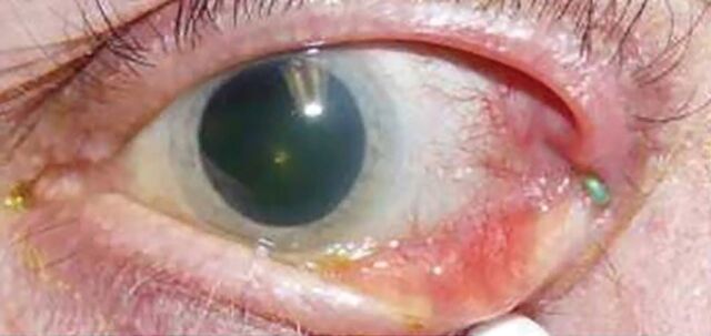
At 3.5 years after the initial LTS procedure, the patient complained of left eye discharge and pus that was unresponsive to lid scrub and polytrim topical treatment prescribed by an optometrist colleague. Slit lamp examination revealed a dark green Polydek suture extruding on the surface of the left lateral canthus (Figure 1). We removed the suture and prescribed maxitrol ointment. One week later, the patient’s left lateral canthus was completely normal.
Figure 1.
The green PolyDek extruded suture is on the surface of the lateral canthus.
Case 2: Recurrent wound abscess and suture extrusion after right side entropion repair
A man, aged 82 years, with no history of obesity or diabetes underwent right side entropion repair. Although the patient’s wound at the lateral canthus area opposed well on postoperative day 2, a small 3-5 mm abscess-like lesion with surrounding erythema and induration was noted 2 weeks later. We performed I&D, but the liquid from the abscess was relatively clear. The patient received topical and systemic antibiotics. One week later, he developed another small abscess at the same location. We collected specimens for culture that revealed a few colonies of S. aureus and confirmed that the bacteria were sensitive to the antibiotics we prescribed. When the patient returned one week later, the abscess was present again. While performing I&D, we explored the deep tissue underneath the wound with gentle dissection. The Polydek suture was not visible, and there was no deep abscess. We prescribed a different antibiotic regimen based on the sensitivity profile from the previous microbiological testing panel. We rechecked the patient twice during the following month. Although his lid was in good position, and there was no new abscess, a small nodular growth had formed at the right canthus area. About 3 months after the initial surgery, the patient’s son, a nurse, noticed a green suture extruding from the lateral canthus area and removed it. Since then, the patient has not had any problems over the wound area.
Case 3: Right side wound infection and granuloma formation after bilateral ectropion repair
A man, aged 73 years, with obesity and diabetes underwent bilateral ectropion repair. Although the wound healed well initially, the patient complained of irritation and scab formation on and off at the right lateral canthus area during his 6-week post-operative visit. We saw a round, small lesion at the right lateral canthus that was excised, and we collected specimens for both microbiology and pathology studies. We placed the patient on a regimen of topical and systemic antibiotics. Microbiological testing revealed a light growth of S. aureus, while the pathology report described the lesion as a “focal ulcer with mixed inflammation including suppurative and rare granulomatous inflammation.” When the patient returned one week later, a new, small, lesion in the same area was noticed. Approximately 3 months after the initial surgery, this persistent lesion was excised along with the underlying Polydek suture. The pathologist noted the “collective features are compatible with foreign body reaction to suture with associated mixed inflammation and granulation tissue.” Since suture removal, the wound has healed completely.
Case 4: Development of right side granuloma after bilateral ectropion repair
A man, aged 88 years, with no history of obesity or diabetes underwent bilateral ectropion repair. During the patient’s one-week post-operative examination, a thickened area at the lateral canthus without tenderness was noticed. One month later, there was neither erythema nor tenderness at the area; however, a “lump” was present. The patient came back 2 months after surgery complaining of a sensitive bump in the temporal area. On examination, there was a solid lesion with a smooth surface superior to the lateral canthus. The lesion was removed as an “eyelid lesion.” However, pathology examination showed “granulation tissue with mixed inflammation with neutrophils predominance, and a few multinucleated giant cells” without mention of any suture, which was not visible while removing the lesion.
Discussion
Out of the 58 cases of LTS procedures (83 eyelids) that we analyzed over a 10-year period, four cases of LTS Polydek suture complications occurred and varied in severity from chronic infection/inflammation and granuloma formation to suture extrusion. Most of these complications were resolved only after the Polydek sutures spontaneously extruded out or were removed surgically. Infection usually occurred within a few weeks after the procedure; and granuloma occurred a few months postoperatively, with suture extrusion occurring anywhere from a few months to years later.
Chronic infection and abscess formation impeded wound healing and were present during suture removal in Cases 1, 2, and 3. In Case 1, the patient had an initial wound infection that responded somewhat to antibiotics. However, the wound did not completely heal until the Polydek suture was surgically removed 1.5 years later. Based on previous literature, it is possible that the braided Polydek sutures facilitated bacterial penetration into the interstices of the wound to promote chronic infection,9-13 though chemical composition of the sutures can also play a role in wound infection.9-12 In contrast to Case 1, the patient in Case 2 did not exhibit purulent pus nor a good response to antibiotic treatment, and the microbiology study only showed light growth of S. aureus, which could result from skin flora contamination. Therefore, this patient might have exhibited a suture-related pseudoinfection due to a tissue foreign-body reaction as reported by Pierannunzii et al.14 However, regardless of pre-existing infection or psuedoinfection, wound complications in Cases 1-4 only resolved when the Polydek sutures were removed.
Complications of wound healing such as granuloma, suture extrusion, and suture abscess may occur if the suture ends are exposed to the skin surface or the permanent suture is not buried deeply within the orbicularis closure.8,15 In the cases we presented, all Polydek sutures were deeply buried and not noticeable while performing I&D or removing the granuloma; they were only visible after deep dissection for suture removal. Therefore, even though the permanent sutures were well-buried, it is still possible to encounter wound complications associated with Polydek sutures. Although our two patients who developed granulomas had bilateral procedures, this complication only occurred on one side. This unilateral presentation of a wound healing complication after a bilateral procedure has also been reported previously for a patient undergoing transconjunctival blepharoplasty.8 Our Case 1 patient also had a bilateral procedure but with two separate instances of complications, specifically chronic infection on the right side and suture extrusion on the left side, which might be related to the amount and duration of inflammation at each site.
Granuloma formation and suture extrusion are extreme tissue reactions to suture material. Studies of suture-related complications in rabbits with corresponding histological sections highlighted more severe tissue reactivity, specifically fibrous capsula formation around non-absorbable multifilament sutures,4,6 like Polydek, compared to non-absorbable monofilament sutures such as nylon4,6 or absorbable Vicryl sutures.6 This is due, in part, to cellular reaction in response to shedding of the polytetrafluoroethylene suture coating and/or filament characteristics of the suture itself.4-7,13 Since multifilament sutures tend to cause more tissue reaction than monofilament sutures,4,6,13 Polydek has a greater likelihood of producing an adverse tissue response overall in patients.5,16 This conclusion is supported by clinical evidence where patients receiving non-absorbable multifilament Polydek sutures for abdominal fascial closures were significantly more likely to develop granulomas in response to suture material than absorbable monofilament nylon or Prolene sutures.5 The Polydek-associated tissue responsibility may occur in response to the polytetrafluoroethylene coating itself, as there is a case where a polytetrafluoroethylene sheet used for congenital ptosis repair produced severe tissue reactivity that resolved only after the sheet was removed.16 Our results support the conclusion that Polydek sutures could produce persistent chronic tissue reaction.
Conclusion
Due to increased risk of tissue reactivity and infection associated with Polydek suture, we suggest the use of nonabsorbable monofilament sutures such as Prolene or absorbable sutures such as Vicryl for LTS procedures. More studies are needed on the Polydek suture complication in non-ophthalmic surgeries before this recommendation could be extended to other surgeries using Polydek sutures.
Acknowledgements
The authors would like to acknowledge Emily A. Andreae, PhD, and Marie A Fleisner for assistance with manuscript preparation.
References
- 1.Medline. Products. Non-absorbable Polydek Sutures by Teleflex Medical. https://www.medline.com/product/Non-absorbable-Polydek-Sutures-by-Teleflex-Medical/Z05-PF41724. Accessed 3 Feb 2020.
- 2.Anderson RL, Gordy DD.. The tarsal strip procedure. Arch Ophthalmol. 1979;97:2192-2196. [DOI] [PubMed] [Google Scholar]
- 3.Mcveigh KA, Harrison R, Ford R.. Entropion and ectropion repair: A snapshot of surgical practice in the United Kingdom. Orbit 2018;37:105-109. [DOI] [PubMed] [Google Scholar]
- 4.Postlethwait RW. Long-term comparative study of nonabsorbable sutures. Ann Surg. 1970;171:892-898. [DOI] [PMC free article] [PubMed] [Google Scholar]
- 5.LoCicero J 3rd, Robbins JA, Webb WR.. Complications following abdominal fascial closures using various nonabsorbable sutures. Surg Gynecol Obstet. 1983;157:25-27. [PubMed] [Google Scholar]
- 6.Setzen G, Williams EF 3rd.. Tissue response to suture materials implanted subcutaneously in a rabbit model. Plast Reconstr Surg. 1997;100:1788-1795. [DOI] [PubMed] [Google Scholar]
- 7.Carr BJ, Ochoa L, Rankin D, Owens BD.. Biologic response to orthopedic sutures: A histologic study in a rabbit model. Orthopedics. 2009;32:828. [DOI] [PubMed] [Google Scholar]
- 8.Soll SM, Lisman RD, Charles NC, Palu RN.. Pyogenic granuloma after transconjunctival blepharoplasty: A case report. Ophthalmic Plast Reconstr Surg. 1993;9:298-301. [DOI] [PubMed] [Google Scholar]
- 9.Alexander JW, Kaplan JZ, Altemeier WA.. Role of suture materials in the development of wound infection. Ann Surg. 1967;165:192-199. [DOI] [PMC free article] [PubMed] [Google Scholar]
- 10.Edlich RF, Panek PH, Rodeheaver GT, Turnbull VG, Kurtz LD, Edgerton MT.. Physical and chemical configuration of sutures in the development of surgical infection. Ann Surg. 1973;177:679-688. [DOI] [PMC free article] [PubMed] [Google Scholar]
- 11.Blomstedt B, Osterberg B, Bergstrand A.. Suture material and bacterial transport. An experimental study. Acta Chir Scand. 1977;143:71-73. [PubMed] [Google Scholar]
- 12.Katz S, Izhar M, Mirelman D.. Bacterial adherence to surgical sutures. A possible factor in suture induced infection. Ann Surg. 1981;194:35-41. [DOI] [PMC free article] [PubMed] [Google Scholar]
- 13.Everett WG. Sutures, incisions, and anastomoses. Ann R Coll Surg Engl. 1974;55:31-38. [PMC free article] [PubMed] [Google Scholar]
- 14.Pierannunzii LI, Fossali A, De Lucia O, Guarino A.. Suture-related pseudoinfection after total hip arthroplasty. J Orthop Traumatol. 2015;16:59-65. [DOI] [PMC free article] [PubMed] [Google Scholar]
- 15.Olver JM. Surgical tips on the lateral tarsal strip. Eye (Lond) 1998;12:1007-1012. [DOI] [PubMed] [Google Scholar]
- 16.Kokubo K, Katori N, Hayashi K, Kasai K, Kamisasanuki T, Sueoka K, Maegawa J.. Frontalis suspension with an expanded polytetrafluoroethylene sheet for congenital ptosis repair. J Plast Reconstr Aesthet Surg. 2016;69:673-678. [DOI] [PubMed] [Google Scholar]


