Abstract
BACKGROUND
Alveolar echinococcosis is a rare condition, but living or working in a rural environment is a substantial risk factor. The liver is the organ primarily affected, with additional extrahepatic manifestations in approximately 25% of cases. Primary extrahepatic disease is rare, and isolated cerebral involvement is extremely unusual.
OBSERVATIONS
The authors described an illustrative case of isolated cerebral alveolar echinococcosis in an immunocompetent farmer. Magnetic resonance imaging of the brain showed a predominantly cystic lesion with perifocal edema and a “bunch of grapes” appearance in the left frontal lobe. Histology revealed sharply demarcated fragments of a fibrous cyst wall accompanied by marked inflammation and necrosis. Higher magnification showed remnants of protoscolices with hooklets and calcified corpuscles. Immunohistochemistry and polymerase chain reaction (PCR) analysis confirmed the diagnosis of cerebral alveolar echinococcosis. Interestingly, serology and thoracic and abdominal computed tomography results were negative, indicative of an isolated primary extrahepatic manifestation.
LESSONS
Isolated, primary central nervous system echinococcosis is extremely rare, with only isolated case reports. As in the authors’ case, it can occur in immunocompetent patients, especially persons with a rural vocational history. Negative serology results do not exclude cerebral echinococcosis, which requires histological confirmation. Immunohistochemical staining and PCR analysis are especially useful in cases without classic morphological findings.
KeyWords: cerebral echinococcosis, Echinococcus, farmer, immunocompetent, brain, neuroimaging
ABBREVIATIONS : CNS = central nervous system, MRI = magnetic resonance imaging, PAS = periodic acid–Schiff, PCR = polymerase chain reaction
Alveolar echinococcosis caused by infection with Echinococcus multilocularis is a rare condition, affecting three to four individuals per million in Switzerland every year.1 The mean age of affected persons is 50 to 60 years, with men being affected slightly more than women.2 Living or working in a rural environment is associated with an increased risk.3 The primary host is the red fox, with a prevalence of more than 50%, but domestic dogs may also be responsible for transmission to humans.4 The liver is the primarily affected organ, with additional extrahepatic manifestations in approximately 25% of cases.1 Secondary involvement of the central nervous system (CNS) occurs in 1% to 3% of cases,2 with only rare cases of primary extrahepatic disease.5 Cerebral lesions are often multiple but can be single and mimic a metastatic or primary brain tumor.6,7 Here, we describe an illustrative case of isolated cerebral alveolar echinococcosis in an immunocompetent farmer.
Illustrative Case
Clinical History
A previously healthy 58-year-old Swiss farmer presented with an episode of hemiplegia and loss of consciousness. Subsequent neurological examination revealed a tongue bite without evidence of neurological deficits, which was indicative of a seizure. The patient’s medical record was unremarkable, especially without B symptoms or tropical destinations in his travel history. Magnetic resonance imaging (MRI) of the brain revealed a predominantly cystic lesion with perifocal edema and a “bunch of grapes” appearance in the left frontal lobe (Fig. 1A and B). The solid areas showed contrast enhancement (Fig. 1C and D). Further diagnostic measures included serological tests for echinococcosis and cysticercosis and computed tomography of the thorax and abdomen, all of which had negative results. The patient was admitted to the neurosurgery unit and underwent complete resection of the lesion. Postoperative recovery was unremarkable, without any remaining clinical signs or symptoms.
FIG. 1.
MRI of the brain. T2-weighted imaging in the sagittal (A) and transverse (B) views. Contrast enhancement in the coronal (C) and transverse (D) views.
Pathology
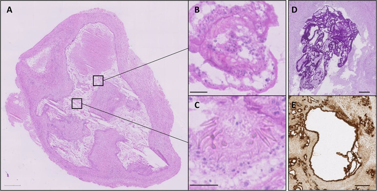
The resected tissue specimen was lobulated and of whitish color, measuring 2.5 × 1.5 × 1.2 cm. Histology revealed gliotic CNS tissue with sharply demarcated fragments of a fibrous cyst wall accompanied by marked inflammation and necrosis (Fig. 2A). Higher magnification showed remnants of protoscolices with hooklets and calcified corpuscles (Fig. 2B and C). The multilaminar membranes were strongly positive for periodic acid–Schiff (PAS) (Fig. 2D) and immunolabeled with an antibody that recognized the Em2G11 protein (Fig. 2E), consistent with the diagnosis of cerebral alveolar echinococcosis. Genetic testing using polymerase chain reaction (PCR) identified E multilocularis haplotype E5, a common and widely distributed haplotype in Europe.8
FIG. 2.
Histology. A: Hematoxylin and eosin stain shows overview section with marked areas magnified (B and C). D: PAS stain. E: Em2G11 (Echinococcus) immunohistochemistry. Scale bars = 1 mm (A), 20 μm (B and C), and 100 μm (D and E).
Discussion
Observations
In this study, we describe an illustrative case of isolated cerebral alveolar echinococcosis in an immunocompetent farmer. Primary extrahepatic manifestation represents 2% to 4% of cases,5 and cerebral involvement is extremely rare, especially in immunocompetent persons.9,10 However, a patient’s vocational history confers a substantial risk.3
Besides alveolar echinococcosis, the radiographic differential diagnosis of the cerebral lesion includes cysticercosis, tuberculosis, toxoplasmosis, fungi or bacterial brain abscesses, and noninfectious diseases such as metastases or even higher-grade gliomas. However, as in the present case, the MRI presentation with a “bunch of grapes” appearance is typical for alveolar echinococcosis.11 In general, serological testing is sensitive for hepatic disease but may produce negative results in cases with a nonhepatic manifestation, as happened with our patient.5 Histological examination usually reveals a sharply demarcated, centrally necrotic lesion with multiple cysts. The cysts are lined with eosinophilic multilaminar membranes, which are strongly PAS-positive. The lesion can be distinguished from other helminthic infections, such as cysticercosis, by the architecture of the cyst wall and the size of the hooklets. However, in our experience, the formation of protoscolices is extremely rare in immunocompetent patients (F. Grimm, personal communication, 2018). Therefore, prototypical hooklets, as seen in the present case, may not be found on histological examination. In that case, immunohistochemical staining for proteins expressed by E multilocularis and PCR analysis are helpful in establishing a diagnosis.
Lessons
Isolated, primary CNS echinococcosis is extremely rare, with few case reports. As in our case, it can also occur in immunocompetent patients, especially persons with a rural vocational history. Negative serology results do not exclude cerebral echinococcosis, which requires histological confirmation. Immunohistochemical staining and PCR analysis are especially useful in cases without classic morphological findings, especially in immunocompetent patients. Here, we describe an extremely rare illustrative case, which is important to consider in the differential diagnosis of a primary cerebral lesion.
Disclosures
The authors report no conflict of interest concerning the materials or methods used in this study or the findings specified in this paper.
Author Contributions
Conception and design: Rushing, Reuss, Wulf, Henzi. Acquisition of data: Rushing, Wulf, Oertel, Henzi, Reinehr, Grimm. Analysis and interpretation of data: Rushing, Reuss, Wulf, Oertel, Bozinov, Henzi, Reinehr, Grimm. Drafting the article: Rushing, Reuss, Wulf, Henzi. Critically revising the article: Rushing, Reuss, Oertel, Bozinov, Kaelin, Reinehr. Reviewed submitted version of manuscript: Rushing, Reuss, Oertel, Kaelin, Grimm. Approved the final version of the manuscript on behalf of all authors: Rushing. Administrative/technical/material support: Rushing, Oertel, Bozinov. Study supervision: Rushing, Bozinov.
References
- 1. Beldi G, Müller N, Gottstein B. Die alveoläre Echinokokkose. Swiss Med Forum. 2017;17(36):760–766. [Google Scholar]
- 2. Kern P, Bardonnet K, Renner E, et al. European echinococcosis registry: human alveolar echinococcosis, Europe, 1982–2000. Emerg Infect Dis. 2003;9(3):343–349. doi: 10.3201/eid0903.020341. [DOI] [PMC free article] [PubMed] [Google Scholar]
- 3. Kern P, Ammon A, Kron M, et al. Risk factors for alveolar echinococcosis in humans. Emerg Infect Dis. 2004;10(12):2088–2093. doi: 10.3201/eid1012.030773. [DOI] [PMC free article] [PubMed] [Google Scholar]
- 4. Oksanen A, Siles-Lucas M, Karamon J, et al. The geographical distribution and prevalence of Echinococcus multilocularis in animals in the European Union and adjacent countries: a systematic review and meta-analysis. Parasit Vectors. 2016;9(1):519. doi: 10.1186/s13071-016-1746-4. [DOI] [PMC free article] [PubMed] [Google Scholar]
- 5. Meinel TR, Gottstein B, Geib V, et al. Vertebral alveolar echinococcosis: a case report, systematic analysis, and review of the literature. Lancet Infect Dis. 2018;18(3):e87–e98. doi: 10.1016/S1473-3099(17)30335-3. [DOI] [PubMed] [Google Scholar]
- 6. Isik N, Silav G, Cerçi A, et al. Cerebral alveolar echinococcosis. A case report with MRI and review of the literature. J Neurosurg Sci. 2007;51(3):145–151. [PubMed] [Google Scholar]
- 7. Tunaci M, Tunaci A, Engin G, et al. MRI of cerebral alveolar echinococcosis. Neuroradiology. 1999;41(11):844–846. doi: 10.1007/s002340050854. [DOI] [PubMed] [Google Scholar]
- 8. Nakao M, Xiao N, Okamoto M, et al. Geographic pattern of genetic variation in the fox tapeworm Echinococcus multilocularis . Parasitol Int. 2009;58(4):384–389. doi: 10.1016/j.parint.2009.07.010. [DOI] [PubMed] [Google Scholar]
- 9. Baldolli A, Bonhomme J, Yera H, et al. Isolated cerebral alveolar echinococcosis. Open Forum Infect Dis. 2018;6(1):ofy349. doi: 10.1093/ofid/ofy349. [DOI] [PMC free article] [PubMed] [Google Scholar]
- 10. Kern P, Menezes da Silva A, Akhan O, et al. The echinococcoses: diagnosis, clinical management and burden of disease. Adv Parasitol. 2017;96:259–369. doi: 10.1016/bs.apar.2016.09.006. [DOI] [PubMed] [Google Scholar]
- 11. Kantarci M, Bayraktutan U, Karabulut N, et al. Alveolar echinococcosis: spectrum of findings at cross-sectional imaging. Radiographics. 2012;32(7):2053–2070. doi: 10.1148/rg.327125708. [DOI] [PubMed] [Google Scholar]


