ABSTRACT
Uvular necrosis is a potential etiology of postesophagogastroduodenoscopy persistent sore throat and odynophagia, and physicians should be alert to the possibility of this potential complication. Diagnosis is clinical and can be made on the basis of symptoms and characteristic findings on oropharyngeal examination. It has a benign course with an overall good clinical outcome. Conservative symptomatic management is the treatment of choice, and full recovery can be expected in 2 weeks. Keeping oropharyngeal instruments and ventilation tubes to the side of the midline, avoidance of blind suctioning, and decreasing the power of suction devices are some of the measures, which might reduce the risk of intraprocedure uvular injury. In addition, it is important to note that the risk of injury is higher in some individuals, for instance, patients with a long uvula, and it might be beneficial to take extra precautions in these patients.
INTRODUCTION
Esophagogastroduodenoscopy (EGD) is generally considered a safe procedure, with sore throat being the most common adverse event. Uvular necrosis is a rare complication after endoscopy but an important differential in patients with a persistent post-EGD sore throat. As most patients who develop a post-EGD sore throat do not present to their physician, uvular injury associated with upper endoscopy is an underdiagnosed condition that requires more attention. Diagnosis is based clinically on characteristic findings on oropharyngeal examination, and treatment is supportive. This article reviews a case of uvular necrosis after uncomplicated intubation and endoscopy in a healthy middle-aged woman. The patient recovered within days and without any long-term negative effects.
CASE REPORT
A 59-year-old woman presented as an emergency department follow-up for evaluation of abdominal pain, diarrhea, and hematochezia. The patient was initially evaluated at a local emergency department for similar complaints 2 days prior when computed tomography imaging demonstrated enteritis involving the terminal ileum, distal descending colon, sigmoid colon, and rectum. A colonoscopy was recommended for further evaluation of colitis, and an EGD because of a family history of celiac disease. The patient had a medical history of renal stones and uterine leiomyomas for which she had undergone hysterectomy 20 years back. She had a body mass index of 31 and had a history of light alcohol consumption (2–3 drinks per week). There was no history of tobacco or any illicit drug use, and she was not taking any routine medication.
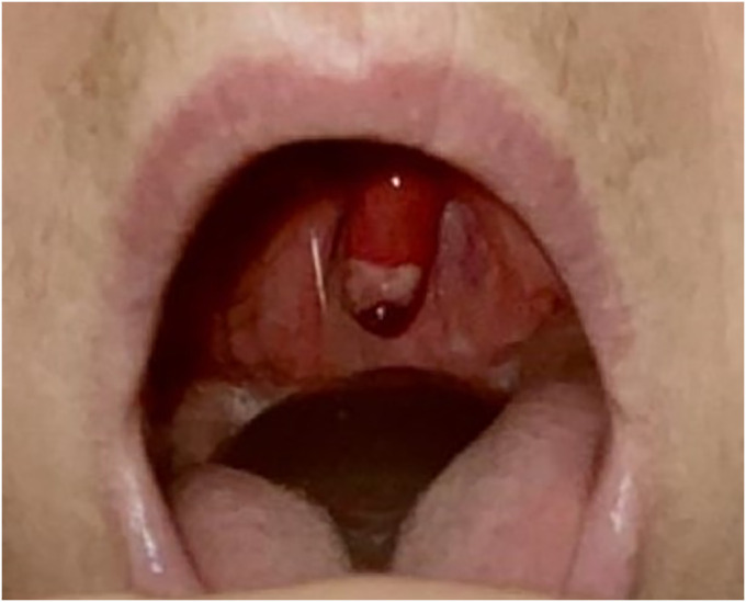
The patient was intubated after starting with endoscopy for airway protection because she was unable to tolerate the insufflation of air in the stomach during endoscopy. The Mallampati score was 2, and propofol was used for sedation. Intubation was successfully performed without any complications. In recovery, extubation was performed without event, and she was discharged home the same day. Colonoscopy revealed ulcerated mucosa, and biopsies were suggestive of ischemic colitis. EGD demonstrated a 2cm hiatal hernia, gastritis, and nonbleeding gastric ulcers. Biopsies from the stomach were positive for Helicobacter pylori, and she was treated with amoxicillin-based therapy. Duodenal biopsies showed small bowel mucosa without significant histopathological abnormality. Two days after procedure, the patient complained of severe throat pain, odynophagia, and dysphagia. On oropharyngeal examination, inflammation and a black, necrotic uvular tip were seen, which is a classic finding of uvular necrosis (Figure 1).
Figure 1.

Inflamed and necrosed tip of the uvula with black discoloration was demonstrated on oropharyngeal examination.
The patient was treated supportively with over-the-counter Tylenol and a full liquid diet. She was offered topical lidocaine for swish and swallow or a short course of steroids. Her symptoms rapidly resolved over 2 days. The necrotic tip eventually got autoamputated. She returned to the clinic for H. pylori eradication testing and reported no long-lasting negative oropharyngeal symptoms.
DISCUSSION
Throat discomfort is a common complaint after orotracheal and nasotracheal procedures and is usually caused by pharyngeal irritation. Although most patients report spontaneous improvement within 1–2 days with minimal requirement for analgesia, persistent sore throat with odynophagia should alert endoscopists to the potential complication of postprocedure uvular injury. The spectrum of post-EGD uvular injury ranges from mild uvular edema to uvulitis and uvular necrosis.
Uvular necrosis has been reported after endotracheal intubation, upper gastrointestinal endoscopy, bronchoscopy through nasal approach, and vigorous suctioning.1 The underlying pathophysiology is believed to be uvular ischemia secondary to the suction-induced pressure or the trauma from oropharyngeal instrumentation. During endoscopy, prolonged ischemia and resultant ulceration and necrosis may be caused by compression of the uvula between the shaft of the endoscope and the hard palate or the posterior pharynx.2 Anatomically, the uvula is a highly vascular structure composed of mucosa, submucosa, and muscle but devoid of the shielding effect of cartilage. This makes it susceptible to ischemic injury with even small amounts of pressure for a short period. Although the underlying pathology is most likely uvular ischemia, there are no studies that show an increase in the incidence of this complication in patients with underlying vascular diseases. However, the differences in oropharyngeal anatomy may predispose certain individuals to this complication. A previous study by Emmett et al showed that males are at a higher risk of developing uvular injury because of the presence of a bulkier tongue and palate and a higher amount of nonfatty tissue in the neck that becomes flaccid with anesthesia, making the uvula more prone to mechanical injury.3,4 Similarly, patients with an elongated uvula are also at a higher risk.5 By contrast, pediatric uvular injury presents more commonly as mild edema, and cases of necrosis seem to be less common in this population.6
Patients may present from 1 hour to 2 days after the procedure. Symptoms may not be present in the immediate post-EGD period, but as the necrosis worsens, the patients may develop sore throat, dysphagia, globus sensation, odynophagia, dyspnea, and gagging. Like the symptoms, the changes in the physical appearance of the injured uvula also follow a progressive course. It may take several days for a visible change to occur, and as the condition progresses, the tip of the uvula becomes elongated and whitish, with a black border.7 Necrotic discharge may also be noted on the surface in some cases. Diagnosis can be made clinically based on the physical examination of the uvula and the temporal relationship between the procedure and the onset of symptoms.
Although optimal treatment remains debated, a general consensus is to follow a conservative course with symptomatic management.8,9 Treatment options include simple observation, acetaminophen, steroids, antihistamines, and topical epinephrine. Corticosteroids have been shown to be beneficial in the empiric treatment of sore throat and may be used in cases when the pain is refractory to standard analgesics.10 They may also be useful in cases of significant edema, leading to airway obstruction. Because there have been no documented cases of infections complicating uvular injury, the role of antibiotics remains unclear and warrants further research.7 They may be used if there are signs of infection on examination or laboratory testing. The symptoms usually resolve in 4–7 days, and the necrotic part of the uvula sloughs off on its own in 14 days.9 The prognosis is excellent, with most patients having a full recovery in 2 weeks.11 Data suggest that surgery is not usually required in this self-limited condition. Surgical excision of the necrosed uvula has been reported in only 2 cases, and in a third case, the patient performed self-excision.6 Faster recovery or symptom resolution was not reported in any of these cases, making surgical management a less-preferred alternative.
Although a designated protocol to mitigate the risk of this complication still requires more study, certain measures may be appropriate to decrease its incidence. These include both procedure and individual-specific measures that reduce uvular compression and trauma. Positioning of instruments and ventilation tubes along the lateral wall of the buccal cavity instead of the midline, decreasing the power of suction devices, and performing suctioning under direct visualization can help prevent uvular injury.7 Further study into the high-risk anatomical parameters of the oropharynx may help in the formulation of a preprocedure screening protocol to identify at-risk individuals. Such patients can be specifically warned about uvular injury and extra precautions, and care should be taken during their procedures.
Familiarity with the pathophysiology, clinical course, and prognosis of this condition may aid clinicians in prevention, early detection, and management while reassuring the patient of its benign course and good clinical outcome with conservative management.
DISCLOSURES
Author contribution: Literature review was performed by all authors. S. Barrett, S. Bajaj, and U. Bhatia drafted the manuscript. The manuscript was critically revised by Y. Bi. Figures acquisition was performed by S. Barrett. All authors approved the final version to be published and agree to be accountable for all aspects of the work. Yan Bi is the guarantor of the article.
Financial disclosure: None to report.
Ethical approval: No institutional review board approval needed.
Informed consent was obtained for this case report. No content of the article has been presented or published previously.
Footnotes
Equal contribution status (joint first authors).
Contributor Information
Unnati Bhatia, Email: unnatibhatia60@gmail.com.
Stevi Barrett, Email: barrett.stevi@mayo.edu.
Yan Bi, Email: bi.yan@mayo.edu.
REFERENCES
- 1.Iftikhar MH, Raziq FI, Laird-Fick H. Uvular necrosis as a cause of throat discomfort after endotracheal intubation. BMJ Case Rep 2019;12(7):e231227. [DOI] [PMC free article] [PubMed] [Google Scholar]
- 2.Tang SJ, Kanwal F, Gralnek IM. Uvular necrosis after upper endoscopy: A case report and review of the literature. Endoscopy 2002;34(7):585–7. [DOI] [PubMed] [Google Scholar]
- 3.Emmett SR, Lloyd SD, Johnston MN. Uvular trauma from a laryngeal mask. Br J Anaesth 2012;109(3):468–9. [DOI] [PubMed] [Google Scholar]
- 4.Whittle AT, Marshall I, Mortimore IL, Wraith PK, Sellar RJ, Douglas NJ. Neck soft tissue and fat distribution: Comparison between normal men and women by magnetic resonance imaging. Thorax 1999;54(4):323–8. [DOI] [PMC free article] [PubMed] [Google Scholar]
- 5.Sunio LK, Contractor TA, Chacon G. Uvular necrosis as an unusual complication of bronchoscopy via the nasal approach. Respir Care 2011;56(5):695–7. [DOI] [PubMed] [Google Scholar]
- 6.Arigliani M, Dolcemascolo V, Passone E, Vergine M, Cogo P. Uvular trauma after laryngeal mask airway use. J Pediatr 2016;176:217. [DOI] [PubMed] [Google Scholar]
- 7.Reid JW, Samy A, Jeremic G, Brookes J, Sowerby LJ. Postoperative uvular necrosis: A case series and literature review. Laryngoscope 2020;130(4):880–5. [DOI] [PubMed] [Google Scholar]
- 8.Fich A, Ligumsky M, Wolnerman JS. Necrosis of the uvula after endoscopy. Gastrointest Endosc 1984;30(5):317. [DOI] [PubMed] [Google Scholar]
- 9.Peghini PL, Salcedo JA, Al-Kawas FH. Traumatic uvulitis: A rare complication of upper GI endoscopy. Gastrointest Endosc 2001;53(7):818–20. [DOI] [PubMed] [Google Scholar]
- 10.Bergeson K, Rogers N, Prasad S. PURLs: Corticosteroids for a sore throat? J Fam Pract 2013;62(7):372–4. [PMC free article] [PubMed] [Google Scholar]
- 11.Evans DP, Lo BM. Uvular necrosis after orotracheal intubation. Am J Emerg Med 2009;27(5):631.e633–4. [DOI] [PubMed] [Google Scholar]


