Abstract
Objective The present study aimed to compare the effects of intraarticular infiltration of platelet-rich plasma with those of hyaluronic acid infiltration in the treatment of patients with primary knee osteoarthritis.
Methods A randomized clinical trial was conducted with 29 patients who received an intraarticular infiltration with hyaluronic acid (control group) or platelet-rich plasma. Clinical outcomes were assessed using the visual analog scale for pain and the Western Ontario and McMaster Universities Arthritis Index (WOMAC) questionnaire before and after the intervention. In addition, the posttreatment adverse effects were recorded. Categorical variables were analyzed using the chi-square and Fisher exact tests, whereas continuous variables were analyzed using the Student t test, analysis of variance, and the Wilcoxon test; all calculations were performed with the Stats package of the R software.
Results An independent analysis of each group revealed a statistical difference within the first months, with improvement in the pain and function scores, but worsening on the 6 th month after the procedure. There was no difference in the outcomes between the groups receiving hyaluronic acid or platelet-rich plasma. There was no serious adverse effect or allergic reaction during the entire follow-up period.
Conclusion Intraarticular infiltration with hyaluronic acid or platelet-rich plasma in patients with primary knee gonarthrosis resulted in temporary improvement of functional symptoms and pain. There was no difference between interventions.
Keywords: osteoarthritis, knee; hyaluronic acid; infiltration
Introduction
Knee osteoarthritis (OA) is a degenerative disease affecting mostly females and resulting in progressive joint cartilage destruction. Osteoarthritis leads to joint deformity, potentially with muscle and ligament imbalance, and most abnormalities occur in regions subjected to greater load. Its typical radiographic signs include bone sclerosis, cysts, and osteophytes. 1 2 3
Knee OA has a great impact on physical performance, and it is considered one of the 10 main causes of disability around the world. Standard conservative treatments for knee OA include weight loss, exercise, non-steroidal antiinflammatory drugs (NSAIDs), analgesic agents, intraarticular injection of hyaluronic acid (HA) and glucocorticoids. 4
Hyaluronic acid is used in the treatment of degenerative joint diseases. It is a glycosaminoglycan that acts on the extracellular matrix providing greater joint lubrication and protection.
Recently, however, orthobiologic injections have emerged as a potentially safe and effective option for knee OA treatment. These injections include bone marrow concentrate (BMC), mesenchymal stem cells (MSC), and platelet-rich plasma (PRP).
Platelet-rich plasma consists in plasma with a high platelet concentration. 5 Depending on the method used for PRP processing, it may also contain white blood cells in abnormally high concentrations. 6 Platelets and white blood cells are sources of high cytokines levels, which play a well-documented role in controlling a number of tissue regeneration processes, including cell movement and proliferation, angiogenesis, inflammation regulation, and collagen synthesis. 6
In addition to their role in local hemostasis, platelets contain an abundance of growth factors and cytokines, which are crucial in soft-tissue healing and bone mineralization. 7 Moreover, they release a number of proteins that attract macrophages, mesenchymal stem cells, and osteoblasts, resulting in necrotic tissues removal and faster tissue regeneration. 4
Recently, some studies investigated the potential beneficial effects of PRP in chronic diseases, including lateral epicondylitis and plantar fasciitis. 4 However, most studies using PRP in the literature are non-randomized and have insufficient samples.
The present study aims to determine the effect on pain and function outcomes of an intraarticular application of PRP in comparison to HA to treat knee OA patients.
Method
This is a randomized clinical trial with 29 consecutively included patients. All patients participating in the present study agreed and signed an informed consent form.
This study complied with the Helsinki Declaration and Guideline for Good Clinical Practice. The research protocol was approved by the local ethics committee (Opinion at Plataforma Brasil , number 3.293.253).
Patient selection
The total sample included 29 patients of both genders, aged between 49 and 75 years old, who met the clinical and radiographic diagnostic criteria of the American College of Rheumatology (ACR) for knee OA and categorized as grade II or III according to the Kellgren-Lawrence classification. 2
The exclusion criteria were the following: previous surgery on the affected knee at any time or orthopedic surgery on the lower limbs within the 12 months prior to the study; previous HA or steroid infiltration within 3 months prior to the study; advanced OA cases (grades IV and V); diagnosis of autoimmune or rheumatological diseases; body mass index (BMI) ≥ 35; secondary OA (i.e., fractures, neoplasms); history of acute or chronic communicable diseases; difficult-to-control or insulin-dependent type I or II diabetes; coxarthrosis diagnosed at the physical or radiographic examination; active infection or history of infection at the affected joint; axial deviation in 10 o varus, 15 o varus or 1 cm discrepancy in lower limbs; use of anticoagulants or immunosuppressants; discontinuation of oral chondroprotective therapy within the last 3 months; and abnormal renal and/or liver function.
All included patients had a confirmed diagnosis of knee OA and underwent conservative treatment with physical therapy, stretching exercises, and analgesic agents for at least 6 months before the start of the study. Osteoarthritis was evaluated with knee radiographies in two views (anteroposterior and lateral) under load.
Tests requested during preselection visits were the following: biochemical blood tests (aspartate aminotransferase, alanine aminotransferase, gamma-glutamyl transferase, fasting blood sugar, creatinine, sodium, potassium, hemoglobin A1C, complete blood count), serology for communicable diseases, magnetic resonance imaging (MRI) of the affected knee, bilateral knee radiography, and panoramic radiography of lower limbs.
Randomization
Patients were randomized using the Research Randomizer System. 8 Thus, the study had two arms: a study group, submitted to an intraarticular application of PRP, and a control group, receiving a HA application.
Application method
Patients from both study arms were scheduled on an outpatient basis for infiltration at the following week. Control group patients underwent a single knee intra-articular infiltration with Synvisc One Hylan G-F20 (Lancaster, Pennsylvania, United States) following specific asepsis and antisepsis protocols.
Upon arrival at the hospital, subjects from the study group were directed to the blood collection sector, where a sample of 15 mL of blood was sterilely collected by peripheral access in a specific tube. The sample was then transported at a controlled temperature for processing at a laboratory from the same hospital.
The sample was centrifuged at 1,500 rotations per minute for 5 minutes at room temperature. Next, the sample was quantified and considered acceptable if it presented a two-fold increase in the number of platelets when compared with the baseline value.
After obtaining approximately 5 mL of PRP, the knee infiltration procedure was performed in a small surgical room. Platelet-rich plasma was applied through an intra-articular puncture on the knee. The entire process was carried out using the Arthrex Autologous Conditioned Plasma system (Arthrex Inc., Naples, FL, USA). The application process was repeated over the next 2 weeks, at 7 and 14 days, respectively, totaling 3 PRP infiltrations.
Clinical follow-up and outcomes evaluation
The subjects' data were collected by the researchers, including age, laterality, BMI, edema, and stiffness in the affected knee. Both groups were followed-up at the same frequency after the control group received the 3 rd application, for a total period of 6 months.
The standardized follow-up consisted in 5 outpatient medical visits over a 6-month period: the 1 st visit occurred after 1 week, and the following visits were at 2 weeks, 1, 3, and 6 months after the treatment. In addition, there were 2 telephone contacts with the patient, at 2 and 4 months after the procedure.
The Western Ontario and McMaster Universities Arthritis Index (WOMAC) score was obtained at the following times: preinfiltration, 1, 3, and 6 months after treatment. The visual analog scale (VAS) for pain was used 2 and 4 months after the procedure.
Statistical analysis
Statistical analysis was performed using the Stats package of the R software (R Foundation for Statistical Computing, Vienna, Austria). 9 Continuous variables were descriptively analyzed using means and standard deviations, followed by a normal distribution evaluation using the Shapiro test. 10 Categorical variables were presented as proportions.
For intra-group comparison, the analysis of variance (ANOVA) and Fisher's least significant difference tests were used 11 to determine any difference at WOMAC scores, whereas the VAS results were analyzed using a Student paired t test.
Intergroup differences were assessed using the Student t tests 12 for parametric variables, and the Mann-Whitney test 13 for non-parametric variables. Categorical variables were assessed between the study and the control groups using the chi-squared test 14 or Fisher exact test. 15
Results
No patient was lost at follow-up. Both the control and study groups were homogeneous, with no statistical difference between parameters, as shown in Table 1 .
Table 1. Patients characteristics.
| Platelet-rich plasma | Hyaluronic acid | P -value | |
|---|---|---|---|
| Number of patients | 14 | 15 | – |
| Body mass index | 28.3 (2.9*) | 28.1 (3.9*) | 0.60 |
| Age (years), Mean (SD) | 62.78 (6.10*) | 63.40 (4.99*) | 0.77 |
| Gender | 11 females 3 males |
13 females 2 males |
0.93 |
| Affected side | Right = 7 Left = 7 |
Right = 11 Left = 4 |
0.36 |
| Radiological Classification | Grade II = 9 Grade III = 5 |
Grade II = 9 Grade III = 6 |
1 |
| Knee swelling | Yes = 1 No = 13 |
Yes = 2 No = 13 |
1 |
| Knee stiffness | Yes = 1 No = 13 |
Yes = 2 No = 13 |
1 |
Abbreviation : SD, standard deviation.
Source: Prevent Senior São Paulo.
Regarding functional outcomes (WOMAC scores), there was no statistical differences between the study and control groups from the preintervention level to 6 months after treatment ( Table 2 ).
Table 2. Western Ontario and McMaster Universities Osteoarthritis Index score in subjects treated with platelet-rich plasma or hyaluronic acid.
| WOMAC, baseline | WOMAC, 1 month | WOMAC, 3 months | WOMAC, 6 months | ||
|---|---|---|---|---|---|
| PRP | Mean | 42.5 | 29.0 | 23.7 | 41.1 |
| Standard deviation | 17.9 | 16.0 | 22.0 | 24.8 | |
| HA | Mean | 41.1 | 24.0 | 26.0 | 35.7 |
| Standard deviation | 15.5 | 14.6 | 22.0 | 35.7 | |
| p -value | 0.82 | 0.39 | 0.78 | 0.73 | |
Abbreviations : HA, hyaluronic acid; PRP, platelet-rich plasma; WOMAC, Western Ontario and McMaster Universities Osteoarthritis Index.
Source: Prevent Senior São Paulo.
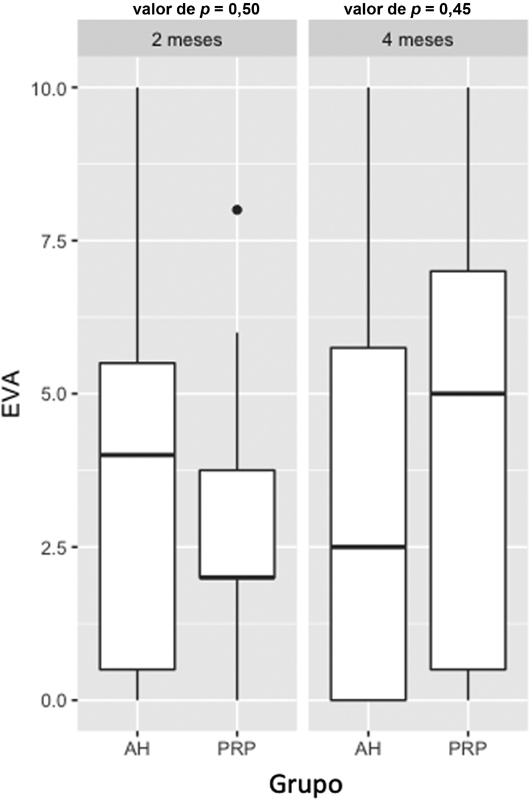
In addition, the VAS score for pain revealed no statistical difference at the 2 nd ( p = 0.50) and 4 th ( p = 0.45) month after the treatment, as shown in Figure 1 .
Fig. 1.
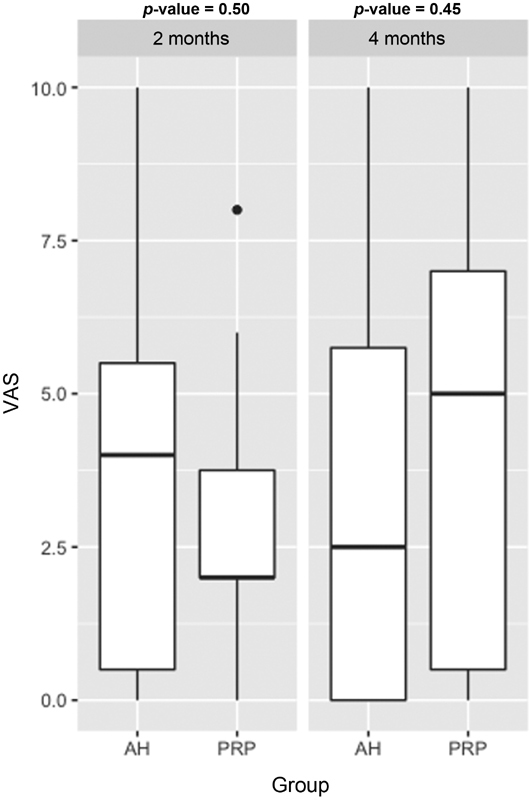
Distribution of the visual analog scale (VAS) for pain score in subjects treated with hyaluronic acid (HA) or platelet-rich plasma (PRP).
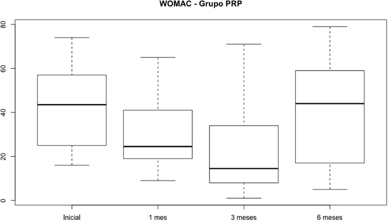
In the intragroup evaluation, function improved after the procedure, but worsened at the last month of evaluation. For the PRP group, there was a statistical difference between the WOMAC score at baseline and 3 months after the procedure ( p < 0.05). In addition, there was a difference between scores from the 1 st and 6 th month after the procedure due to an increased score ( p < 0.05). Pain was also influenced, with a mean difference in VAS score of 1.64 between the 2 nd and 4 th months after the treatment ( p < 0.05). Figure 2 shows WOMAC scores from the study group at different times.
Fig. 2.
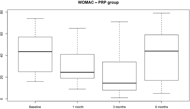
Distribution of the Western Ontario and McMaster Universities Osteoarthritis Index (WOMAC) score in subjects treated with platelet-rich plasma (PRP).
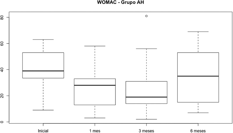
Regarding the HA group, there was a statistical difference in WOMAC scores from baseline and 1 month ( p < 0.05) and baseline to 3 months after treatment ( p < 0.05). There was no statistical difference in the VAS score for pain at the 2 nd and 4 th months after the treatment ( p = 0.49). Figure 3 shows this distribution.
Fig. 3.
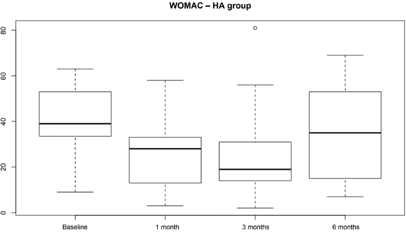
Distribution of the Western Ontario and McMaster Universities Osteoarthritis Index (WOMAC) score in subjects treated with hyaluronic acid (HA).
No infections or allergic reactions were reported during the 6-month follow-up. Pain cases were treated with analgesic agents, cryotherapy, and rehabilitation.
Discussion
The main finding of our study was the lack of difference in functional outcomes and pain assessment at a medium-term follow-up (6 months) between patients undergoing intraarticular infiltration with HA and PRP. However, both treatment methods were effective in improving pain and function over the study period and proved to be safe.
Functional assessment was performed using the WOMAC questionnaire, revealing no differences between the two groups over the 6-month follow-up. The literature is still controversial regarding this outcome. A recently published systematic review using the total WOMAC score for functional assessment concluded that PRP is superior to HA in the medium term (3–6 months). However, the same study found no differences between groups when analyzing fractional WOMAC scores for stiffness and physical function. 16
Another meta-analysis demonstrated the superiority of PRP over HA in pain improvement as assessed by the WOMAC score. However, the study concluded that there is no obvious superiority between PRP and HA in knee OA treatment. 17
Some randomized clinical trials comparing these two methods for OA treatment also found no differences in functional scores after 6 months of follow-up. 18 19
We found no differences regarding pain between the PRP and HA groups. Similarly, these outcomes are quite divergent to the literature. Zhang et al. 17 found no differences in the VAS for pain between both treatments 3 and 6 months after infiltrations. However, Cole et al. 19 demonstrated significant pain improvement according to the VAS in patients treated with PRP 6 and 12 months after the infiltration.
Most systematic reviews on the subject report the challenge in comparing the several published studies due to major variations in the PRP preparation and composition, the number of infiltrations performed, small samples, short follow-up times, and different inclusion and evaluation criteria. 20
Our study used a standardized kit for PRP preparation; in addition, samples were homogeneous, and infiltrations were performed once a week for 3 weeks. Previous studies had shown advantages of multiple PRP applications when compared to a single infiltration, including longer PRP effects when more than one application was performed. 21 22
Our study demonstrated that both PRP and HA were effective in treating pain and improving function. However, their effects deteriorate over time and virtually disappear 5 to 6 months after the treatment. Di Martino et al. 23 found similar outcomes in a randomized clinical trial. According to these authors, patients reported symptom improvement up to 9 months after HA application and up to 12 months after intraarticular PRP infiltration, but with progressive effect loss.
Filardo et al. 18 also observed similar outcomes in a randomized clinical trial with 1 year of follow-up. These authors showed an improvement in pain and function in patients treated with PRP or HA, but these outcomes remained virtually stable 2 months after treatment.
Regarding adverse effects, both PRP and HA proved to be safe in our study. None of the drugs caused severe, lasting side effects. In a meta-analysis, Han et al found no differences between treatment groups regarding adverse effects. 24 Other studies have also concluded that both treatments are safe, and have few side effects during follow-up. 25 26
The study has some limitations. First, despite being a preliminary report, the sample size is small. Second, the follow-up period is relatively short (6 months), and some studies have shown that PRP effects last longer than those of HA. The absence of a sham group (placebo or steroid infiltration) and the lack of group blinding are other limitations from our study.
Conclusion
Knee intraarticular infiltration with HA or PRP in patients with primary gonarthrosis resulted in transient improvement of pain and function. Both treatments proved to be safe. There was no difference between these interventions.
Conflito de interesses Os autores declaram não haver conflito de interesses.
Suporte Financeiro
Não houve suporte financeiro de fontes públicas, comerciais, ou sem fins lucrativos.
Financial Support
There was no financial support from public, commercial, or non-profit sources.
Trabalho desenvolvido no Departamento de Ortopedia e Traumatologia, Instituto Prevent Senior, São Paulo, Brasil.
Study developed at the Orthopedics and Traumatology Department, Instituto Prevent Senior, São Paulo, Brazil.
Referências
- 1.Keyes G W, Carr A J, Miller R K, Goodfellow J W. The radiographic classification of medial gonarthrosis. Correlation with operation methods in 200 knees. Acta Orthop Scand. 1992;63(05):497–501. doi: 10.3109/17453679209154722. [DOI] [PubMed] [Google Scholar]
- 2.Kellgren J H, Lawrence J S. Radiological assessment of osteo-arthrosis. Ann Rheum Dis. 1957;16(04):494–502. doi: 10.1136/ard.16.4.494. [DOI] [PMC free article] [PubMed] [Google Scholar]
- 3.Ahlbäck S.Osteoarthrosis of the knee. A radiographic investigation Acta Radiol Diagn (Stockh) 1968277277–272., 7–72 [PubMed] [Google Scholar]
- 4.Lana J F, Weglein A, Sampson S E. Randomized controlled trial comparing hyaluronic acid, platelet-rich plasma and the combination of both in the treatment of mild and moderate osteoarthritis of the knee. J Stem Cells Regen Med. 2016;12(02):69–78. doi: 10.46582/jsrm.1202011. [DOI] [PMC free article] [PubMed] [Google Scholar]
- 5.Schnabel L V, Mohammed H O, Miller B J. Platelet rich plasma (PRP) enhances anabolic gene expression patterns in flexor digitorum superficialis tendons. J Orthop Res. 2007;25(02):230–240. doi: 10.1002/jor.20278. [DOI] [PubMed] [Google Scholar]
- 6.Fortier L A, Hackett C H, Cole B J. The effects of platelet-rich plasma on cartilage: basic science and clinical application. Oper Tech Sports Med. 2011;19:154–159. [Google Scholar]
- 7.Steinert A F, Rackwitz L, Gilbert F, Nöth U, Tuan R S. Concise review: the clinical application of mesenchymal stem cells for musculoskeletal regeneration: current status and perspectives. Stem Cells Transl Med. 2012;1(03):237–247. doi: 10.5966/sctm.2011-0036. [DOI] [PMC free article] [PubMed] [Google Scholar]
- 8.Research Randomizer . (Version 4.02013) [computer program] http://www.randomizer.org/ http://www.randomizer.org/
- 9.Vienna, Austria: R Foundation for Statistical; 2014. R: A language and environment for statistical computing [computer program] [Google Scholar]
- 10.Shapiro S S, Wilk M B.An analysis of variance test for normality Biometrika 196552(3-4):591–611. [Google Scholar]
- 11.Fisher R A. The Correlation Between Relatives on the Supposition of Mendelian Inheritance. Philos Trans Royal Soc Edinburgh. 1918;52:399–433. [Google Scholar]
- 12.Gosset S WS. The probable error of a mean. Biometrika. 1908;6(01):1–25. [Google Scholar]
- 13.Neuhäuser M. Berlin, Heidelberg: Springer Berlin Heidelberg; 2011. Wilcoxon–Mann–Whitney Test; pp. 1656–1658. [Google Scholar]
- 14.Pearson K X. On the criterion that a given system of deviations from the probable in the case of a correlated system of variables is such that it can be reasonably supposed to have arisen from random sampling. Lond Edinb Philos Mag J Sci. 1900;50(302):157–175. [Google Scholar]
- 15.Fisher R A. On the Interpretation of χ 2 from Contingency Tables, and the Calculation of P . J R Stat Soc. 1922;85(01):87–94. [Google Scholar]
- 16.Chen Z, Wang C, You D, Zhao S, Zhu Z, Xu M. Platelet-rich plasma versus hyaluronic acid in the treatment of knee osteoarthritis: A meta-analysis. Medicine (Baltimore) 2020;99(11):e19388. doi: 10.1097/MD.0000000000019388. [DOI] [PMC free article] [PubMed] [Google Scholar]
- 17.Zhang H F, Wang C G, Li H, Huang Y T, Li Z J. Intra-articular platelet-rich plasma versus hyaluronic acid in the treatment of knee osteoarthritis: a meta-analysis. Drug Des Devel Ther. 2018;12:445–453. doi: 10.2147/DDDT.S156724. [DOI] [PMC free article] [PubMed] [Google Scholar]
- 18.Filardo G, Di Matteo B, Di Martino A. Platelet-Rich Plasma Intra-articular Knee Injections Show No Superiority Versus Viscosupplementation: A Randomized Controlled Trial. Am J Sports Med. 2015;43(07):1575–1582. doi: 10.1177/0363546515582027. [DOI] [PubMed] [Google Scholar]
- 19.Cole B J, Karas V, Hussey K, Pilz K, Fortier L A. Hyaluronic Acid Versus Platelet-Rich Plasma: A Prospective, Double-Blind Randomized Controlled Trial Comparing Clinical Outcomes and Effects on Intra-articular Biology for the Treatment of Knee Osteoarthritis. Am J Sports Med. 2017;45(02):339–346. doi: 10.1177/0363546516665809. [DOI] [PubMed] [Google Scholar]
- 20.Cook C S, Smith P A. Clinical Update: Why PRP Should Be Your First Choice for Injection Therapy in Treating Osteoarthritis of the Knee. Curr Rev Musculoskelet Med. 2018;11(04):583–592. doi: 10.1007/s12178-018-9524-x. [DOI] [PMC free article] [PubMed] [Google Scholar]
- 21.Chouhan D K, Dhillon M S, Patel S, Bansal T, Bhatia A, Kanwat H. Multiple Platelet-Rich Plasma Injections Versus Single Platelet-Rich Plasma Injection in Early Osteoarthritis of the Knee: An Experimental Study in a Guinea Pig Model of Early Knee Osteoarthritis. Am J Sports Med. 2019;47(10):2300–2307. doi: 10.1177/0363546519856605. [DOI] [PubMed] [Google Scholar]
- 22.Görmeli G, Görmeli C A, Ataoglu B, Çolak C, Aslantürk O, Ertem K. Multiple PRP injections are more effective than single injections and hyaluronic acid in knees with early osteoarthritis: a randomized, double-blind, placebo-controlled trial. Knee Surg Sports Traumatol Arthrosc. 2017;25(03):958–965. doi: 10.1007/s00167-015-3705-6. [DOI] [PubMed] [Google Scholar]
- 23.Di Martino A, Di Matteo B, Papio T. Platelet-Rich Plasma Versus Hyaluronic Acid Injections for the Treatment of Knee Osteoarthritis: Results at 5 Years of a Double-Blind, Randomized Controlled Trial. Am J Sports Med. 2019;47(02):347–354. doi: 10.1177/0363546518814532. [DOI] [PubMed] [Google Scholar]
- 24.Han Y, Huang H, Pan J. Meta-analysis Comparing Platelet-Rich Plasma vs Hyaluronic Acid Injection in Patients with Knee Osteoarthritis. Pain Med. 2019;20(07):1418–1429. doi: 10.1093/pm/pnz011. [DOI] [PMC free article] [PubMed] [Google Scholar]
- 25.Chen P, Huang L, Ma Y. Intra-articular platelet-rich plasma injection for knee osteoarthritis: a summary of meta-analyses. J Orthop Surg Res. 2019;14(01):385. doi: 10.1186/s13018-019-1363-y. [DOI] [PMC free article] [PubMed] [Google Scholar]
- 26.Di Y, Han C, Zhao L, Ren Y. Is local platelet-rich plasma injection clinically superior to hyaluronic acid for treatment of knee osteoarthritis? A systematic review of randomized controlled trials. Arthritis Res Ther. 2018;20(01):128. doi: 10.1186/s13075-018-1621-0. [DOI] [PMC free article] [PubMed] [Google Scholar]


