Abstract
Purpose:
To perform -selective excitation using the Bloch-Siegert shift for spatial localization.
Theory and Methods:
A -selective excitation is produced by an RF pulse consisting of two summed component pulses: an off-resonant pulse that induces a -dependent Bloch-Siegert frequency shift and a frequency-selective excitation pulse. The passband of the pulse can be tailored by adjusting the frequency content of the frequency-selective pulse, as in conventional B0 gradient-localized excitation. Fine magnetization profile control is achieved by using the Shinnar-Le Roux algorithm to design the frequency-selective excitation pulse. Simulations analyzed the pulses’ robustness to off-resonance, their suitability for multi-echo spin echo pulse sequences, and how their performance compares to that of rotating-frame selective excitation pulses. The pulses were evaluated experimentally on a 47.5 mT MRI scanner using an RF gradient transmit coil. Multiphoton resonances produced by the pulses were characterized and their distribution across predicted.
Results
With correction for varying across the desired profile, the proposed pulses produced selective excitation with the specified profile characteristics. The pulses were robust against off-resonance and RF amplifier distortion, and suitable for multi-echo pulse sequences. Experimental profiles closely matched simulated patterns.
Conclusion
The Bloch-Siegert shift can be used to perform B0-gradient-free selective excitation, enabling the excitation of slices or slabs in RF gradient-encoded MRI.
Keywords: RF pulse design, Selective RF excitation, Bloch-Siegert shift, RF encoded MRI, Multiphoton, low-field MRI
Introduction
Gradients in the static B0 magnetic field are used in nearly all modern magnetic resonance imaging systems. Although B0 gradients are versatile tools capable of performing a variety of functions in the imaging process, their use also creates substantial challenges. Gradient hardware is expensive and occupies a large portion of the scanner’s bore, constraining scanner design; gradient switching is loud and induces peripheral nerve stimulation, compromising patient comfort; and eddy currents and concomitant non-linear magnetic fields induced by gradients require correction to prevent image artifacts (1, 2).
Imaging with magnitude or phase gradients of the RF transmit field rather than the static B0 magnetic field has long been investigated as an alternative to B0 gradient encoding (3–6). gradients can be switched in near-negligible time without acoustic noise, eddy currents, or peripheral nerve stimulation resulting from large dB/dt (7). However, replacing B0 gradients with RF magnitude gradients in an imaging system introduces a new challenge: how to spatially localize excitation utilizing only the RF transmit field. A number of methods exist which exploit an inhomogeneous transmit field for spatial discrimination, such as early techniques for localized spectroscopy in gradients utilizing composite 180° hard pulses (8–10). These pulse trains achieve a rudimentary degree of spatial localization but require multiple FID acquisitions to define the ROI and do not provide strict profile definition. Another approach, the related depth pulse sequence technique, requires a smaller number of FID acquisitions and is somewhat more robust to off-resonance than composite pulses, but similarly provides only minimal control over the excitation profile (11). A third method proposed by Hoult is rotating-frame -selective excitation (RFSE) by means of an RF pulse with constant B1,x and modulated B1,y (12). These pulses selectively excite magnetization based on the strength of the B1,x component. The selectivity of the rotating frame method was improved over time (13–15), and a later recasting of the problem as a rotated Shinnar-Le Roux (SLR) design enabled pulse designers to meet target slice profile characteristics in (16). Although this most recent method provides fine control of the excitation profile, these pulses face some practical challenges, including unintended excitation at low with off-resonance and sensitivity to RF amplifier distortions due to the pulses’ strict envelope area requirements (16).
This work describes a class of -selective RF pulses that use the Bloch-Siegert shift (17) to localize excitation. The Bloch-Siegert shift is a temporary shift in the resonance frequency of a nucleus, produced by the application of an off-resonant RF field. The size of the frequency shift induced by the off-resonant RF field depends on the frequency offset of the applied pulse and the strength of the RF field. This property has allowed the Bloch-Siegert shift to be used for mapping fields in MRI (18) and calibrating decoupling field strength in NMR (19, 20). More recently, the phenomenon has been exploited as an encoding mechanism for B0 gradient-free imaging (5, 21, 22). In this work, the Bloch-Siegert shift is used to localize excitation. Preliminary aspects of the pulse design algorithm were presented in Ref. (23).
The proposed -selective pulses comprise the summation of 1) a far-off-resonant Bloch-Siegert frequency-shift-inducing pulse, and 2) a frequency-selective excitation pulse (Fig. 1a). The two pulses work in analog to the paired B0 z-gradient and frequency-selective excitation pulse used in conventional imaging slice selection. The Bloch-Siegert pulse creates a nonlinear frequency gradient that varies with field strength, and the frequency-selective pulse is designed with a frequency passband that excites a range of ‘s of interest (Fig. 1b). If the transmit coil generates a spatially inhomogeneous field, the selectivity of the summed pulse translates to spatial selectivity.
Figure 1.
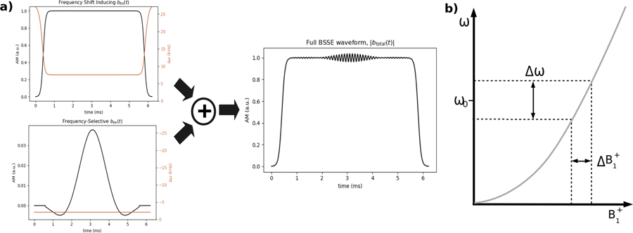
a) Construction of a BSSE RF pulse. The Bloch-Siegert shift producing pulse bbs(t) (top left) and slice-selective pulse bex(t) (bottom left) are designed independently and summed into the full BSSE waveform btotal(t). bbs(t) has Fermi AM (black) and adiabatic frequency sweeps towards and away from a constant frequency offset ωoff (orange). bex(t) has SLR-designed AM (black) and a constant frequency offset (orange). The amplitude of the bbs(t) waveform is generally much larger than that of the bex(t) waveform; in |btotal(t)|, bex(t) is visible as a small ripple in the plateau of the Fermi waveform. b) Bloch-Siegert shift-localized slice selection, in which an off-resonant RF pulse produces an approximately quadratic variation in resonant frequencies across field strengths . When paired with a frequency-selective excitation pulse, this results in the excitation of spins across a range , which can be mapped to space using an amplitude gradient transmit coil.
The primary resonance used for excitation by the proposed pulses is the Bloch Siegert-shifted single-photon resonance. However, the new pulse class is reliant on the simultaneous application of two off-resonant pulses in the xy plane, which also produces additional multiphoton resonances (24, 25). Although imaging techniques that use multiphoton resonances for excitation have recently been demonstrated (26), these additional resonances are here regarded as nuisances and their mitigation is discussed.
In the following, the design of the two component pulses that make up a Bloch-Siegert-localized -selective excitation pulse is described. Although there are many feasible choices for both the off-resonant Bloch-Siegert-shift inducing pulse and the frequency-selective pulse, this work focuses on the promising combination of an amplitude- and frequency-swept off-resonant pulse and an SLR frequency-selective pulse which allows the designer to directly specify and analytically trade off pulse and profile parameters (27). Design of these two pulses is analytic, simple, and fast. Simulations show the pulses’ suitability for selective excitation, inversion, and refocusing. Off-resonance behavior is simulated and shown to be predictable and similar to that of conventional spatially slice-selective excitation pulses, without the out-of-band excitation produced by the RFSE pulses of Ref (16). The pulses are also shown to have improved robustness against RF amplifier distortions over RFSE pulses. Selective excitation is experimentally demonstrated on a 47.5 mT MRI scanner with a gradient solenoid transmit-receive coil.
Theory
The design of a Bloch-Siegert -selective excitation (BSSE) pulse comprises the design of two distinct component pulses: 1) an off-resonant pulse, bbs(t), which produces a -dependent shift in resonant frequencies via the Bloch-Siegert effect, and 2) a frequency-selective SLR pulse, bex(t), which excites a desired passband. Although these two pulses could be transmitted by separate coils, we here assume that they are generated together by the same transmit coil, so they are complex-summed once individually designed, i.e.,
Component Pulse 1: The Bloch-Siegert Shift-Inducing Pulse
In the absence of externally applied RF, the magnetic resonance frequency is the Larmor frequency, ω0 = γB0. If an RF field is applied with amplitude and frequency offset ωoff relative to ω0, then a Bloch-Siegert frequency shift of the resonance is produced, given by (28):
| (1) |
This shift is away from the frequency of the applied off-resonance RF, i.e., a positive ωoff results in a negative ωbs.
The Bloch-Siegert shift-inducing pulse is defined as an amplitude- and phase-modulated function
where is the time integral of the pulse’s time-varying frequency modulation ∆ωrf (t), and T is the pulse duration. For Abs(t) we use a Fermi pulse which was previously used in Ref. (18) to produce a large BS frequency shift with little out-of-band excitation. The Fermi envelope provides an additional benefit for this application: its plateau results in a constant ωbs for the majority of the pulse duration, which simplifies the design of the bex(t) pulse so long as bex(t) is restricted to be nonzero only on the plateau. Thus for Abs(t) a normalized Fermi pulse is used which is centered at t = 0:
| (2) |
where t0 and a are parameters with units of seconds that control the duration of the pulse and the width of the transition, respectively. The duration of this Fermi pulse is T = 2t0 + 13.81a. To ensure that bex(t) is only nonzero during the plateau when Abs(t) ≈ 1, we derive from Eq. 2 that t0 should be set to:
where Tex is the duration of bex(t) required to excite the desired bandwidth and Ath is a user-specified constant. This t0 setting creates a central plateau in the Fermi envelope of duration Tex with Abs(t) ≥ Ath. Herein, all pulse designs used a = 6 × 10⁻⁵ s and Ath = 0.95.
A modification is made to the conventional constant frequency-offset Fermi by starting and ending the pulse’s FM waveform far-off resonance and sweeping adiabatically towards and away from the central constant frequency offset ωoff. This modification spin-locks the magnetization during the Fermi’s amplitude sweeps, reducing undesired excitation by Abs(t) (29, 30). The analytic adiabatic sweep of Ref. (30) is used, with modification to allow for time-varying Abs(t). A frequency sweep adapted from the analytic adiabatic sweep of Ref. (30) is used, with modification to allow for time-varying Abs(t). The first half of the symmetric frequency sweep is given by:
where T is the duration of the full Abs(t) waveform, td is the hardware dwell time, and K is a design parameter which trades off the maximum offset of the frequency sweeps and undesired excitation by Abs(t). K = 0.2 was used for all pulses in this work, which produces a maximum frequency offset of ∼10–30 kHz for a practical ωoff range of 5 − 20 kHz. The full FM waveform with constant frequency plateau ωoff is then given by:
An example of an amplitude- and frequency-modulated bbs(t) is shown in Fig. 1a. Note that the frequency modulation of this pulse is positive for all t, producing a negative Bloch-Siegert shift for all . As a result, the frequency-selective excitation pulse described in the next section is modulated to a negative frequency relative to Larmor, to excite the desired passband.
Component Pulse 2: The Frequency-Selective SLR Excitation Pulse
For a given desired passband between field strengths and , the bandwidth of resonant frequencies that must be excited by the frequency selective SLR pulse bex(t) is:
bex(t) must further be frequency modulated to the center of the passband, which has frequency:
The Shinnar Le-Roux (SLR) pulse design algorithm is used to design bex(t) since it allows for direct design of pulses to meet design constraints including bandwidth (B), duration (Tex), flip angle (θ), transition width, and magnetization profile ripple ( and ) (27). Based on these parameters, SLR designs a pair of polynomials that are transformed to the discretized excitation pulse by the inverse SLR transform. Given the designer’s choice of time-bandwidth product TexB, the duration of the excitation pulse Tex is determined by B. The RF pulse output by SLR is interpolated to the target dwell time and scaled to produce the desired flip angle θ for the at its passband center, based on the relationship:
| (3) |
where ∆t is the dwell time, N is the number of time points in the pulse, and Aex(t) is the baseband RF pulse waveform generated by SLR after interpolating it to the target dwell time. is derived from Eq. 1 after rearranging it to give the producing a Bloch-Siegert shift of ω during an RF pulse with frequency offset ωoff > 0:
| (4) |
After scaling Aex(t) to the desired flip angle, bex(t) is obtained by frequency modulating it to ωcent. The final pulse is then:
Before summing bex(t) with bbs(t), bex(t) is zero-padded to match the full duration T of bbs(t). An example bex(t) with TexB = 4 and θ = 90° is shown in Figure 1a.
However, under the assumption that both bex(t) and btotal(t) are played from the same transmit coil, bex(t) will produce a sloped magnetization profile (or a distorted profile, in the case of 180° excitation) due to the fact that bex(t) is played out slightly weaker than designed at passband locations where and slightly stronger than designed where . The degree of distortion will depend on the width of the passband relative to its center, , and is therefore more pronounced for wide passbands and center positions closer to zero. Modifications can be made to the SLR algorithm to correct for these distortions, and the procedure adopted is outlined in Fig. 2a. For small-tip and 90° excitation, the distortion is approximately compensated by scaling the SLR algorithm’s BN (ω) polynomial by the inverse of the transmit field strength corresponding to each frequency ω in the profile, with normalization to , prior to applying the inverse SLR transform:
Figure 2.
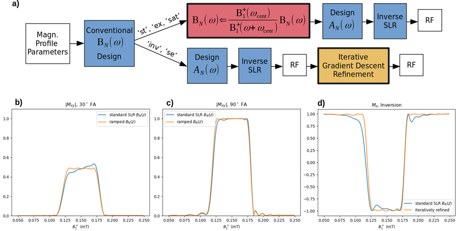
a) SLR design algorithm to compensate for a sloped excitation profile across . In the case of small-tip (‘st’) or 90° (‘ex’, ‘sat’) excitation, a pointwise scaling of the BN(ω) profile is sufficient. For an inversion (‘inv’) or refocusing (‘se’) pulse, iterative refinement is required. b) Uncorrected and corrected 30°, PBC = 0.15 mT, PBW = 0.06 mT, TexB = 8 excitation. c) Uncorrected and corrected 90° excitation. d) Uncorrected and corrected inversion profile after autodifferentiated gradient descent optimization of the pulse with respect to its magnetization profile, Iterative refinement corrected the highly distorted transition bands of the profile.
For 180° inversion or refocusing, profile distortion cannot be corrected with this analytic method. Instead, an iterative optimization of the RF pulse is used to alleviate profile distortion caused by varying (described further in Methods).
Overall Pulse Design Procedure
The overall pulse design procedure is to first select the passband edges and , the excitation pulse’s SLR algorithm parameters and flip angle, and the BS pulse’s frequency offset ωoff. With these parameters, the SLR pulse is designed, interpolated to the target dwell time, scaled to the desired flip angle, and frequency modulated to the center frequency ωcent of the slice, yielding the pulse bex(t). Knowing the duration Tex of bex(t) the BS pulse bbs(t) is calculated, bex(t) is zero-padded to the same length as bbs(t), and the two pulses are complex-summed to obtain the total pulse btotal(t) = bbs(t) + bex(t). It should be noted that the choice of ωoff is critical to the performance of the pulse. Figure 3a shows the relationship between ωoff and total pulse duration. For all BSSE pulses duration is minimized by minimizing ωoff (which maximizes the Bloch-Siegert frequency gradient). However, as shown in Figure 3b, bringing ωoff too close to the Larmor resonance produces undesirable out-of-band excitation.
Figure 3.
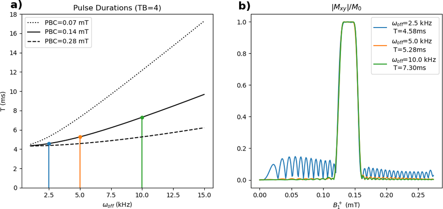
a) Variation in BSSE pulse duration with ωoff and PBC in . Pulse duration increases with increasing ωoff and decreases with increasing PBC. The magnetization profiles of pulses designed at the three points of interest are plotted in (b). b) Magnetization profiles of BSSE pulses with PBC=0.14mT. Out-of band excitation can be large if ωoff is set too small: a pulse with ωoff = 2.5 kHz produces a substantial amount of out-of-band excitation, which is reduced to within design specifications when ωoff → 5.0 kHz. Further increasing ωoff to 10.0 kHz produced only marginal improvement.
As in conventional B0 gradient-selective excitations, at the end of a BSSE excitation pulse there will be a phase ramp across the slice that must be refocused if the slice dimension will not be resolved by subsequent spatial encoding. For BSSE pulses, the phase ramp depends on the bex(t) pulse’s isodelay and the KBS of the bbs(t) pulse (18). A BSSE rewinding pulse can be constructed in the same manner as bbs(t), but with opposite frequency sign and a central constant frequency offset section with duration equal to the isodelay of bex(t). That is, the rewinding pulse should have a KBS equal to the negative of bbs(t)’s KBS, and scaled by bex(t)’s isodelay divided by the total duration of bbs(t).
Motion of the Magnetization Vector
With the full BSSE waveform defined, the trajectory of a magnetization vector during application of a BSSE RF pulse btotal(t) can be described. Figures 4b and 4c show the motion of the normalized magnetization vector in a frame rotating at ωoff for spin isochromats in fields at 0.17 mT (in the stopband) and at 0.14 mT (in the passband), respectively, during application of the BSSE pulse of. During the first adiabatic sweep of the RF pulse, both magnetization vectors rotate down from equilibrium to an axis that is slightly tilted away from the x-y axis. At the end of the initial sweep, when and , is approximately colinear with the tilted effective field
at which point the frequency-selective bex(t) pulse begins. For the stopband isochromat of Fig. 4b this pulse is not resonant, but it is resonant with the isochromat of Fig. 4c and the magnetization is nutated by the component of bex(t) perpendicular to to the opposite side of the unit sphere. After bex(t) is completed, the second adiabatic frequency sweep returns to being approximately colinear with the main magnetic field. The stopband isochromat in Fig. 4b is coaligned and approximately unperturbed from equilibrium, while the passband isochromat of Fig. 4c is successfully inverted. An video illustrating the magnetization motion is included as Supporting Information Video V1.
Figure 4.

a) Magnitude of a T = 3.7 ms BSSE inversion pulse. The pulse is colored red during the adiabatic frequency sweeps when bex(t) = 0, and blue during the constant portion of bbs(t) when bex(t) ≠ 0. b) Motion of the net magnetization vector in the ωoff rotating frame for a stopband isochromat. Simulation timestep was 4 µs. At the end of the pulse, magnetization is essentially unperturbed. c) Motion of the net magnetization vector in the ωoff rotating frame for a passband isochromat. At the end of the pulse, magnetization is successfully inverted.
Compensating flip angle errors
The pulse design algorithm described above is based on the implicit approximation that the effect of applying bbs(t) is to produce a pure -dependent resonance frequency shift. In reality, as illustrated in Fig. 4, bbs(t) rotates magnetization to precess in a plane that is slightly tilted from the x-y plane, at an angle from the z-axis. Ideally, bex(t) would also be applied in this tilted plane, but it remains in the x-y plane, and the actual flip angle produced by bex(t) will be slightly smaller than expected because its projection onto the tilted plane will be elliptical and smaller than its full amplitude. This results in a flip angle attenuation that increases as ωcent/ωoff becomes large. Similar effects have been previously observed in other dual-frequency RF applications (32, 33). Given the difficulty of describing and correcting this effect analytically, a normalized empirical correction is given herein for the amplitude of Aex(t) based on Bloch simulations of slice profiles across a wide range of ωcent and ωoff. The model is shown in Fig. 5a, and the equations for the correction predicted by the model is provided in the Appendix. Deviations are generally small and require minimal correction for pulses where ωcent/ωoff < 1 as suggested by the example profile correction shown in Fig. 5b. This comprises the majority of practical BSSE pulses. Pulses with ωcent substantially larger than ωoff are in general encountered when designing a pulse to select a PBC at very large (which produces a large Bloch-Siegert shift and thus large ωcent relative to ωoff ), or when a very small ωoff is used, which is typically impractical due to the large out-of-band excitation produced.
Figure 5.
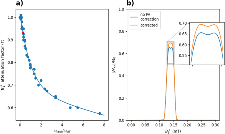
a) Flip angle attenuation factor versus ωcent/ωoff. Data points are empirical correction factors found through simulation, with an exponential fit to the data shown as a continuous line. The red dot indicates the parameters of the pulse simulated in (b). b) Excited slice profile of a PBC =0.14 mT, PBW = 0.03 mT, 45°, ωoff =7.5 kHz pulse with and without the empirical FA correction. For this pulse, ωcent/ωoff = 0.279, which the model predicts will result in a flip angle attenuation of 6.9% if uncorrected. Applying the correction improves the effective flip angle of the pulse, bringing it closer to the anticipated |Mxy|/M0 = 0.7
Multiphoton Resonances
As discussed in the previous section, the simultaneous application of two off-resonance RF pulses in BSSE excitation leads to non-linear effects including small flip angle errors. A second effect is the appearance of subsidiary multiphoton resonances, in addition to the single-photon resonance. These are a consequence of the BSSE pulse’s simultaneous irradiation of nuclei at two frequencies near (generally within 10’s of kHz) but not at the Larmor frequency (24). While unwanted higher-order multiphoton resonances cannot be corrected simply by adjusting the bex(t) pulse’s amplitude or frequency, their positions can be predicted analytically as follows and used to guide the pulse design.
The off-resonance frequencies of the two component pulses bex(t) and bbs(t) are ωcent and ωoff, with an average frequency of the two off-resonant pulses . Multiphoton resonances occur at frequencies ω0,N for which the following is satisfied:
where ω1 is equivalently either ωoff or ωcent (25). N = 1 corresponds to the single-photon resonance while N = 3, 5, 7 . . . correspond to multiphoton resonances. Arbitrarily taking ω1 = ωoff, two solutions exist for ω0,N :
| (5) |
In this application, because ωoff > 0 will produce a negative frequency shift for all , only ω0,N < 0 are of interest. These resonances are given by the solution . To determine the locations of the multiphoton resonances on the axis, we can substitute Eq. 5 into Eq. 4, giving:
| (6) |
Pulses with two combinations of ωoff and ωcent but with the same PBC for the N = 1 excitation are shown in Figure 6a: the red pulses have ωoff =10 kHz and ωcent=−1.655 kHz, and the black pulses have ωoff =7.5 kHz and ωcent=−2.094 kHz. Figure 6b shows the distribution of multiphoton resonances across for the two ωoff. For both ωoff, the combination of RF frequencies place the N = 1 resonance at 0.14 mT. For the 90° and 180° pulses there is a small amount of excitation produced at the N = 3 resonance (maximum |Mxy|/M0 = 0.075 for the excitation and maximum Mz/M0 = 0.918 for the inversion), but excitation at N > 3 is below the stopband ripple level of the pulses. In the case of the pulse scaled up to produce a 720° flip, a greater number of multiphoton resonances can be appreciated. These simulated resonances shown in Figure 7b are at the approximate locations predicted by Equation 6; in the case of the black pulse (ωoff = 7.5 kHz), the resonances (aside from N = 1) are at mT. In the case of the red pulse (ωoff = 10 kHz), the predicted locations of the N = 1 to 11 resonances shift up to mT. Using a larger ωoff can shift multiphoton resonances upward in and out of the imaging bandwidth, although this decreases the Bloch-Siegert frequency gradient and thus requires longer pulse durations.
Figure 6.
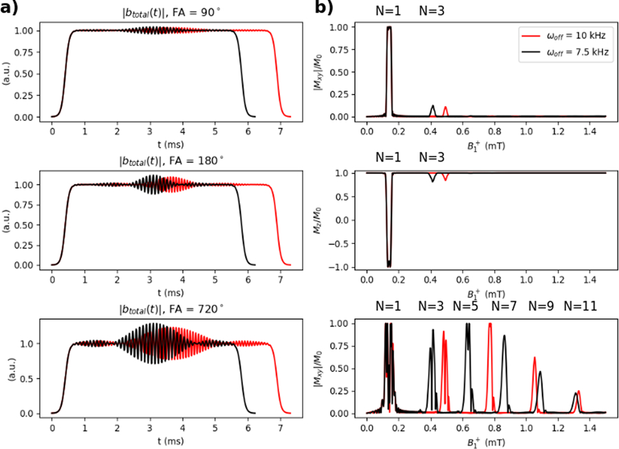
a) Magnitudes of 90°, 180°, and 720° BSSE pulses with ωoff = 7.5 kHz (black) and ωoff = 15.0 kHz (red). b) The corresponding simulated magnetization profiles. Multiphoton resonances are visible at the locations in predicted by Equation 6. Increasing ωoff shifts resonances N ≥ 3 upward in .
Figure 7.
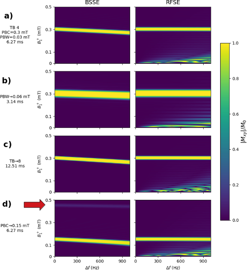
Simulations of BSSE and RFSE pulse off-resonance sensitivity. a) Simulation of the “base” 6.27ms BSSE and RFSE pulses with TB=4, PBC=0.4 mT, PBW=0.03 mT, FA=90°. ωoff was set to 16.37 kHz to match the RFSE pulse duration. b) Same as base pulse but with PBW set to 0.06 mT. ωoff was set to 9.48 kHz to match durations. c) Same as base but with TB set to 8. ωoff was set to 19.25 kHz to match durations. d) Same as base but with PBC set to 0.15. ωoff was set to 8.17 kHz to match durations. In all cases, the BSSE magnetization profiles show a bulk shift in the magnetization profile with increasing off-resonance. The red arrow in d) shows the location of an N = 3 multiphoton resonance. This multiphoton resonance also experiences a bulk shift downward in with increasing off-resonance. The RFSE profiles show no bulk shift, but have substantial unintended excitation at low .
Methods
Simulations
Simulations were performed to demonstrate the effectiveness of SLR BN (ω) ramp correction, to evaluate the pulses’ suitability for refocusing in an RF gradient slice-selective CPMG pulse sequence, and to compare their performance to RFSE -selective excitation pulses (16) in the presence of B0 inhomogeneity and nonideal RF hardware. All simulations were performed using a hard pulse approximation-based Bloch simulator (27) in SigPy.RF (35) with 1 µs dwell time. All BSSE and RFSE pulses were designed using SigPy.RF pulse design functions. Code for BSSE pulse design is available at: https://github.com/jonbmartin/sigpy-rf.
Profile Ramp Correction
Simulations were performed to verify the ability of the BSSE ramp correction algorithm to correct passband distortions caused by varying . A 30° small-tip pulse, a 90° excitation pulse, and a 180° inversion pulse were constructed and their magnetization profiles were simulated across . For all three pulses, PBC = 0.14 mT, PBW = 0.06 mT, TexB = 8, , ωoff = 7.5 kHz; these parameters were chosen to create a profile in which uncorrected distortions were clearly visible. In profiles with smaller TexB or PBW, the distortion is not easily observable. The small-tip and 90° excitation pulses were designed using the analytic BN (ω) ramp correction. In the inversion case, an uncorrected BSSE inversion pulse was designed to meet the profile parameters specified above as well as possible. This pulse was used as the initializer for an iterative gradient descent refinement to eliminate distortions. The following general unconstrained problem was solved:
| (7) |
where is the Bloch-simulated Mz Profile of the BSSE pulse vector btotal across transmit field strengths , and is the desired inversion profile across the same range. The are error weights used to specify the transition regions of the profile as ‘don’t care’ regions for the optimization. The gradient of the loss was calculated across a grid of 250 points in from 0 to 0.25 mT using an automatic differentiation function from the JAX software toolbox (36). Gradient descent steps were taken to minimize this loss, with a fixed step size of 1e−4. Iterations were performed until convergence, defined as a difference in loss between iterations of less than 1%.
Off-Resonance Simulation
Four BSSE pulses and four RFSE pulses were designed, with ωoff of the BSSE pulses set to give the pulses the same duration as the RFSE pulses. The ‘base’ pulse for both pulse classes was a 6.27 ms TexB = 4 pulse at 90° flip angle, centered at PBC = 0.4 mT with PBW = 0.03 mT and Mxy passband and stopband maximum errors of . The other three pulses each had one parameter changed from the base pulse (Pulse 1): Pulse 2 had its’ PBW doubled to 0.06 mT (decreasing the duration to 3.14 ms), Pulse 3 had its’ time bandwidth product doubled to 8 (increasing the duration to 12.53 ms), and Pulse 4 had its’ PBC shifted to 0.15 mT (keeping the duration of the pulses fixed at 6.27 ms). ωoff for the four BSSE pulses were set to 16.370 kHz, 9.485 kHz, 19.250 kHz, and 8.170 kHz, respectively, to match RFSE pulse durations. Magnetization was simulated over a range of 0–0.5 mT and an off-resonance range of 0–1 kHz.
RF Amplifier Droop Simulation
A weakness of RFSE -selective pulses is their requirement that the pulse AM envelope be carefully balanced (16), since the pulses operate by spinning magnetization in the y-z plane while tipping magnetization in the passband towards x, and any errors in the envelope will result in large undesired excitation in y. This is a challenging demand for many RF amplifiers. RF amplifier droop was simulated for both a BSSE and an RFSE pulse to compare their robustness. Both pulses were designed with PBC=0.14 mT, PBW=0.03 mT, TexB = 4, 45° flip angle, and . ωoff of the BSSE pulse was set to 7.608 kHz to match the durations of the two pulses (6.22 ms). Droops of −0.5 dB and −1.5 dB in the relative pulse amplitudes across the total duration were simulated (Figures 8a, 8c).
Figure 8.

Simulation of RF amplifier droop. AM waveforms for RFSE (a) and BSSE (c) RF pulses are shown with −1.5dB of droop. In the case of the RFSE pulse, the magnetization profile (b) is severely degraded by RF amplifier distortion. However, the magnetization profile of the BSSE pulse (d) experiences only a slight shift upward in , with minimal profile distortion.
CPMG Sequence Simulation
A multi-echo spin echo pulse sequence was constructed using BSSE excitation and refocusing pulses. The same BSSE pulse parameters were used as in the previous experiment (PBC = 0.14 mT, PBW = 0.03 mT, TexB = 4, ωoff =7.5 kHz, T = 6.22 ms) to design a 90° excitation pulse and a 180° refocusing pulse. The BSSE refocusing pulse was iteratively refined to eliminate any ramped distortion.
The bbs(t) design algorithm was used to generate a pair of RF pulses serving as phase pre- and rewinder gradients. These pulses were given an equal-magnitude/opposite-signed ωoff as the BSSE pulse to produce a shift in resonant frequency opposite that of the refocusing pulse, with |KBS| half that of the refocusing pulse (32.2 vs 16.1 rad2/G). The 90° BSSE excitation pulse was applied along the x-axis and was followed by eight refocusing pulses applied along the y-axis for an echo train length of 8, with TE = 2τ = 33 ms. The RF pulse sequence diagram is shown in Figure 9a. The pulses were simulated across a range of 0 to 0.3 mT and a range of frequency offsets between −200 and 200 Hz, with T1 = T2 = ∞.
Figure 9.
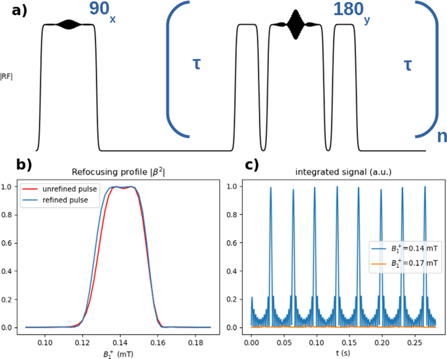
BSSE Multi-echo CPMG pulse sequence. a) Pulse sequence diagram. A 90° excitation pulse was followed by a train of 180° pulses. To satisfy the CPMG phase conditions, RF pre- and re-winders were inserted before and after the 180° refocusing pulses. b) |β2| refocusing efficiency profile for the refocusing pulse. Refocusing was selective and nearly complete across the passband. The gradient descent-refined 180° pulse had a slightly improved profile, although the unrefined pulse also performs well given the narrow PBW. c) Signal timecourse in the passband and stopband. Regularly spaced echos of uniform amplitude were produced in the passband.
Experiments
Phantom experiments were performed to verify the magnetization profiles produced by BSSE pulses. A base BSSE pulse with flip angle 90°, PBC = 0.14 mT, PBW = 0.03 mT, ωoff = 7.5 kHz, TexB = 4, and total duration T = 6.22 ms was designed. Three copies of the pulse were created in which a single parameter was varied: passband center (0.14 mT → 0.11 mT), flip angle (90° → 45°), and time-bandwidth product (4 → 1.5). The pulses were deployed on a 47.5 mT permanent magnet (Fig 10a) (Sigwa MRI, Boston, MA, USA) and image data were acquired using a Tecmag Redstone MRI Console (Tecmag, Houston, Texas, USA). The system used a 1 µs transmit dwell time and amplitude/phase tables for RF waveform definition. A 2 kW BT02000-AlphaS Tomco RF power amplifer (Tomco Rechnologies, Stepney, Australia) was used to drive a 20-turn variable-pitch solenoid RF T/R coil producing a linearly decreasing gradient (Fig 10b). A 50 mL 1 mM CuSO4 vial phantom with 30 mm diameter and 115mm length was imaged using a 3D gradient-recalled echo pulse sequence (TR/TE = 27/425 ms, dx/dy/dz = 2.0/3.5/3.5 mm, matrix size = 128×49×3, 5 signal averages), where the BSSE pulses were used for excitation. Phase variation produced across the excitaton profile by the BSSE excitation pulse was not rewound in this pulse sequence. Conventional B0 gradients were used for image spatial encoding. mapping was also performed using the BS shift method (18) with the same 3D gradient-recalled echo sequence but with the addition of a hard pulse excitation. The 6.22 ms, ωoff =7.5kHz component of the BSSE pulse was kept as the -dependent phase shift-generating pulse (KBS = 32.2 rad2/G) for mapping, producing no excitation without the addition of the bex(t) component pulse. To process the images, a mask was created by thresholding a 90° hard pulse GRE acquisition with the same matrix size and resolution to 20% of the maximum signal intensity. This mask was applied to the phase images of the -mapping sequence and magnitudes of the BSSE-excitation images. The mapping phase image was unwrapped along the coil’s gradient dimension, and background phase was subtracted out using a reference scan without a BS shift pulse. strength was then calculated using the KBS of the off-resonant pulse. The BSSE excitation images were divided by the map to remove the receive sensitivity of the T/R coil, since at this low frequency.
Figure 10.
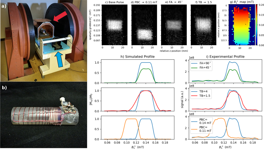
a) 47.5 mT permanent magnet system used for experimental results. A Faraday cage (red arrow) and pickup coil (blue arrow) are used to reduce EMI. b) variable-pitch T/R solenoid coil used in experiments. The tube phantom is placed inside in this image. c-f) middle slice of 3D GRE acquisition with varying BSSE excitation pulse. g) map corresponding to the same slice h) 1D simulated profiles for the pulses, and i) corresponding experimental 1D profiles. The 45° excitation (green) produces reduced signal intensity in relation to the 90° excitation (blue). The TB=1.5 excitation (red) produces a profile with a broader transition region. Designing the pulse with PBC shifted to 1.1 G (orange) produces the corresponding change in the magnetization profile.
Results
Ramp Correction Simulation
Figures 2b–d shows the improved small-tip excitation, 90° excitation, and inversions with ramp correction. The small-tip excitation had a substantial ramp across when uncorrected which was flattened by the analytic correction, with differences between normalized at passband edges reduced from 0.0893 in the uncorrected case to less than 0.001 in the corrected case. The 90° excitation also had substantially reduced distortion in the passband, particularly on the rising edge of the profile. Iterative optimization of the inversion pulse reached convergence after 37 gradient descent iterations. The final optimized pulse produced a substantially improved magnetization profile which met the desired ripple levels and transition width. The substantial ripple produced by over-flipping on the falling edge of the profile was eliminated, and the broad transition on the rising edge of the profile was sharpened.
Off-Resonance Simulations
The results of the off-resonance simulation are shown in Fig. 7. All RFSE pulses produced a triangular pattern of erroneous excitation where the effective size of the off-resonance field was large compared to the field. This effect is absent from the BSSE pulse profiles. At the same time, all four BSSE pulses show a small bulk downward shift in the excitation profile with increasing off-resonance, similar to a conventional B0 gradient-selective excitation pulse. For the four BSSE pulses shown in Figures 8a–d, the shift was −0.033 mT/kHz, −0.019 mT/kHz, −0.038 mT/kHz, and −0.036 mT/kHz. As expected, the slope of the shift correlated with pulse duration: the pulse with the shortest duration (3.14 ms) also had the smallest bulk shift (−0.019 mT/kHz). A faint N = 3 multiphoton resonance is visible in Figure 8d at approximately 0.45 mT. The N = 3 resonance also exhibited a shift downward in with off-resonance.
RF Amplifier Droop Simulation
The effect of RF amplifier droop on the magnetization profile is shown in Figures 8b and 8d. For the RFSE pulse, pulse droop significantly distorted the excitation profile. −0.5 dB of droop reduced the transverse magnetization by approximately 13%; −1.5 dB of droop completely degraded the magnetization profile. In the case of the BSSE pulse, however, the same droop produced minimal distortion, manifesting primarily as a small shift in the magnetization profile. This shift was approximately 0.004 mT (13% of slice width) in the case of −0.5 dB of droop and 0.013 mT (43% of slice width) in the case of the −1.5 dB droop.
CPMG Simulation
Figures 9b and 9c show the results of the multi-echo spin echo sequence simulation. The refocusing profile in Fig. 9b shows close to full refocusing efficiency within the pulses’ 0.03 mT-wide passband. This is supported by Fig. 9c, which shows the signal integrated across off-resonance at a location in the passband (0.14 mT) and the stopband (0.17 mT). In the passband evenly spaced echoes are formed, with near-identical signal amplitude produced at each echo. In the stopband, no appreciable signal was produced at the echo times.
Experimental Results
Figures 10c–f show the measured experimental profiles for the pulses played out in the experiment in the central slice of the 3D acquisition. Figure 10g shows the corresponding map from the same slice. 1D profiles of the simulated (Fig 10h) and experimental (Fig 10i) are shown as well. In the experimental case these were generated by averaging across the z dimension of the profile. For the two-flip angle (90° to 45°) experiment, a decrease in signal intensity was observed consistent with the expected 30% decrease based on the nominal flip angles. The ratio of the signals from the 45° and 90° excitations between the passband edges was 0.705 in simulation and 0.627 in the experiment. In the case of decreasing TexB from 4 to 1.5, the TexB = 1.5 profile had a wider transition width resulting from the decrease in selectivity. The ratio of the full width at half-maximum of the profiles from the TexB = 1.5 excitation and the TexB = 4 excitation was 1.29 in simulation and 1.21 in the experiment, showing close agreement. The pulse with PBC shifted to 0.11 mT produced a magnetization profile centered at the intended locations, and the transition bands of the pulses intersected at approximately 0.125 mT, where each profile was expected to reach its half-maximum.
The phase produced by the BSSE excitation pulses across the magnetization profile is shown in Supporting Information Figure S1. For all four of the BSSE pulses that were examined, the phase of the spins increased in an approximately linear manner across the magnetization profile, with uniform spacing between successive phase wraps over the linearly increasing transmit field.
Discussion
A class of excitation pulses that apply the Bloch-Siegert shift for the purpose of selective excitation was introduced. Although the Bloch-Siegert shift has been widely used in MRI for mapping (18, 30) and in NMR for the calibration of decoupling field power (20, 37), to our knowledge the present work represents its first use to localize selective excitation. Simulation results showed that the pulses are robust against system imperfections and can be used in pulse sequences requiring phase control, making them a viable choice for a wide range of pulse sequences. Overall, BSSE pulses enable the replacement of one axis of B0 gradient encoding with gradient encoding in an imaging system. Most RF encoding systems experimentally implemented thus far including the imaging experiments performed in this work have relied on at least one axis of B0 encoding for 3D imaging (5, 6), and further development is needed to achieve fully RF-encoded 3D imaging. More immediately, BSSE selective excitation could be combined with other pulsed B0 gradient-free imaging systems, such as systems with built-in B0 field inhomogeneities (39–41).
As in the RFSE design algorithm of (16), the proposed BSSE pulse design method is based on the Shinnar-Le Roux algorithm, giving BSSE pulses the same advantage in speed of design and ability to predict and trade off design parameters. However the proposed method also has a number of advantages over RFSE pulses, including the ability to refocus magnetization for spin-echo based pulse sequences, and reduced out-of-band excitation in response to B0 inhomogeneity. RFSE pulses can also be challenging to implement experimentally because they place strict requirements on the integrated area of the RF envelope, which must be accurately balanced between pulse segments to prevent large distortions of the magnetization profile. In this work BSSE pulses were shown to be more tolerant of moderate amplifier errors in the amplitude of the RF pulse, such as amplifier droop, which led primarily to relatively benign slice shifts which could be mostly corrected by transmit gain adjustment.
BSSE pulse designers should keep several practical considerations in mind. First, the BSSE pulses demonstrated here had relatively longer durations than conventional B0 gradient-selective excitations (several ms versus a few ms). For a fixed bex(t) time-bandwidth product TexB, BSSE pulse length is principally determined by two factors: the gradient strength for a given bbs(t) amplitude and ωoff, the central constant frequency offset of bbs(t). These variables determine the magnitude of the Bloch-Siegert frequency gradient produced by bbs(t), which in turn sets the duration of the full BSSE pulse. If the Bloch-Siegert spatial frequency gradient were of the same magnitude as a B0 spatial frequency gradient, a BSSE pulse would have approximately the same duration as an equivalent conventional B0 gradient selective excitation pulse. For example, to create a frequency gradient of 1 MHz/m (produced by a B0 gradient of approximately 23.5 mT/m) using the Bloch-Siegert shift, using the ωoff =7.5kHz value used throughout this work, a gradient of approximately the same strength, 23.5 mT/m, would be required. Though a much weaker gradient of ∼2 mT/m was used here experimentally, this stronger gradient is not difficult to achieve in practice (5, 7). In general, ωoff should be minimized to maximize the Bloch-Siegert frequency gradient. However, excessive reduction of the frequency offset can produce out-of band excitation (Figure 3b, at ωoff =2.5kHz) or move multiphoton resonances closer in to the single-photon resonance (Figure 6) and potentially into the FOV. Although their rapid dropoff in amplitude with N makes their use for imaging challenging, it may be possible to use BSSE pulses for multiband multiphoton excitation in implementations similar to those previously described (26). However in general ωoff should be increased to move multiphoton resonances outside the FOV.
The RF power and consequently the specific absorption rate (SAR) contribution of BSSE pulses is increased compared to an equivalent B0 gradient-selective excitation by the addition of the large, near-constant AM waveform of the off-resonant bbs(t) pulse to the on-resonant bex(t) pulse. Since the pulses are well-separated in frequency, the power increase compared to a conventional pulse is approximately equal to the power of the bbs(t) component. For example, for the 6.22 ms BSSE pulse shown in Figure 1, operating at 47.5 mT, the integrated power of bex(t) normalized by that of the full btotal(t) was 4 × 10⁻⁴ (a.u.), and the integrated power of bbs(t) normalized by that of btotal(t) was 0.9996 (a.u.). Nearly all power is contributed by the bbs(t) pulse. At the same time, SAR is much less a concern in low-field MRI such as the 47.5 mT imager used in this work (42).
Like conventional B0 gradient-selective excitations, as demonstrated experimentally, BSSE pulses leave a phase gradient across the excited slice that should be refocused if subsequent imaging will not resolve the slice dimension. Refocusing can be achieved with a rewinding off-resonant bbs(t) pulse, to cancel phase accrued during the pulse’s isodelay. At the same time, like conventional B0 gradient-selective excitations, there are many situations where this is not necessary, including when using the pulses for saturation, inversion, or for slab selection in 3D imaging sequences. When used for slice-selective excitation, using a minimum-phase bex(t) pulse shape with a small isodelay would also reduce or eliminate the need for a rewinder. When using BSSE pulses for balanced or spin echo pulse sequences, RF pre- and rewinders may be inserted into the pulse sequence.
The experimental setup in this work used a single gradient coil for transmit and receive which is nonideal in practice, since although profiles with uniform flip were excited across the slice, the coil’s nonuniform receive sensitivity would result in spatially varying SNR. To alleviate this, the gradient transmit coil could be paired with a saddle receive coil with uniform receive sensitivity, which has been demonstrated on the 47.5 mT system described here (43).
The pulse design algorithm described here could be extended in numerous ways. Applying the VERSE algorithm (44) may allow the frequency-selective excitation pulse to extend onto the time-varying portion of the Bloch-Siegert shift inducing pulse, to shorten overall pulse duration. Additionally, BSSE pulse designs may benefit from joint gradient-based optimization of the two component pulses, in analogy to previously demonstrated joint optimization of RF and gradient waveforms (45). Further optimizing the waveforms this way could be used to, e.g., reduce multiphoton effects, decrease pulse duration, produce uniform excitation across a broad range of , or constrain the pulses to have a flat amplitude so they can be more easily generated by current-mode amplifiers(46). Finally, the pulses could be extended to produce Hadamard or other multiband excitations, by designing the excitation pulse bex(t) using multiband pulse designers developed for B0 gradient-based selective excitation (47–49).
Conclusion
In this work, we presented a novel class of pulses for -selective excitation which localize excitation using frequency gradients produced by the Bloch-Siegert shift. Simulations demonstrate that these pulses produce magnetization profiles of excellent quality with phase control, and that the pulses are robust against off-resonance. Experimental results on an ultra-low-field 47.5 mT verified the design algorithm’s ability to control magnetization profile parameters. BSSE pulses may help enable new methods of B0-gradient-free acquisition and novel imaging approaches.
Supplementary Material
Supporting Information Video V1 a) Magnitude of a T = 3.7 ms BSSE inversion pulse. The red trace is 10 µs long. b) Stopband magnetization trajectory in the ωoff rotating frame. The Simulation timestep was 4 µs. c) Passband magnetization trajectory in the ωoff rotating frame.
Supporting Information Figure S1 a-d) Top: middle slice of experimental 3D GRE acquisition with varying BSSE excitation pulse. Bottom: corresponding phase across profile. Several wraps in phase are visible across all profiles, since a phase rewinder was not applied following the BSSE pulse. However the phase behavior is regular and can be rewound. Note that in d), in which the TB was decreased to 1.5, the phase increases more slowly due to the reduced isodelay of the excitation pulse. e) map corresponding to the same slice.
Acknowledgments
This work was supported by NIH grants R01 EB 030414 and R01 EB 016695. The authors would like to thank Benjamin Hardy for many helpful conversations about this work.
Appendix
The models used to correct for flip angle inaccuracy is as follows. The observed flip angle attenuation was fit by a biexponential model, using MATLAB’s fit() function. This attenuation model is:
where x = ωcent/ωoff. The correction is applied by accounting for Γ(x) in the flip angle relation of Eq. 3:
The goodness-of-fit of this model was R2 = 0.9899.
References
- [1].Bernstein MA, Zhou XJ, Polzin JA, King KF, Ganin A, Pelc NJ, Glover GH. Concomitant gradient terms in phase contrast MR: Analysis and correction. Magnetic Resonance in Medicine 1998; 39(2):300–308. [DOI] [PubMed] [Google Scholar]
- [2].Spees WM, Buhl N, Sun P, Ackerman JJ, Neil JJ, Garbow JR. Quantification and compensation of eddy-current-induced magnetic-field gradients. Journal of Magnetic Resonance 2011; 212(1):116–123. [DOI] [PMC free article] [PubMed] [Google Scholar]
- [3].Hoult DI. Rotating frame zeugmatography. Journal of Magnetic Resonance 1979; 33(1):183–197. [DOI] [PubMed] [Google Scholar]
- [4].Sharp JC, King SB. MRI using radiofrequency magnetic field phase gradients. Magnetic Resonance in Medicine 2010; 63(1):151–161. [DOI] [PubMed] [Google Scholar]
- [5].Kartäusch R, Driessle T, Kampf T, BasseLüsebrink TC, Hoelscher UC, Jakob PM, Fidler F, Helluy X. Spatial phase encoding exploiting the Bloch–Siegert shift effect. Magnetic Resonance Materials in Physics, Biology and Medicine 2014; 27:363–371. [DOI] [PubMed] [Google Scholar]
- [6].Torres E, Froelich T, Wang P, DelaBarre L, Mullen M, Adriany G, Pizetta DC, Martins MJ, Vidoto ELG, Tannús A, Garwood M. B1-gradient–based MRI using frequency-modulated Rabi-encoded echoes. Magnetic Resonance in Medicine 2021; 87(2)674–685:. [DOI] [PMC free article] [PubMed] [Google Scholar]
- [7].Canet D Radiofrequency field gradient experiments. Progress in Nuclear Magnetic Resonance Spectroscopy 1997; 30(1–2):101–135. [Google Scholar]
- [8].Tycko R, Pines A. Spatial localization of NMR signals by narrowband inversion. Journal of Magnetic Resonance 1984; 60(1):156–160. [Google Scholar]
- [9].Shaka AJ, Freeman R. Spatially selective radiofrequency pulses. Journal of Magnetic Resonance 1984; 59(1):169–176. [Google Scholar]
- [10].Garwood M Localized MRS Employing Radiofrequency Field (B1) Gradients. eMagRes 2016; 5(1):1015–1025. [Google Scholar]
- [11].Bendall M, Gordon R. Depth and refocusing pulses designed for multipulse NMR with surface coils. Journal of Magnetic Resonance 1983; 53(3):365–385. [Google Scholar]
- [12].Hoult DI. NMR imaging. Rotating frame selective pulses. Journal of Magnetic Resonance 1980; 38(1):369–374. [Google Scholar]
- [13].Karczmar GS, Lawry TJ, Weiner MW, Matson GB. Shaped pulses for slice selection in the rotating frame-A study using computer simulations. Journal of Magnetic Resonance 1988; 76(1):41–53. [Google Scholar]
- [14].Maffei P, Elbayed K, Brondeau J, Canet D. Slice selection in NMR imaging by use of the B1 gradient along the axial direction of a saddle-shaped coil. Journal of Magnetic Resonance 1991; 95(2):382–386. [Google Scholar]
- [15].Hedges LK, Hoult DI. The techniques of rotating frame selective excitation and some experimental results. Journal of Magnetic Resonance 1988; 79(3):391–403. [Google Scholar]
- 16.[] Grissom WA, Cao Z, Does MD. -selective excitation pulse design using the Shinnar-Le Roux algorithm. Journal of Magnetic Resonance 2014; 242(1):189–196. [DOI] [PubMed] [Google Scholar]
- [17].Bloch F, Siegert A. Magnetic Resonance for Nonrotating Fields. Physical Review 1940; 57(6):522. [Google Scholar]
- [18].Sacolick LI, Wiesinger F, Hancu I, Vogel MW. B1 mapping by Bloch-Siegert shift. Magnetic Resonance in Medicine 2010; 63(5):1315–1322. [DOI] [PMC free article] [PubMed] [Google Scholar]
- [19].Hosur RV, Ernst RR, Wothrich K. A simple two-dimensional measurement of the decoupler power during continuous homonuclear irradiation for the correction of Bloch-Siegert shifts. Journal of Magnetic Resonance 1983; 54(1):142–145. [Google Scholar]
- [20].Hung I, Gor’kov P, Gan Z. Using the heteronuclear Bloch-Siegert shift of protons for B1 calibration of insensitive nuclei not present in the sample. Journal of Magnetic Resonance 2020; 310(1):1–5. [DOI] [PubMed] [Google Scholar]
- [21].Zhipeng C, Chekmenev EY, Grissom WA. Frequency encoding by Bloch-Siegert shift. In: Proc. Intl. Soc. Mag. Reson. Med, Milan, Italy: p. 4220 (2014). [Google Scholar]
- [22].Wan Y, Qiu M, Galiana G, Constable R. Phase Encoding with Bloch-Siegert effect using Parallel Transmit. In: Proc. Intl. Soc. Mag. Reson. Med, Honolulu, USA: p. 1498 (2017). [Google Scholar]
- [23].Martin J, Vaughn C, Griswold MA, Grissom WA. Bloch-Siegert -Selective Excitation Pulses. In: Proc. Intl. Soc. Mag. Reson. Med, Virtual p. 3337 (2021). [DOI] [PMC free article] [PubMed]
- [24].Zur Y, Levitt MH, Vega S. Multiphoton NMR spectroscopy on a spin system with I=1/2. The Journal of Chemical Physics 1983; 78(9):5293. [Google Scholar]
- [25].Krauss EM, Vega S. Four-field excitation of multiphoton NMR resonances in spin I=1/2. Physical Review A 1986; 34(1):333. [DOI] [PubMed] [Google Scholar]
- [26].Han V, Liu C. Multiphoton magnetic resonance in imaging: A classical description and implementation. Magnetic Resonance in Medicine 2020; 84(3):1184–1197. [DOI] [PubMed] [Google Scholar]
- [27].Pauly J, Nishimura D, Macovski A, Roux PL. Parameter Relations for the Shinnar-Le Roux Selective Excitation Pulse Design Algorithm. IEEE Transactions on Medical Imaging 1991; 10(1):53–65. [DOI] [PubMed] [Google Scholar]
- [28].Ramsey NF. Resonance transitions induced by perturbations at two or more different frequencies. Physical Review 1955; 100(4):1191–1194. [Google Scholar]
- [29].Garwood M, DelaBarre L. The Return of the Frequency Sweep: Designing Adiabatic Pulses for Contemporary NMR. Journal of Magnetic Resonance 2001; 153(2):155–177. [DOI] [PubMed] [Google Scholar]
- [30].Khalighi MM, Rutt BK, Kerr AB. Adiabatic RF Pulse Design for Bloch-Siegert Mapping. Magnetic Resonance in Medicine 2013; 70:829–835. [DOI] [PMC free article] [PubMed] [Google Scholar]
- [31].Hannaford P, Pegg DT, Series GW. Analytical expressions for the Bloch-Siegert shift. Journal of Physics B: Atomic and Molecular Physics 1973; 6:L222–L225. [Google Scholar]
- [32].Mccoy MA, Mueller L. Selective Decoupling. Journal of Magnetic Resonance, Series A 1993; 101(2):122–130. [Google Scholar]
- [33].Steffen M, Vandersypen LMK, Chuang IL. Simultaneous Soft Pulses Applied at Nearby Frequencies. Journal of Magnetic Resonance 2000; 146:369–374. [DOI] [PubMed] [Google Scholar]
- [34].Vega S Multiple-quantum cross-polarization NMR on spin systems with I=1/2 and S=3/2 in solids. Physical Review A 1981; 23:3152. [Google Scholar]
- [35].Martin J, Ong F, Ma J, Tamir J, Lustig M, Grissom W. SigPy.RF: Comprehensive Open-Source RF Pulse Design Tools for Reproducible Research. In: Proc. Intl. Soc. Mag. Reson. Med 28th Annual Meeting, Virtual p. 1045, (2020).
- [36].Frostig J, Johnson MJ, Leary C. Compiling machine learning programs via high-level tracing, In: SYSML, Stanford, USA: (2018). [Google Scholar]
- [37].Claridge TD. High-Resolution NMR Techniques in Organic Chemistry 3rd Ed. Cambridge, MA:Elsevier; 2016;1–541. [Google Scholar]
- [38].Duan Q, Gelderen Pv, Duyn J. Improved Bloch-Siegert Based B1 Mapping by Reducing Off-Resonance Shift. NMR in Biomedicine 2013; 26(9):1070. [DOI] [PMC free article] [PubMed] [Google Scholar]
- [39].Stockmann J, Cooley C, Guerin B, Rosen M, Wald L. Transmit Array Spatial Encoding (TRASE) using broadband WURST pulses for RF spatial encoding in inhomogeneous B0 fields. Journal of Magnetic Resonance 2016; 268:36–48. [DOI] [PMC free article] [PubMed] [Google Scholar]
- [40].Cooley C, Stockmann J, Armstrong B, Sarracanie M, Lev M, Rosen M, Wald L. Two-dimensional imaging in a lightweight portable MRI scanner without gradient coils. Magnetic Resonance in Medicine 2015; 73(2):872–883. [DOI] [PMC free article] [PubMed] [Google Scholar]
- [41].Sarty G Cyclic generalized projection MRI. Magnetic Resonance Imaging 2015; 33(3):304–311. [DOI] [PubMed] [Google Scholar]
- [42].Van Speybroeck CDE, O’Reilly T, Teeuwisse W, Arnold PM, Webb AG. Characterization of displacement forces and image artifacts in the presence of passive medical implants in low-field (<100 mT) permanent magnet-based MRI systems, and comparisons with clinical MRI systems. Physica Medica 2021; 84:116–124. [DOI] [PubMed] [Google Scholar]
- [43].Srinivas S, Vaughn C, Martin J, Grissom W. EMI-Suppressed Gradient-Free Phase-Encoded Imaging at 47.5mT Using an Optimized Square-Root Solenoid with Bucking Coil for Encoding and a Nested Saddle Coil for Imaging. In: Proc. Intl. Soc. Mag. Reson. Med, London, UK: (2022). [Google Scholar]
- [44].Hargreaves B, Cunningham C, Nishimura D, Conolly S. Variable-rate selective excitation for rapid MRI sequences. Magnetic Resonance in Medicine 2004; 52:590–597. [DOI] [PubMed] [Google Scholar]
- [45].Luo T, Noll DC, Fessler JA, Nielsen JF. Joint Design of RF and gradient waveforms via auto-differentiation for 3D tailored excitation in MRI. IEEE TMI 2021; 40:3305–3314. [DOI] [PMC free article] [PubMed] [Google Scholar]
- [46].Bolding R, Vaughn C, BladesThomas C, Grissom W, Griswold M. Low-cost Modular RFPA Platform for Gradient-Free Quantitative Imaging. In: Proc. Intl. Soc. Mag. Reson. Med, London, UK: (2022). [Google Scholar]
- [47].Cunningham CH, Wood ML. Method for Improved Multiband Excitation Profiles Using the Shinnar-Le Roux Transform. Magnetic Resonance in Medicine 1999; 42:577. [DOI] [PubMed] [Google Scholar]
- [48].Sharma A, Lustig M, Grissom WA. Root-flipped multiband refocusing pulses. Magnetic Resonance in Medicine 2016; 75(1):227–237. [DOI] [PMC free article] [PubMed] [Google Scholar]
- [49].Ma J, Witzel T, Grissom WA, Setsompop K. Minimum peak power root-flipped gSlider-SMS RF pulses for high-resolution in vivo diffusion imaging. In: Proc. Intl. Soc. Mag. Reson. Med, Honolulu, USA: p. 0523 (2017). [Google Scholar]
Associated Data
This section collects any data citations, data availability statements, or supplementary materials included in this article.
Supplementary Materials
Supporting Information Video V1 a) Magnitude of a T = 3.7 ms BSSE inversion pulse. The red trace is 10 µs long. b) Stopband magnetization trajectory in the ωoff rotating frame. The Simulation timestep was 4 µs. c) Passband magnetization trajectory in the ωoff rotating frame.
Supporting Information Figure S1 a-d) Top: middle slice of experimental 3D GRE acquisition with varying BSSE excitation pulse. Bottom: corresponding phase across profile. Several wraps in phase are visible across all profiles, since a phase rewinder was not applied following the BSSE pulse. However the phase behavior is regular and can be rewound. Note that in d), in which the TB was decreased to 1.5, the phase increases more slowly due to the reduced isodelay of the excitation pulse. e) map corresponding to the same slice.


